Regeneration


Recent Advances in Tissue Bioengineering

Ashraf Ayoub, Kurt Busuttil Naudi

Regeneration, Reconstruction & Restoration (Triple R), Vol. 5 (2020), 24 March 2020, Page e1
https://doi.org/10.22037/rrr.v5.30419

There is a considerable demand for tissue bioengineering to eliminate the need for the associated autogenous grafts which are still considered the gold standard for maxillofacial reconstruction following trauma and cancer treatment. Contemporary advances in the bioengineering of the jawbone depend on the production of three-dimensional scaffolds that facilitate the vascularisation and encourage targeted cellular adhesion for the reconstruction of the critical-size defect. In this respect, three-dimensional (3D) printing technology is promising in that it can create complex composite tissues. Various technologies have been utilised to achieve this target. The shape of the printed scaffold can be obtained from the 3D radiographic image of the patient where the defect is digitally reconstructed using mirror imaging techniques. Using Computer Aided Design (CAD) Computer Aided Manufacturing (CAM) technology, the digital model is then converted into a printed scaffold. This is usually followed by dispensing cells into discrete locations within the scaffold.  Therefore, the structure of the 3D-printed bio-scaffold should fulfil the following criteria; the incorporation of microchannels to facilitate diffusion of nutrients, and the microporosity of 100–200 µm for cell survival.

3D printing, also known as additive manufacturing, is revolutionizing the practice in the tissue engineering (TE) field. Various types of 3D printing methods are available, the main types will be discussed in the following section.
Periodontal Complex Regeneration: From Hard to Soft Tissue Interface

Farahnaz Fahimipour, Farshid Bastami, Tahereh Jafarzadeh Kashi, Sepanta Hosseinpour, Lobat Tayebi

Regeneration, Reconstruction & Restoration (Triple R), Vol. 5 (2020), 24 March 2020, Page e3
https://doi.org/10.22037/rrr.v5.29856

Periodontal disease, as a prevalent public health problem, could leads to bone resorption and ultimate tooth loss. Due to the nature of interfacial hard to soft tissue structure in periodontal complex, its wound healing is more complicated than normal oral mucosa after surgical procedures. To improve the success rate of surgical regenerative therapies in periodontal tissue, having insight to the natural tissue dissimilarities and biomechanical concepts in periodontium is crucial. Optimal periodontal regeneration contains restoration of the all three components of periodontal ligament, cementum and alveolar bone. In recent decades, many researchers have developed and characterized several strategies according to interfacial tissue engineering for periodontal regeneration. In the current review, the bio-clinical principles and current strategies for periodontal regeneration and future perspective are discussed.

Identification of the Crucial Regulatory Elements Modulating the Host Respiratory Response to SARS-CoV-2 Using Motif Detection, A Systems Biology Approach

Seyed Amir Mirmotalebisohi, Zahra Molavi, Sara Razi, Marzieh Sameni, Farshid Karami, Mohsen Yazdani, Mohammad Mehdi Ranjbar, Vahid Niazi, Ameneh Jafari, Amir Jafar Adibi, Payman Firouzabadi, Hakimeh Zali

Regeneration, Reconstruction & Restoration (Triple R), Vol. 5 (2020), 24 March 2020, Page e4
https://doi.org/10.22037/rrr.v5i1.30069

Introduction: SARS-CoV-2, as a major threat to human health and economy, has brought in uncertain consequences in the early decade of the 21st century. Since no antiviral therapy or effective vaccine against SARS-CoV-2 is currently available, deciphering the possible mechanisms by which the host responds to the virus seems critical, as it may affect the scientific community around the world toward the development of novel therapeutics. Here, we identified the key regulatory molecules modulating the host response to SARS-CoV-2 that affected the transcriptional profiles of respiratory infections in vitro.

Materials and Methods: We used the data recently published on the effect of SARS-CoV-2 on two lung cell lines. We selected the shared differentially expressed genes (DEGs) between the two cell lines. To find the key regulatory molecules, we used transcription factors-miRNA-gene interaction databases and analyzed the data using the FANMOD software to detect the crucial regulatory motifs. Cytoscape was then applied to construct the network. We used the KEGG pathway and Gene Ontology (GO) enrichment analysis to predict the probable intermediating biochemical pathways and biological processes.

Results: Our data demonstrated that four triangle-shaped (3edge) feed-forward loop motifs (FFLs) played significant roles, and the integrated FFLs subnetwork was constructed. STAT1, IRF9, IRF7, and PRK12 were the genes shared among them. The most important biological processes relating to the effect of the new virus were linked to response to cytokine, innate immune response, and adaptive immune response. Besides, significantly enriched pathways associated with other different viral infections included the nuclear factor κ-light-chain-enhancer of activated B cells (NF-kappa B) signaling pathway, the nucleotide-binding oligomerization domain-like (NOD-like) receptor signaling pathway, and the Jak-STAT signaling pathway.

Conclusion: Most of the pathways were related to the cytokines storm that may contribute to different levels of lung injury. These regulatory motifs shed light on the transcriptional signature of the respiratory cells and may be responsible for the development of COVID-19 or can also be used as a potential target for further drug therapies or vaccines.

Effect of Vitamin K2 as A Stimulants of Bone Regeneration in Osteoporosis Therapy

Mahboubeh Bohlouli, Hanieh Nokhbatolfoghahaei, Amir Alireza Rasouli-Ghahroudi, Arash Khojasteh

Regeneration, Reconstruction & Restoration (Triple R), Vol. 5 (2020), 24 March 2020, Page e5
https://doi.org/10.22037/rrr.v5i1.30269

Introduction: Osteoporosis stems from misbalance between bone forming and bone resorption, which lead to increased risks of bone fractures. In recent years, stem cell therapy introduced as a promising strategy for bone regeneration in osteoporosis due to their bone regeneration potential.  However, stem cells require different stimulator to accelerate bone regeneration and repair processes. Previous Studies showed that Vitamin K2, as an osteoprotective factor, promote and inhibit proliferation and activity osteoblast and osteoclasts cell line, respectively. We aimed to elucidate effect vitamin K2 on dental pulp stem cells (DPSCs) proliferation and their differentiation into osteoblast, and evaluation effect of this vitamin on the process of differentiating peripheral blood mononuclear cells (PBMNCs) into osteoclast and the activity of these cells.

Material and Methods: DPSCs and PBMNCs were used for induction towards the osteoblast and osteoclasts, respectively in the presence of various concentrations of vitamin K2. Cell viability was assessed by MTT assay. Osteogenesis assayed by alizarin red S staining and osteogenic gene expression as well as osteoclastogenesis by tartrate-resistant acid phosphatase (TRAP) staining, Annexin V/PI assay, pit formation and NF-κB gene expression.

Results: Our data showed that vitamin K2 at a concentration of 10µM increased both proliferation and osteogenesis activities of DPSCs and also increased the incidence of apoptosis in TRAP-positive cells as well as decrees in an expression of NF-κB and pit formation.

Conclusion: These results suggest that simultaneous use of vitamin K2 and DPSCs can be a purpose of stem cell therapy in osteoporosis and conducting further pre-clinical studies.

Cell Therapy Based SARS-cov 2-2019 Managements: A Literature Review

Bahareh Pourjabbar, Parham Latifi, Masoud Soleimani, Saeed Heidari keshel, Hamidreza Jamaati, Seyed Mohammadreza Hashemian, Seyed Mehdi Mortazavi

Regeneration, Reconstruction & Restoration (Triple R), Vol. 5 (2020), 24 March 2020, Page e7
https://doi.org/10.22037/rrr.v5i1.30058

In early December 2019, an outbreak of coronavirus disease 2019 (COVID-19) occurred in Wuhan City, Hubei Province, China, affirmed to be the result of a novel coronavirus (later named Severe acute respiratory syndrome coronavirus 2 (SARS-CoV-2)). Coronavirus disease-19, a kind of viral pneumonia, is caused by SARS-COV2. This virus infects angiotensin-converting enzyme 2 (ACE2) and transmembrane serine protease 2 (TMPRSS2) positive cells and causes influenza like symptoms ranging from mild disease to severe type with multi-organ involvement. In patients with more sever conditions, dysregulation of immune system, due to either virus activity or immune-based issues, leads to cytokine storm and unwanted inflammatory changes further followed by tissue damage. COVID-19 has spread rapidly by human-to-human transmission, caused an outbreak worldwide with considerable morbidity rates and mortality, which forced WHO to officially classified it as pandemic and a global concern on March 11. Without any signs of amelioration and with confirmed cases that are expected to increase further as they are updated daily as of May 8, the outbreak has stressed global socioeconomic and health systems and posed governments to clinical management challenges. Finding appropriate clinical strategy to control and even prevent is nowadays top priority issues globally. Recent studies and investigation considerably improve our understanding of disease pattern of distribution, pathology and immune-related changes in both mild and sever cases. Thus, many promising therapeutic approaches for clinical intervention and treatment of the disease are now under further investigations and their safety and efficiency are yet to be proved in clinical trials. One of these therapeutic approaches is cell therapy based interventions. Mesenchymal stem cells (MSCs) have commonly applied clinical trials and possess two positive roles, immunomodulatory effect and differentiation ability. MSCs immunomodulatory effects which mediated by anti-inflammatory cytokine release prevent or reduce the overactivation of the immune system and cytokine storm. So, MSCs therapy and other immune-based cell therapies can be promising in severe COVID-19 management and treatment. In this review the aim is investigation the impact of these kind of interventions in treatment of COVID-19 patients.

Chitosan-based Scaffolds, Suitable Structures for Wound Healing Dressing: A Short Review

Niloofar Taghipour, Niloofar Deravi, Mohsen Rahimi

Regeneration, Reconstruction & Restoration (Triple R), Vol. 5 (2020), 24 March 2020, Page e11
https://doi.org/10.22037/rrr.v5i1.31130

Introduction: Different kinds of substances has been used for wound dressing, however, some disadvantages such as unsatisfactory mechanical stability, poor flexibility, severe shrinkage, low porosity, hard separation from the wound site, and non-antibacterial activity, has been reported. Over the last two decades, much effort has been made to find suitable biopolymer materials for wound healing applications. Chitosan has revealed various biological properties like biodegradable, biocompatible, non-toxic and non-allergenic, antibacterial effects thus can be used for the production of biofilms and nano-scaffolds. The poor solubility and thermal properties of chitosan restrict its widespread uses, but this polysaccharide is highly compatible with other biopolymers, and researchers are using this property to improve the limitations of chitosan and produce various types of chitosan-based hybrids materials. The purpose of this study is to provide an overview of various chitosan-based nanoscaffolds as wound healing dressings.

Materials and Methods: This narrative review was performed using ISI Web of Science, PubMed, SID, Scholar, Scopus, and Science Direct and articles published up to Jan 2020 were included. The keywords of chitosan, chitosan-based scaffolds, chitosan-based composite, and wound dressing were  used.

Results: Many researches have been accomplished to obtain chitosan-based scaffolds, including the construction of chitosan based blends and composite scaffolds and etc. The results of most of these researches showed positive effects of chitosan, and its nanocomposite scaffolds/biofilms in blood clotting, activated platelet activity, facilitated tissue regeneration and wound healing process. Conclusion: The use of chitosan-based scaffolds is effective in biological dressings and wound healing. Futuristic and innovative approaches in chitosan derivatives and nanocomposites can lead to the preparation of suitable co-polymers and the production of wound dressings with the desired properties. the authors hope that this review will help  for researchers.

Local and Systemic Administration of Mesenchymal Stem Cells in Regeneration of Mandibular Defects: An Experimental Study

Arash Khojasteh, Mohammad Mehdi Dehghan, Saeed Reza Motamedian, Saeed Farzad-Mohajeri, Pantea Nazeman, Hanieh Nokhbatolfoghahaie, Marzieh Alikhasi

Regeneration, Reconstruction & Restoration (Triple R), Vol. 5 (2020), 24 March 2020, Page e10
https://doi.org/10.22037/rrr.v5i1.30375

Introduction: Mesenchymal stem cells (MSCs) have been utilized as an aid in regeneration of bone defects however due to the poor homing ability via systemic injection, local administration, genetic manipulation has been used more. The present study aimed to compare the local and systemic application of MSCs in regeneration of bone defects.

Materials and Methods: Sixty Wistar male rats were used in this study. Circular 5×5 mm bone defects were created in the angle of the mandible. Autogenous bone marrow mesenchymal stem cells (BMMSCs) were obtained and culture expanded. Green fluorescent positive (GFP+) transfection has been performed for the possible detection of the cells in the healed bone. The rats were randomly treated in four groups; 1. Beta tricalcium phosphate (β-TCP) scaffold seeded with MSC (β-TCP+MSC), 2. Intravenous MSC injection with β-TCP scaffold in the defect (MSC), 3. β-TCP without cell therapy (β-TCP) and 4. Control defects that left empty. The rats were euthanized after 8 weeks. New bone formation (BF) were investigated by hematoxylin and eosin staining.

Results: Localized cell therapy (β-TCP+MSC group) showed the highest healing rate (BF= 54.05±2.39%) followed by MSC systemic injection (BF= 22.69±3.87%) (P≤0.05). Inflammatory cells infiltration was evident dominantly in systemic injection cell group. Immunohistchemical analysis showed existence of the MSCs around the defects. GFP+ cells were mainly detected in the defect in β-TCP+MSC group while few GFP+ cells were detected in the MSC systemic group.

Conclusion: local application of the MSCs with synthetic scaffold showed better results than intravenous administration of MSCs in treatment of rat mandible bone defects. 

Effect of Curcumin on Odontogenic/Osteogenic Differentiation of Dental Pulp Stem Cells

Masoud Seifi, Shirin Farivar, Malihe Sadat Mirhosseini, Atefe Ahmadvand

Regeneration, Reconstruction & Restoration (Triple R), Vol. 5 (2020), 24 March 2020, Page e13
https://doi.org/10.22037/rrr.v5i.31127

Introduction: Stem cells have gained great attention in tissue engineering and curcumin is a natural phenolic product that had showed some positive effects on these cells. The aim of this study was to evaluate the effect of curcumin on the differentiation of human dental pulp stem cells (hDPSCs).

Materials and Methods: In this experimental study, hDPSCs were isolated from human third molars and treated without and with different concentrations (5, 10 and 15 µM) of curcumin and dimethyl sulfoxide (DMSO) as solvent for curcumin. Proliferation of the cells was measured by methyl-thiazol-tetrazolium (MTT) assay. Osteo/odntogenesis were assessed by alkaline phosphatase (ALP) assay and alizarin red staining. The collected data were subjected to statistical analysis (two-way ANOVA and Bonferroni correction) at a significant level of 0.05 by using SPSS software version 21.

Results: MTT assay showed that addition of curcumin at 5μM concentration to the medium had no significant effect on cell proliferation compared with control group. Higher concentrations significantly inhibited cell proliferation at days 2 and 14.  ALP showed reduced cell activity at all concentrations compared with control group. However, curcumin at 5μm concentration increased the ALP activity compared with DMSO group. Alizarin red staining showed that curcumin had no effect on mineralization.

Conclusion: Curcumin did not induce osteo/odontogenic differentiation of hDPSCs. However, low concentration of curcumin was not toxic and increased the ALP activity of the cells compared with the DMSO group.

Cytotoxicity Evaluation of Madrepora Coral on Peripheral Blood Mononuclear Cells

Hamid Tofighi, Hamidreza Azimi, Tooba Ghazanfari, Baharak Eshghipour, Seyed Hossein Bassir

Regeneration, Reconstruction & Restoration (Triple R), Vol. 5 (2020), 24 March 2020, Page e14
https://doi.org/10.22037/rrr.v5i.31185

Introduction: The mineral skeleton of corals possesses physical and chemical properties that could resemble the matrix of human bone. It is crucial to evaluate the cytotoxicity of the coral before utilizing it in clinical settings. The present study aimed to assess the cytotoxicity of Madrepora coral on peripheral mononuclear blood (PBM) cells.

Materials and Methods: Different concentrations (50, 20, 10, 5, 2, 1 and 0.5 mg/ml) of coral powder were prepared. 96-well plate containing PBM cells, culture medium, and different concentrations of the coral powered was incubated in 37° C  with 5% CO2 for  24, 48, 72 hours. The cell viability was evaluated using MTT assay.

Results: After 24 h, only 50 mg/ml dose of the coral significantly decreased the viability of PBM cells compared to the control group. After 48 h, 20 mg/ml and 50 mg/ml doses significantly decreased the viability of PBM cells (P < 0.05). After 72 h, the viability of PBM cells was significantly decreased with 10 mg/ml, 20 mg/ml, and 50 mg/ml doses (P < 0.05).

Conclusion: It can be concluded that the Madrepora coral has low toxicity for mononuclear peripheral blood cells in high doses, and it can be a candidate for implantation in human as a bone substitute. 

Photobiomodulation Therapy and Dental-derived Mesenchymal Stem Cells: a Review of Literature

Farshid Vahdatinia, Reza Fekrazad

Regeneration, Reconstruction & Restoration (Triple R), Vol. 5 (2020), 24 March 2020, Page e17
https://doi.org/10.22037/rrr.v5i.30479

Introduction: The role of PBMT in regulating cellular activity as well as its biostimulation effect on cell proliferation or inhibition has led a group of studies to investigate its effect on stem cells. The aim of this review study was to investigate the effect of photobiomodulation therapy (PBMT) on the function of Dental Derived Mesenchymal Stem Cells (DDMSCs).

Materials and Methods: The study was done by reviewing laboratory and clinical studies conducted until 2020 according to the following keywords: Photobiomodulation therapy, Low Level Laser Therapy, Regeneration, Dental Derived Mesenchymal Stem Cells using “and” conjunction in Pub med, Medline, Springer, Elsevier, and Science direct databases.

Results: The findings of 9 related articles indicated the role of PBMT in improving proliferation and viability of DDMSCs. Also, the functional improvement of stem cells in the regeneration of dental pulp can be one of the most crucial effects of PBMT.

Conclusion: In general, the most important finding of this study was the positive effect of PBMT in proliferation and differentiation of Mesenchymal stem cells (MSCs). However, the insufficient clinical trials remain an obstacle in achieving definitive results in examining the relationship between PBMT and MSCs.

Design of an Epitope Candidate Vaccine Against Iha Protein in Escherichia Coli: an in Silico Approach

Fateme Sefid , Armina Alagheband Bahrami , Masoumeh Rajabibazl , Mohammad Rahmati , zahra payandeh, Seyed Mehdi Kalantar , Siavoush Dastmalchi , Mohsen Nabi Afjadi

Regeneration, Reconstruction & Restoration (Triple R), Vol. 5 (2020), 24 March 2020, Page e19
https://doi.org/10.22037/rrr.v5i.29954

Introduction: Iron-regulated outer membrane virulence protein (IrgA) involved in the initial step of iron uptake by binding ferric- iron chelation siderophore that allows the bacterium to extract iron from the environment. IrgA homologue adhesion (Iha) revealed as a novel adherence conferring molecule. In this study, homology modeling, fold recognition and Ab-initio approaches along with their combination were invoked to determine the tertiary structure of Iha.

Material and Methods: Specific bioinformatics methods were used to forecast their immunological, biochemical and functional properties.

Results: The results showed that IrgA constitutes beta barrel structures. The immunological, biochemical and functional analyzes led us to pick a region of every antigen with the highest immunogenic properties. Comparison of antigenicity scores for selected regions and the whole proteins showed that the antigenicity of the selected regions is considerably higher than the whole antigen.

Conclusion: Our strategy for prediction of the 3D structure and epitopes could be deemed as an amenable approach for efficient vaccine design. These approaches used could provide the basis for future functional studies to design and development of a suitable vaccine. In this regard, a region includes residues 200-340, covering a part of barrel, was chosen as vaccine candidate against Iha protein in Escherichia Coli.

Antibody Engineering Towards Enhancement of Spike Protein SARS-COV-2 -m396 Binding Affinity

Fateme Sefid, Zahra Payandeh, Ghasem Azamirad, Iraj Rasooli

Regeneration, Reconstruction & Restoration (Triple R), Vol. 5 (2020), 24 March 2020, Page e24
https://doi.org/10.22037/rrr.v5i.32838

Introduction: The SARS-COV-2 is a non-segmented positive-sense RNA virus that belongs to the genus Beta Coronavirus. The envelope-anchored trimeric spike protein on the virus surface is considered to be the key protein for the viral entry into the host cells. The angiotensin-converting enzyme 2 (ACE2) is reported to be the effective human receptor for SARS-COV-2.  ACE2 receptor can be prevented by neutralizing antibodies such as m396 targeting the virus receptor-binding site.

Material and Methods: Considering the importance of computational docking, and in silico affinity maturation we aimed at finding the important amino acids of the m396 antibody. These amino acids were then replaced by other amino acids to improve antibody-binding affinity to receptor-binding domain (RBD) of the SARS-COV-2 spike protein. Finally, we measured the binding affinity of antibody variants to the antigen.

Result: Our findings disclosed that several variant mutations could successfully improve the characteristics of the antibody binding compared to the normal antibodies.

Conclusion: the antibodies developed may be possible candidates for stronger affinity binding to antigens.

Effect of Non-Surgical Therapy on Receptor Activator Level of Salivary Nuclear Factor Kappa B Ligand and Osteoprotegerin in Chronic Periodontitis: A Prospective Cohort Study

Zahra Alizadeh Tabari, Mohammad Amir Alizadeh Tabrizi, Fatemeh Baharak Ghaedi, Ali Nohekhan, Hamidreza Moslemi, Shervin Shafiei

Regeneration, Reconstruction & Restoration (Triple R), Vol. 5 (2020), 24 March 2020, Page e25
https://doi.org/10.22037/rrr.v5i.32784

Introduction: The receptor activator of NF-kB ligand (RANKL) and osteoprotegerin (OPG) are molecules that play a significant role in bone resorption. The aim of this study was to evaluate the effect of periodontal therapy on the salivary levels of RANKL, OPG, and their relative ratio in patients with chronic periodontitis.

Materials and Methods: This prospective cohort study was conducted and twenty-five patients with chronic periodontitis and 25 volunteers with healthy periodontium were recruited in this study. All the patients received scaling and root planing (SRP) combined with oral hygiene instructions (OHI).  Five mL unstimulated saliva sample was collected from all individuals at baseline and four weeks after periodontal therapy. Salivary RANKL and OPG concentrations were determined by enzyme -linked immunosorbent assays (ELISA). Demographic and clinical vari¬ables were compared between the groups using Fisher’s exact test. Comparisons of indices and analyses between the test and control groups were performed by independent t-test and Mann-Whitney U test. The correlations between the salivary and periodontal parameters were evaluated by Spearman correlation coefficient test.

Results: SRP improved all examined clinical parameters, and the RANKL concentration and sRANKL/OPG ratio decreased significantly in the saliva after the treatment (p=0.012 and p=0.011). However, no significant change was found for OPG (p=0.840).

Conclusion: Our observations indicated that non-surgical periodontal treatment significantly reduced the RANKL and sRANKL/OPG levels. Salivary levels of RANKL and sRANKL/OPG reflected the disease response to therapy, which is suitable for monitoring the results of periodontal treatment.

The Evaluation of Antioxidant Activity and Cytotoxicity of Leaf, Orange Fruit, and Calyx Extract of Physalis alkekengi on Human Lung Cancer A549 Cell Line

Babak Babakhani, Mahdieh Houshani, Sogol Motallebi Tala Tapeh, Seyed Afshin Hosseini Boldaji, Maryam Shoja Shafiee, Mitra Arman, Saeed Heidari keshel

Regeneration, Reconstruction & Restoration (Triple R), Vol. 5 (2020), 24 March 2020, Page e26
https://doi.org/10.22037/rrr.v5i.32950

Introduction: Lung cancer is the most common cancers, and chemotherapy treatment as one of the methods leads to the occurrence of intolerable side effect. Thus recently, natural products extracted from medicinal plants can play an important role in cancer treatment. It was aimed in this study, based on medicinal effect of the Physalis alkekengi, to determine the content and activity of some antioxidant compounds of leaves, orang fruits, and calyx of P. alkekengi and also the inhibitory effects of their crude extract on lung cancer A549 cell line.

Materials and Methods: Leaf, Orange Fruit, and Calyx Extract of P. alkekengi were harvested from the region of Tonekabon city and A549 cells line was cultivated and proliferated. Then, the cells exposed to different concentrations of the leaves, fruits ,and calyx extract of P. alkekengi (31.25 to 2000 μg/ml) were incubed for 24, 48, and 72 hours. After the incubation period, the colorimetric MTT method was used to determine cytotoxicity. Also, total phenol, flavonoid, anthocyanin, and carotenoid contents were determined as well as antioxidant activity of extract using DPPH method.

Results: The results showed that the highest total phenol, flavonoids, and anthocyanins were in leaves and the highest antioxidant activity and total carotenoids were seen in fruits. The results have also shown that the extracts of  leaves, fruits, and calyxes brought the highest inhibition of cancer cell growth at the concentration  level of 2000 μg/ml that were 88.55, 91.81, and 94.97% respectably.

Conclusion: These results suggest that the leave, fruit and calyx of ethanolic extract of P. alkekengi have the most antioxidant and antimicrobial activity and cytotoxicity against A549 cell line. It seems to come with further research, and utilizes its compound in cancer treatment.

Long-term Evaluation of the Effect of Platelet-rich Fibrin on Cartilage Tissue Regeneration: An Animal Model Study

Ali Goljanian Tabrizi, Hamzeh Hashemi , Zhaleh Mohsenifar , Mahboubeh Bohlouli

Regeneration, Reconstruction & Restoration (Triple R), Vol. 5 (2020), 24 March 2020, Page e27
https://doi.org/10.22037/rrr.v5i.33067

Introduction: Autologous cartilage graft in rhinoplasty is associated with common challenges, such as resorption, inflammation, and bone metaplasia.  This study aimed to assess platelet-rich fibrin's long-term effect, as a superphysiological source of biomolecules, on cartilage regeneration in an animal model.

Material and Methods: Cartilage and fascia were excised autologously from the ear and leg of thirty-three Wistar rats. They then transplanted with cartilage alone (control group), cartilage wrapped with fascia (fascia group), and cartilage wrapped with PRF (fibrin group) autologously and subcutaneously.

Result: The histological results demonstrated no significant difference in chondrocyte viability, fibrosis, and resorption between three groups after six months. However, other parameters, including calcification, bone metaplasia, infection, and basophilia, were lower in the fibrin group.  

Conclusion: This study concluded that PRF could be an appropriate autologous cell source for cartilage regeneration and nasal augmentation in the long term.

The Blood Glucose Changes Following Third Molar Surgery

Reza Sharifi, Mahboube Hasheminasab , Farzaneh Bolandparva, Zahra Aboutalebi, Tayebeh Ghasemi

Regeneration, Reconstruction & Restoration (Triple R), Vol. 5 (2020), 24 March 2020, Page e28
https://doi.org/10.22037/rrr.v5i.33138

Introduction: Surgical removal of third molars has been well- known for causing stressful situation for the majority of the patients. Increasing serum glucose level is expected following acute rise in epinephrine level or probably local anesthetic injection. This study was designed to compare blood glucose levels before and after third molar surgery.

Materials and Methods: In this prospective clinical trial study, candidates for third molar removal surgery were enrolled. Blood glucose level was measured by glucometer before and 10 minutes after surgery in each patient and the result were compared.

Results: The mean blood glucose in 92 patients before the procedure were 110.82±15.38 mg/dl and after surgery, it increased significantly to 123.95±16.87 mg/dl (P<0.001). The only variable that effects the blood glucose level in this study was the type of local anesthetic (P = 0.019).

Conclusion: The current study showed that there is a significant increase in blood glucose level in patients undergoing surgical removal of impacted third molar teeth.

Reconstruction


Triage and Prioritize Procedures in Oral and Maxillofacial Surgery during COVID-19 Pandemic and a Review of Literature

Mehrdad Dehghanpour Baruoj, Arash Khojasteh

Regeneration, Reconstruction & Restoration (Triple R), Vol. 5 (2020), 24 March 2020, Page e8
https://doi.org/10.22037/rrr.v5i1.30673

Introduction: The novel coronavirus disease (COVID-19) pandemic, caused by severe acute respiratory syndrome coronavirus, is rapidly expanding. Due to the rapid spread of this disease through respiratory droplets, oral and maxillofacial surgery can be correlated with a high risk of Covid 19 transmission.

Materials and Methods: A comprehensive review of the literature has been done for this purpose. Interventions were prioritized based on their necessity and urgency for the well-being of the patients.

Results:  Although Elective surgery must be delayed until COVID-19 pandemic situation has been insolvent, urgent surgery can be done in negative pressure operative room on the patients with Covid 19 and with the adequate personal equipment protection in healthy patients.

Conclusion: Triage and prioritize of procedures in oral and maxillofacial surgery during Covid 19 pandemic is of great importance.


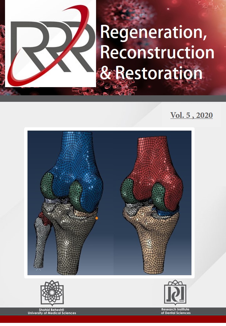
Evaluating the Partial Fibulectomy on Stress Distribution in Tibiofemoral Joint

Mohammad Tabatabaei

Regeneration, Reconstruction & Restoration (Triple R), Vol. 5 (2020), 24 March 2020, Page e9
https://doi.org/10.22037/.v5i1.26885

Introduction: Partial fibulectomy has been suggested for patients who encounter with severe varus/valgus or ununited fractures of the tibia. This study develops a finite element (FE) model of partial fibulectomy to study stress distribution in the tibiofemoral joint.

Materials and Methods: A 3D magnetic resonance imaging (MRI)-based of tibiofemoral joint and FE model was developed for study from a man volunteer with the normal left knee. Components consisted of the exact geometry of femur, tibia, fibula, meniscus, and articular cartilages. Firstly, geometries were constructed in Mimics and then exported to Rapid Forming XOR2 and finally, the Computer-aided design (CAD) model was analyzed in ABAQUS 6.10. Mechanical properties of the model for soft tissues were considered to be linear elastic, isotropic and homogenous and for bony parts were considered to be rigid.

Results: Model predictions were compared with normal one and used to derive stress distribution under physiological loading for standing in quasi- static condition. The results showed load transferring toward lateral condyle due to partial fibulectomy. The variation of stress distribution would increase the risk of osteoarthritis.

Conclusion: Our results have been predicted that partial fibulectomy could be an unknown risk factor for osteoarthritis and the model could be used for extended similar studies.

Comparison of Fixation Strength of Mandibular Angle Fracture with Different Lengths of Miniplate and Screw Using the Champy Technique in Sheep

Alireza Pournabi, Ramin Alijannia, Mansoureh Mohammadi

Regeneration, Reconstruction & Restoration (Triple R), Vol. 5 (2020), 24 March 2020, Page e12
https://doi.org/10.22037/rrr.v5i.31026

Introduction: This study sought to compare the fixation strength of mandibular angle fracture with 4- and 6-hole miniplates and different screw lengths using the Champy technique in sheep.

Materials and Methods: This is an in vitro study in 10 sheep mandibles. The soft tissue was removed and the mandibles were sectioned at the symphysis to obtain 20 hemimandibles. An artificial fracture was induced at the mandibular angle 1 cm farther from the most distal tooth using a saw. The hemimandibles were then randomly divided into two groups (n=10) for fixation with 4- or 6-hole miniplates without a bar. Each group was then randomly divided into 2 subgroups (n=5) for using 6 or 8-mm screws. The plates were adapted over the external oblique ridge. The distance from the closest hole to the fracture line was minimally 1 mm. Next, the hemimandibles were fixed to the holding arm of the universal testing machine by self-cure acrylic resin. Compressive load was applied to an occlusal jig prepared on the occlusal surface of mandibular molars at 3 cm distance from the fracture line at a crosshead speed of 1 mm/min until fracture. Data were analyzed using SPSS version 23.

Results: The mean fixation strength was not significantly different between 6-mm and 8-mm screws (P=0.243). The mean fixation strength in 6-hole miniplate group was significantly higher than that in 4-hole miniplate group (P=0.028).

Conclusion: The mean fixation strength in 6-hole miniplate group was significantly higher than that in 4-hole miniplate group. It seems that 6-hole miniplates provide higher fixation strength than 4-hole miniplates, irrespective of screw length. The mean fixation strength was not significantly different between 6-mm and 8-mm screws.

Reconstruction of Ramus-Condyle Unit with Two Rib Grafts: Report of a Case

Mehrdad Dehghanpou Barouj, Hassan Mirmohammad Sadeghi, Pedram Bakhshaei

Regeneration, Reconstruction & Restoration (Triple R), Vol. 5 (2020), 24 March 2020, Page e18
https://doi.org/10.22037/rrr.v5i.29930

Introduction: Ramus-condyle unit (RCU) reconstruction is one of most challenging procedures in maxillofacial surgery. Autologous graft, custom made prosthesis and transport disc distraction are some treatment options.

Case Report: In this study, we reported a mandibular DF in a 9-year-old child with condylar involvement. Our treatment plan was segmental resection and reconstructed RCU with 2 rib grafts.

Results: After one year of follow-up, there is no evidence of recurrence. Mandible has symmetrical appearance and normal centric and eccentric movement.

Conclusion: Rib graft is recommended for RCU reconstruction, especially in children in the growing stage.

Clinical Pearls in Treatment of Zygomaticomaxillary Complex Fractures

Mehrnoush Momeni Roochi, Mahboube Hasheinasab, Neda Afsar

Regeneration, Reconstruction & Restoration (Triple R), Vol. 5 (2020), 24 March 2020, Page e20
https://doi.org/10.22037/rrr.v5i.29335

Zygomaticomaxillary complex (ZMC) fractures are common facial injuries that can significantly change the structure, function and esthetics of the midface. Proper and timely management of these fractures can reduce the rate of late complications. In this paper, we review the current literature regarding ZMC fracture treatment and discuss our experience from surgical treatment of multiple ZMC fractures in the course of 10 years.

Surgery-First Technique for a Patient with Major β-Thalassemia

Milad Soleimani, Mohammad Behnaz

Regeneration, Reconstruction & Restoration (Triple R), Vol. 5 (2020), 24 March 2020, Page e22
https://doi.org/10.22037/rrr.v5i.30698

Introduction: Thalassemic patients commonly present malocclusions, which require both orthognathic treatments and surgury. This study aimed to assess prioritizing the surgery over orthodontic treatment.

Case report: Surgery-first method was performed to treat severe gummy smile and mild retrognathic mandible and prognathic maxilla in a 24-year-old Iranian girl with major β-thalassemia. Brackets and Ni-Ti wires were adjusted on all teeth to align their levels for one month. Then, the patient underwent the surgery, and the soft tissue was excised for Lefort dissection by two cutting lines, followed by osteotomy. Afterwards, down fracture and bilateral septoplasty were carried out, and excessive bones were removed.  The surgery retruded the maxilla by 10 mm.

Results: The 2-year follow-up revealed no overgrowth of the maxilla or malocclusion.

Conclusion: Surgery-first method can be successfully used for treatment of malocclusions in patients with major thalassemia.

Mechanical Behavior of Lumbar Spine Functional Units in Response to Cement Augmentation of Vertebra Body

Seyed Mohammad Tabatabaei, Mohammad Haghpanahi

Regeneration, Reconstruction & Restoration (Triple R), Vol. 5 (2020), 24 March 2020, Page e21
https://doi.org/10.22037/rrr.v5i.31495

Introduction: Cement augmentation in vertebrae is used to promote mechanical strength after spinal fracture and recently vertebroplasty gaining popularity as a treatment for patients. The numerical simulation could be helpful to enhance the quality of treatments such as vertebroplasty via exact modeling of the lumbar spine.

Materials and Methods: In this study, a three-dimensional finite element model created from CT images of L1-L3. According to clinical observation and recent studies, we consider that L2 augmented with two different volumes in 10 different distributions. Loadings were assumed to be pure momentum which applied in three anatomical directions (axial rotation, flexion, and lateral bending).

Results: Our results were validated with experimental data which shows segments range of motion, ligaments forces, and intradiscal pressure had good agreement with our results. Cement augmentation increases max Von Misses stress in L2 cancellous bone and Increment in Cement volume has the same result. Cement augmentation increases L1-L2 intradiscal pressure. Cement augmentation decreases segments range of motion. Finally, Cement augmentation increases total stiffness of model.

Conclusion: Taken together, vertebroplasty as a well-known method to treat the fractured vertebra, could be optimized to enhance patients' range of motion and decrease the complication of treatment.

Restoration


Floating Teeth Appearance: A Diagnostic Radiographic Alarm

Hamed Mortazavi, Yaser Safi, Hamid Reza Khalighi, Gelareh Forouzani, Mohammad Behnaz

Regeneration, Reconstruction & Restoration (Triple R), Vol. 5 (2020), 24 March 2020, Page e6
https://doi.org/10.22037/rrr.v5i1.29629

 Introduction: Floating teeth is a condition when teeth lose their supporting alveolar bone as a result of a destructive process, which is most frequently occur in the molar/ premolar region of both jaws. The aim of this study was to review the common conditions associated with floating teeth.

Materials and methods: We used general search engines and specialized databases including Google Scholar, Pub Med, Pub Med Central, Science Direct, and Scopus to find relevant studies by using keywords such as "jaw lesion", "jaw disease", "floating tooth", “floating teeth”, "tooth mobility", and “tooth loss”. About 100 articles were found, of which approximately 80 were broadly relevant to the topic, and finally 30 articles closely related to the above topic were chosen.

Results: When the data were compiled, the following 9 lesions were found to have more relationship with floating teeth appearance: Aggressive periodontitis, Langerhans histiocytosis, Burkitt’s lymphoma, multiple myeloma, Metastatic tumors, Primary intraosseous carcinoma (intraosseous squamous cell carcinoma), Ewing’s sarcoma, Hyperparathyroidism, and Cherubism.

Conclusion: When clinicians encounter a lesion associated with floating teeth, they should consider these entities especially malignant disorders in differential diagnosis to make more accurate diagnoses and develop better treatment plans according to patients’ imagings.

Which Mouthwashe is Appropriate for Eliminating Coronaviruses? A Mini Literature Review

Arash Farzan, Parsa Firoozi

Regeneration, Reconstruction & Restoration (Triple R), Vol. 5 (2020), 24 March 2020, Page e2
https://doi.org/10.22037/rrr.v5i1.29543

Aim and background: Coronaviruses can lead to severe respiratory disease and have a significant fatality rate. Dental professionals are high-risk groups because of too many exposures to patients in dental practice. Various mouthwashes have been used for different goals in dentistry. The aim of this study is to assess the appropriate mouthwash to eliminate coronaviruses for pre-procedural rinsing in dental practice.

Materials and methods: Electronic databases: PubMed, Scopus, and MEDLINE searched systematically. 5 different keyword combinations used based on MeSH (Medical Subject Headings) database. The search language and time period were English and 1990 to 2020, respectively.

Results: Finally, 4 studies included in this review. According to the studies substances that were tested against coronaviruses as a mouthwash were Povidone-iodine (PVP-I) and Chlorhexidine (CHX). 3 studies indicated that PVP-I is a promising substance to eliminate coronaviruses such as SARS-CoV, MERS-CoV and also, influenza virus A (H1N1) and rotavirus. Virucidal effect of CHX against coronaviruses was insufficient.

Conclusion: PVP-I gargle/mouthwash is the only approved mouthwash for pre-procedural rinsing in dental practice to eliminate coronaviruses according to the available literature.

Prosthetic Complications in a Patient with Papillon-Lefevre Syndrome Treated with Dental Implants: A Case Report

Michel Tanous, Marziyeh Alikhasi

Regeneration, Reconstruction & Restoration (Triple R), Vol. 5 (2020), 24 March 2020, Page e15
https://doi.org/10.22037/rrr.v5i.30530

Introduction: Patients with Papillon-Lefevre syndrome (PLS) lose their teeth because of periodontal disease followed by alveolar bone resorption. On the other hand, this complicate implant treatment, force the surgeon to insert implants more palatally, or to do more extensive surgical procedure.

Case report: A 24-year-old female diagnosed with PLS received an implant supported metal-acrylic prosthesis which was failed due to the dissatisfactory design and unpleasant influence on the patient function and esthetics. The prosthesis was substituted by a new designed one, fabricated by CAD/CAM technology to compensate the implants positions and fulfill patient function and esthetics.

Results: The patient followed up the day after delivery, one week, and each 6-month, without any reported prosthetic complications or bone loss after three-year follow-up appointment.

Conclusion: We presented the ability to restore esthetics and functions of a patient suffering from severe bone loss due to PLS by using the bone grafts and dental implants.

Surgical Endodontic Parameters in the Mandibular Premolars Area: An Anatomic Evaluation with Cone -Beam Computed Tomography

Pouya Jabbari Moghadam , Maryam Sanei

Regeneration, Reconstruction & Restoration (Triple R), Vol. 5 (2020), 24 March 2020, Page e16
https://doi.org/10.22037/rrr.v5i.31266

Introduction: This study aimed to evaluate the relationship between mandibular premolars and the surrounding critical anatomical structures for endodontic surgery using cone-beam computed tomography (CBCT).

Materials and Methods: In this cross sectional study, 170 CBCT images were evaluated in the sagittal, coronal, and axial planes to measure the buccal bone thickness (BBT) covering each root, the diameter of the premolars in the buccolingual and mesiodistal dimensions, besides the total amount of tissue needed to be resected for endodontic surgery; these measurements were performed at the 3mm level of the apex. Also, the distances from the superior border of mental foramen (MF) to premolars apex and alveolar crest (AC) were measured. Moreover, the position of MF in relation to adjacent teeth was evaluated. Descriptive statistics, independent-sample T-test and Wilcoxon test were used in the SPSS 26 to analysis of the data (α=.05).

Results: The mean values of BBT for the first and second premolars were measured 1.18 mm and 1.57 mm, respectively. The root diameters of the first and second premolars were measured 4.02 mm and 3.98 mm in the buccolingual dimension, and 2.74 mm and 2.98 mm in the mesiodistal dimension. The total amount of tissue needed to be resected for endodontic surgery in the second premolar has a higher amount with a mean of 5.55 mm. The second premolar was recorded as the tooth with the closest root apex to MF with a mean value of 3.97 mm. The mean distance between MF and AC was measured 13.88 mm, and MFs in most cases (44.11 %) were positioned in line with the second premolar long axis.

Conclusion: According to findings of the present study, the CBCT imaging evaluation is recommended for each patient before the surgical endodontic operation.

Short Dental Implants: An Umbrella Review of Current Systematic Reviews and Meta-analysis on Survival Rate of Short Dental Implants

Shaqayeq Ramezanzade, Frank Tuminelli, Duarte Marques, Seied Omid Keyhan, Parisa Yousefi, Jose Lopez-Lopez

Regeneration, Reconstruction & Restoration (Triple R), Vol. 5 (2020), 24 March 2020, Page e23
https://doi.org/10.22037/rrr.v5i.32850

Introduction: This study aimed to report on systematic reviews assessing the survival/failure rate of short implants. Additionally, marginal bone loss (MBL) and factors affecting survival rates was assessed.

Materials and Methods: PubMed-Medline, Google Scholar, Latin American and Caribbean Health Sciences Literature (LILACS) and the Cochrane Database of Systematic Reviews were searched up to January 2020.  Also reference lists of included systematic reviews were hand-searched. The AMSTAR tool was used for quality assessment.

Results: 659 studies were reviewed and 37 studies (25 metanalysis) were included. According to the AMSTAR tool, 19 of the 37 reviews were classified as high, 13 as medium and 5 as low quality. The mean score (±SD) was 7.10 (±2.74). The survival rate reported in studies varied from 84% to 100% for follow up periods of 1-5 years (more than 80% of included studies had a minimum follow-up period of 1-5 years), while failure rates ranged from 0% to 37.5% (the same as survival rate range from 62.5% to 100%). Included Systematic reviews reported no significant difference between survival rate/failure of Short implants (SIs) and standard implants with bone augmentation. Two high-quality metanalysis revealed a statistically significant lower survival rate for SIs as well as 2 systematic reviews which found SIs had significantly increased failures.  Surface characteristics and implant location showed some association with survival rate. In some studies, SIs experienced lower MBL than standard implants while in others no differences were observed.

Conclusion: Current knowledge from systematic reviews demonstrated acceptable survival rates for SIs. No significant difference between survival rate/failure of SIs and standard implants with bone augmentation was reported in the majority of studies. Systematic review registration number: PROSPERO Number: CRD42020163931.