Regeneration


Evaluation of Periodontal Status in Diabetes Mellitus Type 2 Patients Based on HbA1c and CRP

Maryam Zohary, Anahita Ashouri Moghaddam, Heydar Ali Balou, Melika Moghaddam

Regeneration, Reconstruction & Restoration (Triple R), Vol. 6 (2021), 13 March 2021, Page e1
https://doi.org/10.22037/rrr.v6i1.32654

Introduction: During the last decades, there has been an increasing interest in the relationship between diabetes mellitus (DM) and periodontitis. Some evidence has suggested that inflammatory factors like C-reactive protein (CRP) can be contributing factors to both periodontitis and diabetes. This study was aimed at assessing periodontal position in diabetes mellitus type 2 patients based on HbA1c and CRP.

Materials and Methods: 76 patients with diabetes mellitus2 (DM2) were divided based on glycemic control: 35 subjects with HbA1c less than 7% and 41 subjects with HbA1c≥7%. The following measurements were conducted: Serum HbA1c and C-reactive protein (CRP), gingival Index (GI), plaque Index (PI), clinical attachment loss (CAL), bleeding on probing (BOP), and probing depth (PD). Moreover, age, gender and duration of diabetes of the patients were also analyzed.

Results: In this study, 24 women and 11 men by mean age of 55/31±8/37 were in a good diabetic patients’ group (HbA1c<7%) and 30 females and 11 males by the mean age of 53/76±9/91 were in poor control diabetic patients (HbA1c≥7%). A significant correlation between the elevation of CRP and increased level of HbA1c was observed (P<0/001). The patients` age was associated with the duration of diabetes (P=0/024) and women had significantly more duration of diabetes than men (P=0/012). Regarding PD, CAL, BOP and PI, there was no significant difference between the analyzed groups. Also, no significant relationship between CRP and periodontal parameters has been found.

Conclusion: CRP was found as a predictor of HbA1c in patients with poor glycemic control. This implies higher infection rates due to diabetes.
Harvesting Tissue from Teeth-related Structures as a Source of Stem Cells

Fazele Atarbashi-Moghadam

Regeneration, Reconstruction & Restoration (Triple R), Vol. 6 (2021), 13 March 2021, Page e2
https://doi.org/10.22037/rrr.v6i.33483

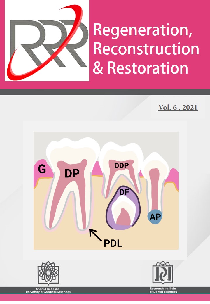
The tooth bud comprises of aggregation of cells demarcated as enamel organ, dental papilla, and dental follicle. They produce different parts of tooth and its supporting structures. More attention is paid to the teeth and its related structures as sources of mesenchymal stem cells (MSCs). Using dental sources has many advantages, such as easy access and less ethical consideration. MSCs from these tissues, besides In vitro achievements, showed effective results in clinical applications, especially in oral and maxillofacial regeneration.  The case selection and how to harvest the tissue from teeth structures are rarely described. In this concise review, the criteria of subject and teeth selection as a source of MSC isolation were assessed and how to harvest the considered tissue was also discussed.

Preparation and in vitro Evaluation of Injectable Alginate/Thiolated Chitosan Hydrogel Scaffold for Neural Tissue Engineering

Fatemeh Saadinam , Mahmoud Azami , Mir Sepehr Pedram , Javad Sadeghinezhad , Massoumeh Jabbari Fakhr, Mohammad Mehdi Dehghan

Regeneration, Reconstruction & Restoration (Triple R), Vol. 6 (2021), 13 March 2021, Page e4
https://doi.org/10.22037/rrr.v6i.33902

Introduction: Spinal cord injuries are one of the main causes of disability with devastating neurological consequences and secondary conflicts in other organs. Tissue engineering and regenerative medicine have been recognized as novel, promising methods in the treatment of tissue injuries, especially in neurological damage in recent decades. Hydrogels have the advantage of compatibility with damaged tissue, and injectable hydrogels can be applied in minimally invasive surgeries. This study aimed to evaluate an injectable hydrogel-based scaffold consisting of thiolated chitosan and alginate for neural tissue regeneration.

Materials and Methods: In the present study, an injectable hydrogel-based containing thiolated chitosan and alginate was prepared. Microbiology and pH tests were performed. Microstructural properties and porosity of scaffold were evaluated by scanning electron microscope (SEM). The swelling /shrinkage ratio and rates of biodegradation were also conducted. Finally, the viability of L929 cells on the scaffold was assessed using 3-(4,5-dimethylthiazol-2-yl)-2,5-diphenyltetrazolium bromide (MTT) assay.

Results: Thiolated chitosan/ alginate hydrogel had low pH with no contamination. SEM showed hydrogel had a porous microstructure with a mean pore diameter of 21.89 ± 0.32 μm which is suitable for cell culture. Furthermore, according to MTT test results, this hydrogel was biocompatible.

Conclusion: Thiolated chitosan/ alginate hydrogel is convenient for application in neural tissue engineering based on its structural properties and its ability to support cell proliferation. According to the in vitro analysis, it can also be used as a scaffold to create a suitable environment for increasing cell viability.

Effect of Long-time Mild Heat Stress on Proliferative, Differentiation and Bone Regeneration Capabilities of Dental Pulp Stem Cells

Hamidreza Moslemi, Zahrasadat Paknejad , Saeed Farzad Mohajeri, Arash Khojasteh

Regeneration, Reconstruction & Restoration (Triple R), Vol. 6 (2021), 13 March 2021, Page e13
https://doi.org/10.22037/rrr.v6i.40431

Introduction: The application of various strategies, including heat stress, has been attempted to maintain and improve mesenchymal stem cells (MSCs) plasticity and efficiency for bone regeneration. Although cell responses to heat stress are one of the most examined cellular stress responses, most studies, in this context, investigated the effect of that for a short period. Hence, the current study aimed to investigate the properties of MSCs, derived from dental pulp stem cells (DPSCs) following long-term heat stress.

Materials and Methods: DPSCs was loaded on β-tricalcium phosphate scaffold blocks and cultivated at elevated temperature up to 39 ° C. In vitro, DPSC properties, including proliferation, osteogenic differentiation, and bone regeneration in vivo, have been evaluated.

Results: The results of in vitro study showed that mild heat stress for a lengthy period could maintain and improve both the proliferative and differentiation potential of DPSCs during in vitro expansion and differentiation. Also, bone regeneration in vivo showed increased bone regeneration in the cells cultivated at a higher temperature.

Conclusion: This study provided evidence for the beneficial effects of the administration of mild heat stress for an extended period to maintain and improve DPSC properties, which may serve as a starting point for developing clinically compliant procedures for MSC treatment before transplantation.

Reconstruction


Maxillary Sinusitis due to Dental Infection: A Case Series Study

Fahimeh Akhlaghi, Masoud Kazemi, Amin Shams, Mohammad Esmaeelinejad

Regeneration, Reconstruction & Restoration (Triple R), Vol. 6 (2021), 13 March 2021, Page e3
https://doi.org/10.22037/rrr.v6i.33163

Introduction: Odontogenic infection is one of the most common causes of maxillary sinusitis which can lead to severe complications like cerebral abscess and orbital cellulitis. This study aims to evaluate the frequency of acute and chronic maxillary sinusitis by odontogenic source in 120 patients.

Materials and Methods: One hundred and twenty patients (240 sinuses) were examined by clinical examinations and their sinuses were assessed for the extent of fluid or mucosal thickening in computed tomography (CT) images and water's views. A dental examination has been performed to detect dental involvement and priapical radiography has been obtained to confirm the findings. Combination treatments were performed on five patients with refractory odontogenic sinusitis. Chi-square analysis has been used to identify the correlation between the prevalence of odontogenic sinusitis, age, and gender.

Results: Sinus involvement caused by dental origin has been found in 11.2% of the cases. Periapical involvement of maxillary first molar has been the most common cause of sinusitis with dental source (68%). The incidence of chronic apical periodontitis has been significantly higher than the other periapical infections (75% versus 25%). The signs and symptoms of sinusitis in all five cases were eliminated after the combination treatment after four months.

Conclusions: Odontogenic sources especially chronic apical periodontitis may lead to inflammation of the maxillary sinus membrane which usually is ignored because of its silent progression.

Morphometric Evaluation of Frontal Sinus and Investigating the Role of Gender in Anatomical Variations in Cone-beam Computed Tomography Images of an Iranian Population

Anita Mahaseni aghdam, Sandra Mehralizadeh, Ahmadreza Talaipour, Amirreza Mehdizadeh, Niloofar Ghadimi

Regeneration, Reconstruction & Restoration (Triple R), Vol. 6 (2021), 13 March 2021, Page e6
https://doi.org/10.22037/rrr.v6i.35535

Introduction: The frontal sinus has been of great interest to surgical and forensic specialists for human identification due to anatomical variations. The purpose of this study was to investigate the role of gender in the dimensions and anatomical variations of frontal sinus in cone-beam computed tomography (CBCT) images.

Materials and Methods: In this historical cohort study, CBCT images of 40 patients i.e, 20 males and 20 females, older than 18 years of age were reviewed. CBCT images with 1-mm-thick sections and NNT Viewer software for image analysis were used to measure frontal sinus height. The mean width, depth, thickness of the anterior cortex of the frontal bone, and the number of septa were measured in the axial sections, and the height was measured in the coronal sections of the CBCT images in both sexes. The data were analyzed by Student's t-test.

Results: The mean frontal sinus height was 20.5 mm in women and 25.9 mm in men; this difference was statistically significant (P<0.05). The mean frontal sinus width was 48.3 mm in women and 57.1 mm in men (P<0.002). The mean frontal sinus depth was 16.5 mm for women and 22.1 mm for men (P<0.05). The mean number of frontal sinus septa was 2.1 in females and 3.1 in males (P<0.05).

Conclusion: The results of this study showed the higher height, width, depth, and number of septa in males than in females, but the difference in the number of the frontal sinuses and the thickness of the anterior cortex of the frontal sinus was not sufficient to accurately determine gender.

Misdiagnosis of Adenoid Cystic Carcinoma of Palate: A Case Report

Hossein Daneste, Niloofar Ghadimi, Ladan Hafezi, Afsoon Jalali Ara, Mohsen Mardani, Amirreza Mehdizadeh

Regeneration, Reconstruction & Restoration (Triple R), Vol. 6 (2021), 13 March 2021, Page e11
https://doi.org/10.22037/rrr.v6i.37356

Introduction: Neoplasms of the salivary glands are uncommon, accounting for approximately 3 to 5 percent of all head and neck tumors. Adenoid cystic carcinoma (ACC) is a rare salivary gland malignancy with a poor survival rate because of a high rate of late local recurrence and distant metastasis.  

Materials and Methods: This paper reports a misdiagnosis of palatal ACC via fine needle aspiration cytology (FNAC). A 27-year-old female with painful swelling in the right side of the palate underwent FNAC and was misdiagnosed with pleomorphic adenoma. During surgery, hemi-maxillectomy was performed due to the fragile consistency of the tissues.

Result: The tumor was found to be ACC after histopathologic evaluations. Hence, the patient underwent radiotherapy and chemotherapy postoperatively.

Conclusion: Given specific limitations and diagnostic pitfalls, it seems that pre neural and pre vascular invasion of tumors as a sign of malignancy should be noticed.

Incidence of Maxillofacial Fractures and Their Care Proceedings in Patients Referred to Shariati Hospital in 2019-2020: A Prospective Study

Abbas Karimi, Alireza Nasir Pour, Hassan Mir Mohammad Sadeghi, Ramtin Dastgir, Meysam Mohammadi Khah

Regeneration, Reconstruction & Restoration (Triple R), Vol. 6 (2021), 13 March 2021, Page e12
https://doi.org/10.22037/rrr.v6i.38570

Introduction: Trauma to maxillofacial structures can cause growth disorders and deformities in craniofacial bones, permanently affecting patient’s quality of life. Moreover, a significant increase in incidence of maxillofacial fractures due to more widespread use of motor vehicles and interpersonal assaults cannot be overlooked. This study evaluated the incidence of maxillofacial fracture types and the employed management course in patients referred to Shariati Hospital, Tehran, Iran over 2019-2020.

Materials and Methods: In this cross sectional study, patients with maxillofacial traumas referred to this center were selected using census sampling method. Age, gender, etiology of trauma, location of fractures, drug and/or alcohol addiction, duration of referral until hospitalization, opt surgical treatments and recruitment were documented using clinical examinations and interview. Obtained results were reported by means of the descriptive statistics methods.

Result: Two hundred and twenty-three (223) patients (83.4% males and 16.6% females) were enrolled. The mean age of patients was 29.95 years. The etiology of fractures were motor vehicle accidents (85; 38.1%), motorcycle accidents (36; 16.1%); interpersonal assaults (33; 14.8%), falls (56; 25.1%), sport related injuries (6; 2.7%) and industrial incidents (5; 2.2%). In 181 patients (81.5%), there was no fracture in the other parts of the body while in 41 patients (18.4%) concurrent fractures in other sites were observed as well. The most anatomic site of the fracture was mandible. The mean time passed from the referral until hospitalization, surgery and recruitment day was 2.09, 6.89 and 10.56 days respectively.

Conclusion: In patients with maxillofacial fractures referred to Shariati Hospital during 2019-2020, the fractures predominantly occurred in males,in the mandible and mostly due to traffic accidents on motor vehicles, consistent with reported data obtained in other developing countries.

Restoration


How to Find Abutment Screw Channel of a Cement-retained Implant Supported Restoration? A Literature Reviews

Hesan Shafaei, Marzieh Alikhasi

Regeneration, Reconstruction & Restoration (Triple R), Vol. 6 (2021), 13 March 2021, Page e5
https://doi.org/10.22037/rrr.v6i.34097

Introduction: Screw loosening is the most common technical complication in cement-retained dental implants. To retrieve screw loosened dental abutment, making abutment screw channel (ASC) is a useful method to access the abutment screw. This article reviews the methods for preparing ASC of cement-retained implant supported restoration.

Materials and Methods: The search was conducted on PubMed, Google Scholar, and Scopus. The keywords were screw loosening, cement-retained implant restoration, screw access hole, abutment screw channel and retrieving.

Results: Having applied the inclusion and exclusion criterion, 24 articles, 21 technical notes, 1 case report, and 2 reviews were found.

Conclusion: The techniques reviewed were categorized into 3 groups of marking techniques, customized guides, and imaging techniques with different outcomes.

Evaluation of Temporomandibular Disorders after Orthognathic Surgeries: A Literature Review

Farnoosh Razmara, Akram Ghanadpour , Zainab Abdulkareem Aleedawi, Mina Khayamzadeh

Regeneration, Reconstruction & Restoration (Triple R), Vol. 6 (2021), 13 March 2021, Page e7
https://doi.org/10.22037/rrr.v6i.35100

Introduction: Maxillofacial orthognathic surgery is performed to repair or correct the skeletal anomalies of the jaw and its associated dental and facial structures. There is a conflict on whether orthognathic surgery has a negative or positive effect on temporomandibular disorders (TMD). The aim of this study is to review disorders of temporomandibular joint (TMJ) after orthognathic surgery.

Materials and Methods: Data for this review was obtained from the articles published between 2010-2020 via PubMed, Google Scholar, Web of Sciences, and Scopus. The content keywords matched those used in PubMed and Mesh engines.

Results: Based on the inclusion and exclusion criteria, 27 articles were included. Most of the selected articles were retrospective reviews and performed on class II and class III patients. Ages ranged from 19- 47 years. Pain reduction was reported in 11 studies, while 8 studies reported a click reduction post orthognathic operation. In two studies, decreased joint noises were reported after theorthognathic operation, and seven articles reported a decrease in maximum mouth opening. Three studies reported a bilateral sagittal split osteotomy (BSSO) and in one study, reduced and improved symptoms were reported after Le Fort I + (BSSO) surgery.  One study exhibited that in retrognathic patients BSSO orthognathic surgery is less predictable in reducing TMD symptoms. Three articles showed that orthognathic patients with TMJ click have a high predictive value.

Conclusion: To accomplish accurate results regarding TMD post orthognathic surgery, a larger number of subjects, as well as extended long term follow-up are required.

Biomechanical Stability of Two Different Maxillofacial Screws in a Rabbit Model

Farzad Nouri, Mohammadjavad Einafshar, Ata Hashemi, Fahimeh Akhlaghi

Regeneration, Reconstruction & Restoration (Triple R), Vol. 6 (2021), 13 March 2021, Page e8
https://doi.org/10.22037/rrr.v6i.37069

Introduction: Internal rigid fixation techniques are commonly used to treat maxillofacial fractures by stabilizing bone segments using titanium plates and screws. The current study aimed to compare the biomechanical stability of two maxillofacial screws with different tip designs in vivo.

Materials and Methods: Six male rabbits were randomly divided into two experimental periods of 0 and 4 weeks. Under general anesthesia, the screws were randomly placed in the tibia bone on both sides of each animal. The pullout test was conducted using a Sentam test device. Data were statistically analyzed using ANOVA.

Results: The average insertion torque for non-self-tapping and self-tapping screws amounted to 4.2±1.7 and 4.8±1.4 Newton/centimeter, respectively (P-value >0.05). The calculated measures for the pullout test demonstrated a significant increase of secondary stability after 4 weeks in comparison to 0 week (P-value<0.001). However, the results among the two screws showed no statistically significant difference in each time point (P-value >0.05).

Conclusion: No significant differences were demonstrated among self-tapping or non-self-tapping maxillofacial screws.

Introduction: The formation of localized periodontal pockets on the distal surface of adjacent molars is known as one of the asymptomatic lesions associated with impacted wisdom teeth. In this study, the effect of different factors on the prevention of periodontal pocket formation on the distal surface of the second molar following impacted wisdom tooth removal surgery was investigated.

Materials and Methods: The present study was a review of the related literature published over the last 10 years. The main tool recruited for this purpose was a researcher-made checklist, developed based on the main objectives of the study. Accordingly, wisdom tooth occlusion, suture technique, flap type, and periodontal envelope, extracted from the articles, were recorded in the relevant checklist. The extracted information was finally imported into the SPSS (ver. 20) software package and analyzed, using descriptive and analytical statistics.

Results: In total, 22 articles, reflecting on the effect of different factors on the prevention of periodontal pocket formation on the distal surface of the second molar following impacted wisdom tooth removal surgery, and meeting the inclusion criteria in this study were retrieved and then reviewed. Based on the preliminary analyses, 15 articles (68.18%) had mentioned periodontal pocket formation after impacted tooth surgery. As well, nine studies had referred to attachment loss in patients. In this review using surveys among patients undergoing surgery, had not demonstrated a significant relationship between periodontal pocket formation and attachment loss. Moreover, eight articles (36.36%) examined flap type employed during the surgery.

Conclusion: It was concluded that impacted wisdom tooth removal surgery had been thus far introduced directly in many studies as a factor affecting the reduction of periodontal complications, including periodontal pocket formation in adjacent teeth.