Green Synthesis of Silver Nanoparticles Mediated by Plant Extracts: Food Science and Technology, Synthesis Pathways, and Applications in Nanobiotechnology
食品生物技术的应用,
卷 13 编号 1 (2026),
7 十二月 2025
,
第 1-25 (e28) 页
https://doi.org/10.22037/afb.v13i1.53432
摘要
Background and Aim: The green synthesis of silver nanoparticles (AgNPs) using plant extracts has emerged as a sustainable, economical, and eco-friendly alternative to conventional chemical and physical approaches. This narrative review links the phytochemical composition of plant extracts (polyphenols, flavonoids, tannins, terpenoids, alkaloids, proteins, and reducing sugars) to the physicochemical properties of the resulting AgNPs; compares plant parts, extraction conditions, and reaction parameters; and critically evaluates food-related applications, particularly antimicrobial packaging, together with reproducibility, safety, toxicity, migration, and regulatory considerations.
Material and Methods: The literature was searched in Web of Science, Scopus, and Google Scholar for the period from 2002 to 2026, using combinations of the following keywords: “green synthesis,” “silver nanoparticles,” “plant extract,” “phytochemicals,” “food packaging,” “migration,” and “toxicity
Results and Conclusion: Characterization of plant extracts (e.g., HPLC, LC–MS, GC–MS, NMR, total phenolic and flavonoid assays) and characterization of AgNPs (UV–Vis, XRD, FTIR, TEM, SEM, DLS/zeta potential, and XPS where applicable) answer different questions and should be reported separately; a color change alone is not sufficient evidence of nanoparticle formation. Green-synthesized AgNPs show antimicrobial, antioxidant, anticancer, wound-healing, biosensing, environmental, and food-packaging potential, but most of the evidence is in vitro. Lack of standardization, batch-to-batch variability of extracts, limited control over particle size and shape, and scarce data on long-term toxicity and migration remain the main barriers to industrial and food-contact use. Standardized protocols, chemically characterized extracts, and comprehensive safety evaluation are required before large-scale application.
- INTRODUCTION
Nanotechnology is a broad interdisciplinary field of research, development, and industrial activities that has been rapidly growing worldwide over the past decade [1]. Metallic nanoparticles, especially silver nanoparticles (AgNPs), due to their very high surface-to-volume ratio and unique physical, chemical, and biological properties—including high specific surface area, excellent optical characteristics, and strong antimicrobial activity—have become one of the most widely used materials in various fields, including drug delivery, cancer therapy, tissue engineering, biosensors, wound healing, and environmental technologies [2,3]. Reducing the size to the nanoscale increases the active surface area. It enhances reactivity, such that these nanoparticles are able to exhibit significant antimicrobial and optical effects even at low concentrations [4].
The antibacterial and antiviral activity of AgNPs is mainly attributed to the disruption of the cell membrane and interference with metabolic processes [5,6]. The high chemical stability of these nanoparticles allows their use in biological, aqueous, and polymeric environments [7]. Their optical properties arise from surface plasmon resonance (SPR), which provides wide applications in optical sensors and bioimaging [8].
However, conventional methods for producing AgNPs, which rely on chemical and physical approaches, have serious limitations, including high energy consumption, the use of hazardous reducing and stabilizing agents, the generation of toxic waste, and the potential for biological toxicity, especially in medical applications [9].
Meanwhile, the green synthesis of silver nanoparticles has emerged as a sustainable and environmentally friendly approach [10]. The use of biological resources, especially plant extracts, rich in phytochemicals such as polyphenols, flavonoids, terpenes, alkaloids, and tannins, provides the possibility of reducing silver ions, natural stabilization, and controlling the morphology of nanoparticles [11,12]. Plant extracts may act both as reducing agents and as stabilizing agents in nanoparticle synthesis [13]. These compounds may not only help prevent aggregation and oxidation of the particles, but also influence the size, shape, and biological properties of the nanoparticles [14,15]. This is because different extracts contain varying concentrations and compositions of organic reducing agents [16].
This review differs from other articles by focusing on the following specific aspects: linking phytochemical classes to the physicochemical properties of silver nanoparticles (AgNPs), including size, shape, and stability; comparing plant parts, extraction conditions, and reaction parameters; critically evaluating food-related applications, particularly antimicrobial packaging and nanoparticle migration; discussing reproducibility and standardization; and integrating considerations of safety, toxicity, regulations, and food contact. Accordingly, the review is organized around three main axes: (i) evaluation of some compounds in plant extracts and their role in the reduction process, stabilization, and morphology control of AgNPs; (ii) a detailed review of synthesis pathways, influential parameters, and methodological differences in producing silver nanoparticles using plant extracts, including their relevance to food nanotechnology; and (iii) an examination of biotechnological, biomedical, and food-related applications, together with an analysis of safety issues, existing challenges ,and limitations. Finally, the review summarizes the current state of research on the use of nanotechnology in food systems and identifies the evidence gaps that must be closed before food-contact application.
- METHODOLOGY: LITERATURE SEARCH STRATEGY
This article is a narrative (non-systematic) review and is not presented as a systematic review. The literature was searched in Web of Science, Scopus, and Google Scholar for the period from 2002 to 2026, using combinations of the following keywords: “green synthesis,” “silver nanoparticles,” “plant extract,” “phytochemicals,” “food packaging,” “migration,” and “toxicity.”
Inclusion criteria: (1) Peer-reviewed English-language research and review articles; (2) studies reporting the synthesis, characterization, or applications of plant-mediated AgNPs; and (3) studies providing characterization data, such as UV–Vis spectroscopy, TEM/SEM, XRD, DLS, and FTIR.
Exclusion criteria: (1) Articles lacking characterization data; (2) conference abstracts; (3) studies based on non-plant biological sources; and (4) non-English articles.
After removing duplicate records, titles and abstracts were screened, followed by a full-text assessment of the eligible articles.
- MECHANISTIC PRINCIPLES OF THE GREEN SYNTHESIS OF AGNPS
The green synthesis of silver nanoparticles using plant extracts is generally described as involving three mechanistic stages: reduction, nucleation, and growth and stabilization (Fig. 1) [9].
Fig. 1. Schematic illustration of the green synthesis process of silver nanoparticles (AgNPs) using plant extract, including the reduction, nucleation, and final growth and stabilization stages.
In the reduction stage, specific bioactive compounds present in plant extracts reduce Ag⁺ ions to metallic silver (Ag⁰). The rate of this process depends on the type and concentration of reducing compounds, temperature, and the pH of the medium [4,15].
The nucleation stage involves the aggregation of silver atoms to form initial nuclei. The size and number of these nuclei influence the final nanoparticle size [1].
In the growth and stabilization stage, the nuclei grow and are coated by extract constituents that limit agglomeration and support colloidal stability. For example, polyphenols are often proposed to control the growth rate, At the same time, proteins and other biomolecules are thought to form protective layers that enhance the stability and biological properties of the nanoparticles [7].
A precise understanding of this mechanism enables prediction of nanoparticle behavior and optimization of synthesis to achieve particles with desired size and morphology, which is essential for biotechnological applications and materials engineering [9,10].
- NANOBIOTECHNOLOGY AND PLANT EXTRACTS IN SILVER NANOPARTICLE SYNTHESIS
4.1. The Role of Phytochemicals in Silver Nanoparticle Synthesis
Plant extracts contain a diverse set of secondary metabolites such as flavonoids, polyphenols, tannins, terpenes, alkaloids, proteins, amino acids, reducing sugars, and vitamins, each of which is reported to contribute to the reduction, nucleation, growth, and stabilization stages of AgNP formation [1,10].
Therefore, precisely identifying of the major phytochemical compounds and their functions is essential for designing controlled synthesis and producing AgNPs with optimal quality and performance [4,13].
4.2. Main Phytochemical Groups and Their Roles in Reduction and Stabilization
4.2.1. Polyphenols and flavonoids
Polyphenols and flavonoids are considered the most important reducing compounds in plant extracts; with their multiple hydroxyl groups, they have a high capacity for electron transfer. This property allows the reduction of Ag⁺ ions to metallic silver (Ag⁰) through the formation of temporary complexes [3,11]. In addition to their reducing role, these compounds are thought to act as stabilizing agents by adsorbing onto the surface of nanoparticles and limiting aggregation, and this surface coating can increase colloidal stability and yield nanoparticles of small size and uniform morphology [2,4].
4.2.2. Tannins
Tannins, high-molecular-weight phenolic compounds, are reported to play a dual role in the synthesis of silver nanoparticles; their multiple hydroxyl groups facilitate the reduction of Ag⁺ ions while simultaneously forming a protective coating on the surface of nanoparticles [15]. This feature ensures the production of AgNPs with long-term stability, small size, and uniform dispersion, and tannins, especially in extracts with high phenolic content, play a key role in controlling nanoparticle morphology [2].
4.2.3. Alkaloids
Alkaloids, which often possess active nitrogen-containing structures, mainly interact with Ag⁺ ions as stabilizing ligands. Although a slight reducing role has been observed in certain plant species [12,13], their main function is thought to be surface stabilization of nanoparticles; this characteristic may enable the formation of particles with specific morphology and long-term stability [10].
4.2.4. Terpenes and terpenoids
Terpenes and terpenoids, major components of essential oils and plant extracts, are reported to act as biological reducing agents and to convert Ag⁺ ions into metallic silver (Ag⁰) through electron transfer [10]. The presence of these compounds in aromatic plants is often associated with the formation of non-spherical morphologies such as triangular or polyhedral nanoparticles, and in addition to their reducing role, they may contribute to stabilization by forming a surface coating [12].
4.2.5. Proteins and amino acids
Proteins and amino acids present in plant extracts form complexes with Ag⁺ ions and, after reduction to Ag⁰, are believed to act as natural coatings on the surface of nanoparticles [7, 16]. The –NH₂/–NH₃⁺ and –COOH groups in amino acids can enhance colloidal stability and reduce particle aggregation, which may provide long-term stability and improve functional properties such as antimicrobial activity and drug-delivery capability of the nanoparticles [5,13].
4.2.6. Reducing sugars and vitamins
Reducing sugars such as glucose and vitamin C (ascorbic acid) can reduce Ag⁺ ions to metallic silver (Ag⁰) without producing toxic compounds [15,16]. Although their reducing power is generally lower than that of polyphenols and tannins, they may play a complementary role in some extracts and contribute to initial nanoparticle stabilization; their contribution is probably most relevant in extracts with low phenolic content [2].
Table. 1. Summary of plants used in the green synthesis of silver nanoparticles, including scientific names, utilized plant parts, probable active compounds, and reported nanoparticle sizes based on various references, which have been investigated by several researchers.
4.3. Analytical Techniques: Plant-Extract Characterization versus AgNP Characterization
Analytical techniques used in plant-mediated AgNP synthesis provide complementary information and should be selected according to the material being investigated. Importantly, characterization of the plant extract and characterization of the synthesized nanoparticles represent two distinct analytical levels and should therefore be reported and interpreted separately. Extract-level analysis identifies the phytochemical constituents potentially involved in silver-ion reduction and nanoparticle stabilization, whereas nanoparticle-level characterization determines the physicochemical properties of the resulting AgNPs [43,44].
4.3.1. Characterization of plant extracts
HPLC and HPLC–MS/LC–MS: These techniques are widely used for the separation, identification, and quantification of non-volatile phytochemicals, particularly phenolic compounds, flavonoids, and other secondary metabolites. HPLC provides chromatographic profiles and quantitative information, while coupling with mass spectrometry enables more reliable identification based on molecular mass and fragmentation patterns [10]. This information can help establish relationships between extract composition and AgNP formation [45].
GC–MS: Gas chromatography–mass spectrometry is particularly suitable for volatile and thermally stable compounds, including terpenes, terpenoids, and other low-molecular-weight constituents [1]. It can provide a detailed chemical profile of volatile components that may contribute to the reducing or stabilizing capacity of the extract [46].
NMR: Nuclear magnetic resonance spectroscopy provides structural information about major metabolites and can be used to characterize relatively abundant compounds in plant extracts. It may also provide information about molecular interactions between phytochemicals and nanoparticle surfaces when appropriate experimental approaches are applied. NMR is therefore valuable for investigating the molecular mechanisms underlying reduction and surface functionalization [47].
Total Phenolic and Total Flavonoid Assays: Colorimetric methods such as the Folin–Ciocalteu assay provide rapid estimates of the total phenolic content, while aluminum chloride-based assays are commonly used for total flavonoid estimation [39]. These measurements can serve as useful indicators of the overall reducing potential of an extract; however, they are non-specific and should not be considered substitutes for compound-specific chromatographic analysis [48,49].
TLC: Thin-layer chromatography is a rapid and relatively inexpensive screening technique for separating major classes of plant metabolites. Although its quantitative and structural resolution is limited compared with HPLC or LC–MS, TLC can be useful for preliminary comparison of extracts and monitoring changes in phytochemical profiles [50].
4.3.2. Characterization of AgNPs
UV–Vis Spectroscopy: UV–Vis spectroscopy is commonly used as an initial method for monitoring AgNP formation through the characteristic surface plasmon resonance (SPR) band. For many approximately spherical AgNP dispersions, the SPR maximum occurs within the approximate range of 400–460 nm. However, the position and width of the band depend on particle size, shape, aggregation, and the surrounding medium. Therefore, UV–Vis provides evidence of nanoparticle formation and information about optical behavior but cannot independently confirm crystallinity, exact particle size, morphology, or purity [51].
X-Ray Diffraction (XRD): XRD is used to determine the crystalline structure of AgNPs and identify the characteristic reflections of metallic silver with a face-centered cubic (FCC) structure [44,47]. The diffraction pattern can also provide information about crystallinity and, when appropriate models are applied, an estimate of crystallite size. However, crystallite size obtained from XRD should not be directly equated with the physical particle diameter measured by electron microscopy [51,52].
Fourier-Transform Infrared Spectroscopy (FTIR):
FTIR identifies functional groups associated with organic molecules present on the nanoparticle surface and can provide evidence for their involvement in reduction or capping processes [4]. Comparison of the spectra of the original extract and purified AgNPs can help identify changes in functional groups associated with nanoparticle formation. FTIR, however, generally provides functional-group-level information rather than definitive identification of individual phytochemicals [53].
TEM and SEM–EDX: Transmission electron microscopy (TEM) provides high-resolution information on nanoparticle size, shape, morphology, and aggregation state, making it particularly useful for determining the primary particle size distribution [9,47]. Scanning electron microscopy (SEM) provides complementary information on surface morphology and particle distribution. When coupled with energy-dispersive X-ray spectroscopy (EDX), these techniques can also confirm the presence and elemental composition of silver. Sample preparation and aggregation should be considered when interpreting microscopy-based size measurements [54].
DLS and Zeta Potential: Dynamic light scattering (DLS) determines the hydrodynamic diameter of nanoparticles dispersed in a liquid and is useful for evaluating particle aggregation and colloidal behavior [6]. The hydrodynamic diameter generally exceeds the particle diameter measured by TEM because it includes the associated solvent layer and surface-associated molecules. Zeta-potential measurements provide an indicate of surface charge and electrostatic colloidal stability, although stability may also depend on steric and other interparticle interactions [55].
X-Ray Photoelectron Spectroscopy (XPS): XPS provides surface-sensitive information on elemental composition and chemical states. In AgNP systems, it can help distinguish metallic Ag⁰ from oxidized silver species such as Ag⁺ or Ag₂O and characterize the chemical environment of surface-associated elements. This technique is particularly useful for investigating surface chemistry and oxidation processes [56].
Thermogravimetric Analysis (TGA): TGA measures changes in sample mass as a function of temperature and can be used to estimate the organic fraction associated with the nanoparticle surface [47]. In plant-mediated AgNPs, this information can provide an approximate indication of the amount of phytochemical or other organic capping material present. TGA results should be interpreted carefully because different organic components may decompose over overlapping temperature ranges [57].
A visible color change in the reaction mixture, typically from pale yellow to brown, can serve as an initial indicate of AgNP formation due to changes in optical properties associated with surface plasmon resonance. However, color change alone is insufficient to confirm nanoparticle formation or determine their physicochemical properties. At minimum, complementary evidence from techniques such as UV–Vis, XRD, and electron microscopy should be considered. Similar multi-technique characterization strategies have also been applied to plant-mediated gold nanoparticles [36]. Overall, integrating extract-level phytochemical analysis with comprehensive nanoparticle characterization provides a stronger basis for understanding the mechanisms of plant-mediated synthesis, correlating phytochemical composition with nanoparticle properties, and developing more reproducible and controlled AgNP production processes[51,52].
4.4. Influence of Plant Diversity on the Properties of Silver Nanoparticles
The diversity of plants causes the nanoparticles synthesized from each extract to exhibit different characteristics. Extracts rich in flavonoids have frequently been associated with smaller AgNPs of uniform dispersion, as these compounds may enhance nucleation rates and control particle growth [1,10].
Soluble proteins in the extracts may increase the colloidal stability of nanoparticles and reduce agglomeration, while aromatic plants rich in terpenes and terpenoids have been reported to yield nanoparticles with diverse morphologies such as polyhedral or triangular particles [12, 10]. This biological diversity offers the potential to engineer AgNP properties [2].
- GREEN SYNTHESIS PATHWAYS OF SILVER NANOPARTICLES AND INFLUENTIAL PARAMETERS
Fig.2. Schematic overview of the plant-mediated green synthesis of silver nanoparticles (AgNPs), covering the topics discussed in Section 4: (1) plant source and its characteristic phytochemicals (leaves, roots/rhizomes, bark/stem, flowers, fruits, and seeds); (2) extraction conditions (solvent, temperature, time, particle size, pH, and solid-to-liquid ratio) and their effect on the composition of the plant extract, including reducing and stabilizing/capping agents; and (3) the synthesis stages (reduction, nucleation, growth, and stabilization), reaction parameters (pH, temperature, [AgNO₃], extract amount, reaction time, and light intensity), assisted synthesis methods (ultrasound, microwave, photo-mediated, and hybrid), and separation and purification.
5.1. Classification of Synthesis Methods Based on Plant Source Type
The choice of the plant part used as the extract source plays a fundamental role in determining phytochemical content and reducing capability of the extract [1,10], and consequently influences the final characteristics of the synthesized AgNPs, including particle size, morphology, stability, and uniformity [4,13].
5.1.1. Leaves
Leaves, due to their high concentration of polyphenols, flavonoids, and vitamin C, are often capable of producing very small and highly stable AgNPs [4,6]. Additionally, leaves are the most common source in green nanoparticle synthesis because of their easy availability, simple extraction, and rich bioactive content. Even slight variations in leaf type or extraction conditions can significantly affect nanoparticle size, uniformity, and functional activity [1, 10].
5.1.2. Roots and rhizomes
Roots and rhizomes, which often contain high levels of alkaloids, saponins, and polysaccharides, usually produce of nanoparticles with diverse morphologies [12,13]. The reducing activity of compounds in these plant parts increases under low pH conditions, enabling more precise control over particle size, shape, and distribution [13].
5.1.3. Bark and stem extracts
Extracts derived from bark and stems, typically rich in tannins and other phenolic compounds, have been reported to yield highly stable nanoparticles [2,15]. However, the particle size is often slightly larger than that of AgNPs synthesized using leaves.
5.1.4. Flowers
Flowers, due to their content of pigment flavonoids, may show strong reducing ability and can produce nanoparticles with intense optical absorption and diverse colors. This feature makes flowers attractive for synthesizing AgNPs for optical sensors, bioimaging, and optoelectronic technologies [8].
5.1.5. Fruits
Fruits often accelerate the reduction rate due to their organic acids and reducing sugars, resulting in rapid nanoparticle formation. However, the stability of the produced nanoparticles is sometimes lower, and maintaining uniform size distribution requires precise control of reaction parameters and extraction conditions [15,18,20].
5.1.6. Seeds
Seeds, typically rich in oils and soluble proteins, are thought to play an important role in stabilizing nanoparticle surfaces [5,20], and by forming a natural coating on AgNPs, they enhance colloidal stability and particle uniformity. Using these plant sources improves the physicochemical properties of nanoparticles and enhances their antibacterial activity and drug delivery capability [5,19].
5.2. Main Stages of Plant-Mediated Synthesis of Silver Nanoparticles
Green synthesis of silver nanoparticles primarily involves three stages: reduction, nucleation, and growth and stabilization. Each step influences the final characteristics of AgNPs.
5.2.1. Preparation of plant extract
In the first stage researchers typically prepare the plant extract using common methods such as boiling, soaking, or extraction in aqueous, alcoholic, or aqueous–alcoholic solvents. The choice of solvent, temperature, and extraction duration directly affects the type and concentration of phytochemical compounds present in the extract, which in turn influence the reduction reaction rate and the stability of the produced nanoparticles [36-38].
5.2.2. Reduction reaction
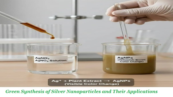
Fig.3. Green synthesis of silver nanoparticles (AgNPs): adding a plant extract (reducing agent) to a silver nitrate (AgNO₃) solution and observing the yellow-brown color change as a preliminary indication of AgNP formation.
After preparing the extract, adding it to a silver nitrate (AgNO₃) solution initiates the reduction reaction. The color change of the solution from pale yellow to dark brown is a visual but only preliminary indicator of the formation of metallic silver nanoparticles (Ag⁰) (Fig3.); The reaction time, which depends on the concentration of reducing compounds, temperature, pH, and light intensity, may vary from a few seconds (microwave-assisted) to several hours and affects the size, morphology, and distribution of nanoparticles [1,6,9].
5.2.3. Separation and purification
After the reaction is completed, the nanoparticles are separated from the solution using centrifugation and washed with water or alcohol to remove remaining compounds. Subsequently, drying at low temperatures or freeze-drying can help preserve the structure and stability of the nanoparticles and prepare them for applications in biological, pharmaceutical, and industrial fields [7,13,39].
5.3. Parameters Affecting the Synthesis Process
The physical and chemical parameters of the reaction environment, including pH, temperature, silver nitrate concentration, extract amount, reaction time, and light intensity, play a decisive role in the size, morphology, stability, and uniformity of nanoparticles. The trends below are those most commonly reported; their direction and magnitude depend on the extract and should be optimized experimentally [38-40].
5.3.1. Reaction pH
The pH of the reaction solution is one of the most sensitive controlling factors. Under alkaline conditions, the hydroxyl groups in phenolic compounds become more ionized, increasing their reactivity, which generally leads to the production of smaller and more stable AgNPs. In contrast, acidic pH often slows nanoparticle growth, increases particle size, and raises the likelihood of aggregation and agglomeration. pH changes can also shift the SPR peak position and the color of the solution in the UV–Vis spectrum [6,8,11].
5.3.2. Temperature
Increasing the reaction temperature enhances the reduction rate and nucleation, often resulting in smaller and more uniform nanoparticles. However, excessively high temperatures may alter the particle structure and increase aggregation; therefore, determining the appropriate temperature range for each plant extract is essential [15,10].
5.3.3. Silver nitrate concentration
The concentration of Ag⁺ ions determines the number of initial nuclei and consequently the size and quantity of the final nanoparticles. A high concentration of AgNO₃ may lead to excessive growth and aggregation of particles, whereas a low concentration tends to give smaller but fewer particles. The optimal ratio of extract to silver nitrate solution should be determined experimentally based on the plant type [38-40].
5.3.4. Concentration and amount of plant extract
Increasing the amount of extract raises the concentration of reducing compounds, leading to enhanced reaction rate and formation of more nuclei. However, excessively high concentrations may form a thick coating on the nanoparticle surface and increase their size; thus, precise optimization of extract amount is necessary for each system [12,15].
5.3.5. Reaction time
The reaction duration determines the growth and development of nanoparticles. Longer reaction times can cause particle growth, SPR band broadening, and increased risk of aggregation, while insufficient time results in incomplete nanoparticle formation and reduced stability [8,1].
5.3.6. Light intensity and irradiation
In some plant extracts, exposure to visible or UV light accelerates the reduction reaction, probably because active plant compounds gain more energy to transfer electrons to Ag⁺ ions. This approach, known as photo-assisted green synthesis, enables rapid production of AgNPs with more controlled morphology [8,9].
5.4. Improved Green Synthesis Methods
Advanced green synthesis approaches, utilizing technologies such as ultrasound, microwave irradiation, and light exposure, enable better control over nanoparticle size and morphology and can improve the uniformity and quality of the final product.
5.4.1. Ultrasonic-assisted synthesis
Ultrasonic waves, by generating cavitation, increase the interaction between phytochemical compounds and Ag⁺ ions, enhancing the reduction rate and the formation of initial nuclei. This process can result in nanoparticles with more uniform size distribution and improved physicochemical stability [4,13].
5.4.2. Microwave-assisted synthesis
The microwave method, due to uniform heat distribution, high reduction rate, and efficient energy utilization, enables the production of smaller and more uniform AgNPs while significantly reducing reaction time. This technique is highly suitable for rapid and controlled nanoparticle synthesis on both laboratory and industrial scales [1,9].
5.4.3. Photo-mediated synthesis
The use of light irradiation activates plant compounds and increases the reduction reaction rate. This method is suitable for heat-sensitive compounds and enables the production of nanoparticles with controlled morphology and desirable optical properties, allowing tuning of SPR features and nanoparticle color [20,26]
5.4.4. Hybrid methods
Combining plant extracts with other biomaterials such as polysaccharides, peptides, or other biogenic nanoparticles provides more precise control over the shape, size, and stability of nanoparticles, resulting in AgNPs with optimized and application-specific functional properties [2,13].
5.5. Comparison of Extraction from Different Plant Parts and Extraction Conditions
The selection of the appropriate plant part and extraction conditions is one of the key factors determining the phytochemical composition and, consequently, the performance of plant extracts in the green synthesis of silver nanoparticles (AgNPs). Different plant organs vary considerably in terms of the type, amount, and stability of their bioactive compounds. Therefore, a single extraction method cannot be considered equally suitable for all plant parts and plant species. Compounds such as polyphenols, flavonoids, tannins, terpenoids, alkaloids, reducing sugars, organic acids, proteins, and amino acids may participate in the reduction of silver ions as well as in the stabilization of the nanoparticle surface. Consequently, the extraction yield and preservation of these compounds directly affect the rate of nanoparticle formation, particle size, morphology, and stability [58,59].
Leaves are among the most widely used plant parts for the green synthesis of silver nanoparticles because they are generally readily available and contain considerable amounts of phenolic compounds, flavonoids, terpenoids, tannins, proteins, and pigments. For leaf extraction, methods such as maceration, mild boiling, and extraction with water or water–ethanol mixtures are commonly employed. In green synthesis applications, aqueous extracts are particularly common because of their greater environmental compatibility and because they eliminate the need for toxic organic solvents. However, excessive temperature or prolonged extraction time may lead to the degradation of certain sensitive compounds and alter the composition of the extract [58-60].
Roots and rhizomes generally contain phenolic compounds, flavonoids, alkaloids, terpenoids, and other secondary metabolites, the levels of which vary depending on the plant species and growth stage. Due to the denser structure of these tissues, chopping or grinding prior to extraction is more important, and longer extraction times or the use of more suitable solvents may enhance the transfer of bioactive compounds into the extraction phase. When the target compounds have medium or low polarity, the use of ethanol or water–ethanol mixtures may provide more efficient extraction than pure water [59,60].
Barks and stems are also important sources of phenolic compounds, tannins, flavonoids, and certain aromatic compounds. Their harder structure and the presence of structural components such as lignin and cellulose make pretreatment, particle-size reduction, and increasing the solid–solvent contact area particularly important. Extraction of these plant parts may require longer contact times or controlled temperatures. However, excessive heating may cause the degradation or modification of certain phenolic compounds; therefore, extraction conditions should be optimized according to the target compounds [58-60].
Flowers are phytochemically important because they contain flavonoids, anthocyanins, phenolic acids, and volatile and aromatic compounds. Compounds present in flowers are often more sensitive to heat, light, and oxidation. Therefore, extraction at mild temperatures and under controlled time conditions can help preserve bioactive compounds. In the extraction of colored compounds such as anthocyanins, pH is also an important factor, as inappropriate conditions may cause structural changes and reduce their stability [59-61].
Fruits generally contain polyphenols, flavonoids, vitamins, sugars, organic acids, and other antioxidant compounds. In this plant part, the high water content and matrix composition can influence extraction efficiency. Water or hydroalcoholic solvents are commonly used to extract polar and moderately polar compounds. In contrast, when lipophilic compounds are the target, solvents with lower polarity may be more appropriate. However, the use of safe and environmentally compatible solvents is prioritized for green nanoparticle synthesis [59,62].
Seeds differ from other plant organs in terms of their bioactive composition and may contain phenolic compounds, flavonoids, proteins, amino acids, sugars, and particularly oils and lipophilic compounds. Therefore, solvent selection is especially important for seed extraction. When the objective is to utilize water-soluble compounds for the reduction of silver ions, aqueous or hydroalcoholic extraction may be suitable, whereas the isolation of nonpolar compounds requires different methods and solvents. The presence of oils and fatty substances may also affect extract stability and the nanoparticle formation process [62].
5.6. Effect of Extraction Conditions on Extract Composition
In addition to the plant part, solvent type is one of the most important factors determining extraction efficiency. Water is suitable for extracting polar compounds such as certain phenolic compounds, flavonoid glycosides, sugars, and proteins. In contrast, ethanol and water–ethanol mixtures can extract a broader range of polar and moderately polar compounds. In studies involving green synthesis, the use of ethanol and water is generally preferred over toxic organic solvents because of their lower toxicity and greater compatibility with the principles of green chemistry [63].
Temperature should also be carefully controlled. Increasing the temperature generally enhances solvent penetration into plant tissues and accelerates the transfer of compounds into the extraction medium. However, excessively high temperatures may cause the degradation of heat-sensitive compounds, oxidation, or structural changes in certain metabolites. Therefore, the optimum temperature should be determined according to the plant type and the target compounds [63,64].
Extraction time exhibits a similar behavior. Increasing the contact time initially generally increases the amount of extracted compounds. However, after equilibrium is reached, further prolongation of the extraction time may have little significant effect on the extraction yield. It may even increase the likelihood of degradation or oxidation of sensitive compounds. Therefore, prolonged extraction does not necessarily result in a higher-quality extract [64].
Plant particle size is another important factor. Appropriate chopping or grinding increases the contact area between the plant tissue and the solvent, thereby enhancing mass transfer. However, excessive grinding may increase sample temperature, promote oxidation of sensitive compounds, or cause undesirable changes in the extract [63,64].
The pH of the extraction medium can also affect the solubility and stability of phytochemical compounds. Certain phenolic compounds and pigments exhibit greater stability within specific pH ranges; therefore, pH adjustment may be useful for enhancing the extraction of target compounds. However, changes in pH should be performed cautiously because they may alter the structure of certain compounds and ultimately affect the reducing activity of the extract [64].
5.7. Relationship between Extraction Conditions and Silver Nanoparticle Synthesis
In green synthesis, the objective is not merely to achieve the highest possible extraction yield but rather to preserve compounds capable of reducing Ag⁺ to Ag⁰ and stabilizing the resulting nanoparticles. Therefore, an extract with a high concentration of phytochemicals does not necessarily provide the best performance; rather, the type and relative proportions of these compounds are also important. For example, phenolic compounds, flavonoids, and certain sugars may participate in the reduction of silver ions. In contrast, polyphenols, proteins, and other biomolecules may contribute to the coating and stabilization of nanoparticles through interactions with their surfaces, thereby preventing aggregation [58,59,62].
Accordingly, differences in the plant part used, extraction method, plant-to-solvent ratio, temperature, extraction time, and pH can result in differences in the rate of silver-ion reduction, nanoparticle size and morphology, size distribution, colloidal stability, and surface characteristics of AgNPs. Therefore, even the use of the same or a similar plant species under different extraction conditions may result in nanoparticles with different physicochemical properties. This highlights the necessity of standardizing extraction conditions and accurately reporting process parameters in green synthesis studies [59,62].
Overall, the selection of the plant part should be based on its phytochemical profile, the availability of reducing and stabilizing compounds, the thermal stability, and the solubility of the target compounds. Subsequently, extraction conditions, including solvent type and concentration, solid-to-liquid ratio, temperature, extraction time, pH, and particle size, should be systematically optimized. Such an approach can lead to the production of more uniform extracts and, ultimately, more controllable and reproducible synthesis of silver nanoparticles [58,56,62].
- GREEN-SYNTHESIZED SILVER NANOPARTICLES: APPLICATIONS IN NANOBIOTECHNOLOGY
Silver nanoparticles (AgNPs) synthesized using plant extracts, due to their high biocompatibility, colloidal stability, and the presence of a natural layer that acts both as a reducing and stabilizing agent, have gained widespread attention in nanobiotechnology. The main applications of green AgNPs include targeted drug delivery, biosensors, tissue regeneration, food packaging, and environmental remediation. Fig.4. From plant to package: green-synthesized silver nanoparticles (AgNPs) embedded in biopolymer films create an antimicrobial barrier for food. By disrupting cell membranes, generating reactive oxygen species (ROS), and blocking DNA replication and protein synthesis, AgNPs curb microbial growth, reduce contamination, and extend shelf life.
6.1. Food Nanotechnology, Antimicrobial and Antifungal Activity
Green-synthesized silver nanoparticles, due to their small size, stable surface charge, and natural biomolecular coating, can effectively disrupt the structure and function of bacterial and fungal cells. These nanoparticles penetrate the cell membrane and stimulate the production of reactive oxygen species (ROS), disrupt cellular metabolism, and, by binding to DNA and vital enzymes, inhibit cell division and biochemical processes, ultimately leading to cell death. The active biomaterials coating on the particle surface makes their effectiveness equal to or higher than conventional chemically synthesized nanoparticles [67,39,40].
In the context of food nanotechnology, green-synthesized AgNPs have attracted attention because of their potential to improve food safety and extend shelf life. Their incorporation into food-contact materials, particularly polymeric films and coatings, can provide antimicrobial and antifungal properties and help control the growth of spoilage microorganisms. In addition, AgNP-based packaging systems may improve packaging protection and reduce microbial deterioration during storage. However, their application in food-contact materials requires careful evaluation of nanoparticle and Ag⁺ migration, exposure levels, and consumer safety [41,42].
Food nanotechnology also includes the use of nanoscale systems for improving food processing, preservation, and the delivery of bioactive compounds. Nanocarriers such as nanoemulsions and nanocapsules can enhance the stability and functionality of sensitive compounds against environmental factors such as light, oxygen, and temperature. These approaches demonstrate the potential of nanotechnology to support the development of safer, more stable, and higher-quality food products [65,66]
It emphasizes the characteristics of food nanotechnology, as well as its current and emerging uses within the field of food science. In the following, the present review discusses the potential applications of nanotechnology in improving strategies related to the processing, packaging, as well as antimicrobial and antifungal activity [41, 42].
6.2. Anticancer and Antitumor Applications
Silver nanoparticles synthesized with plant extracts, due to their biological coating and active surface compounds, exhibit high cellular uptake in cancer cells and exert selective toxicity toward these cells, without significant damage to healthy cells. These nanoparticles stimulate apoptotic pathways such as cytochrome c release, Caspase-3 activation, and increased ROS production, inducing programmed cell death and preventing uncontrolled proliferation of cancer cells. Extracts rich in flavonoids and polyphenols enhance the anticancer effects of AgNPs and increase selectivity while reducing side toxicity [3,10].
6.3. Antioxidant Applications
AgNPs synthesized with antioxidant compounds can neutralize free radicals, prevent lipid peroxidation, and reduce cellular oxidative stress. These nanoparticles enhance cellular defense pathways and improve antioxidant performance, reducing oxidative damage. The natural biomaterials coating also increases the adsorption of radicals and oxidized compounds and improves their antioxidant activity in complex biological environments [1, 13].
6.4. Application in Biosensing
The unique optical properties of green AgNPs, particularly the localized surface plasmon resonance (LSPR) phenomenon, enable the development of precise, sensitive, and selective biosensors. The biomaterials surface coating facilitates effective binding of biological molecules such as DNA, enzymes, and antibodies, ensuring high sensitivity and rapid response of the sensors [6,8].
6.5. Anti-inflammatory and Wound Healing Applications
AgNPs synthesized with plant extracts play a vital role in wound healing by inhibiting infectious bacterial growth, stimulating collagen production, enhancing tissue formation, and reducing inflammation. These features have led to the extensive use of AgNPs in skin patches, hydrogels, and antimicrobial dressings, increasing the rate of damaged tissue repair [1,7,36].
6.6. Environmental Applications
Green silver nanoparticles can act as water purification agents for removing pollutants (dyes, heavy metals, and nitrates), biocompatible catalysts in redox reactions, and stable antibacterial agents, collectively helping reduce environmental pollution and maintain water and soil quality [1,2].
6.7. Application in Food Packaging
One of the most important applications of nanotechnology in the food industry is food packaging. In nanostructured packaging systems, incorporating nanoparticles or nanomaterials into packaging polymers can improve the mechanical and barrier properties of films and reduce the penetration of oxygen, water vapor, and other factors that contribute to food deterioration. In addition, some nanomaterials possess antimicrobial activity and can inhibit the growth of spoilage-causing bacteria and fungi on or within the packaging material. Among these materials, silver nanoparticles (AgNPs), owing to their broad-spectrum antimicrobial activity, relatively high stability, and ability to be incorporated into polymeric materials, are considered one of the most extensively studied nanomaterials for the development of antimicrobial packaging. However, their application in food-contact materials requires careful evaluation of nanoparticle or silver-ion migration, exposure levels, and consumer safety [66-68].
Another important application is the delivery and protection of bioactive compounds. Many beneficial food compounds, such as vitamins, antioxidants, plant essential oils, and certain phenolic compounds, are unstable when exposed to light, oxygen, temperature, and moisture. Nanocarrier systems, including nanoemulsions, nanocapsules, and nanoparticles, can protect these compounds from unfavorable environmental conditions and enhance their bioavailability and functional performance. This approach is particularly important for developing functional foods and food products containing bioactive ingredients [67-70].
Overall, nanotechnology in the food industry is not limited to the production of materials at the nanoscale; rather, it represents a multidimensional approach for controlling food spoilage, extending shelf life, improving the delivery of bioactive compounds, developing innovative packaging systems, and enabling rapid monitoring of food safety. Despite the considerable potential of this technology, the translation of nanomaterial-based applications from laboratory-scale research to commercial food products requires standardization of production methods, precise control of the physicochemical properties of nanomaterials, evaluation of their interactions with food components, and comprehensive assessment of safety and potential long-term exposure effects. Therefore, the development of nanotechnology in the food industry should simultaneously focus on technological performance, environmental sustainability, and consumer safety [67-70].
- SAFETY, TOXICITY, AND REGULATORY CONSIDERATIONS
7.1. Cytotoxicity and In Vivo Toxicity
Although plant-mediated silver nanoparticles (AgNPs) are often considered more environmentally friendly than chemically synthesized nanoparticles, their biological safety remains a major concern. Cytotoxic effects may result from the release of Ag⁺ ions, oxidative stress, mitochondrial dysfunction, membrane damage, and interactions with cellular proteins and DNA. Toxicity depends strongly on particle size, morphology, surface chemistry, concentration, exposure duration, and the phytochemicals present on the nanoparticle surface. In vivo studies have also reported potential effects on the liver, kidneys, immune system, and gastrointestinal tract. Therefore, comprehensive dose-dependent and long-term toxicological evaluations are required before their use in food and biomedical applications [71,72].
7.2. Environmental Effects and Bioaccumulation
After environmental release, AgNPs may undergo aggregation, dissolution, transformation, and interaction with natural organic matter and microorganisms. Their antimicrobial activity can adversely affect beneficial microorganisms and aquatic organisms. AgNPs or released Ag⁺ ions may enter aquatic and terrestrial food webs and potentially accumulate in organisms. However, their environmental fate and bioaccumulation depend heavily on physicochemical and environmental conditions. Consequently, assessment of their persistence, transformation, trophic transfer, and ecotoxicity is essential for evaluating the environmental risks associated with large-scale applications [73,74].
7.3. Migration and Food Safety
The incorporation of plant-mediated AgNPs into food packaging and food-contact materials raises important concerns regarding nanoparticle and silver-ion migration into foods. Migration can be influenced by particle characteristics, polymer composition, food matrix, temperature, pH, storage time, and ionic strength. Excessive migration may increase dietary exposure and consequently pose toxicological risks. Therefore, food-contact applications require reliable evaluation of total silver and nanoparticle-specific migration, together with assessment of consumer exposure under realistic storage and processing conditions [66].
7.4. Regulatory Considerations
The regulatory status of AgNPs varies among jurisdictions and depends on their intended application, physicochemical characteristics, and potential exposure. Regulatory authorities generally require nanoparticles characterization assessment of toxicological and environmental risks, evaluation of migration or exposure, and evidence of efficacy for specific applications. In food-related uses, particular attention is given to exposure through food, food-contact materials, labeling, and compliance with established limits or safety requirements. The lack of internationally harmonized regulations specifically addressing plant-mediated AgNPs remains an important challenge for their commercialization and widespread application [75].
7.5. Antimicrobial Resistance
The extensive use of AgNPs as antimicrobial agents has raised concerns regarding the possible development of microbial resistance or tolerance. Although AgNPs act through multiple mechanisms, including membrane disruption, oxidative stress, and interference with cellular proteins and genetic material, microorganisms may adapt through mechanisms such as changes in membrane permeability, enhanced efflux, biofilm formation, and metal-resistance systems. The potential co-selection of resistance to silver and clinically relevant antibiotics is particularly important. Therefore, the long-term and repeated use of AgNPs should be carefully monitored, and their antimicrobial application should be accompanied by appropriate risk assessment and resistance surveillance [76].
- CHALLENGES IN RESEARCH AND DEVELOPMENT
8.1. Lack of Standardization of Protocols
Variations in plant species, plant parts, extraction methods, solvent type, and synthesis conditions result in inconsistent and difficult-to-compare outcomes [9]. In plant-mediated AgNP synthesis, parameters such as extract and AgNO₃ concentrations, extract-to-precursor ratio, pH, temperature, reaction time, and mixing conditions strongly affect nanoparticle formation and properties. These variations can alter particle size, morphology, surface charge, crystallinity, stability, and biological activity. Standardized procedures for extract preparation, synthesis, purification, and characterization are therefore essential to improve reproducibility and support industrial scale-up, particularly for food-related applications [58,59,62].
8.2. High Diversity of Plant Metabolites
The composition of plant extracts varies with species, growth stage, climatic conditions, and harvest time, complicating the control of nanoparticle formation and stability [10,13]. Phytochemicals such as phenolics, flavonoids, terpenoids, proteins, and sugars can simultaneously participate in reduction and surface stabilization. Standardized extract preparation combined with phytochemical profiling is therefore important for obtaining more predictable AgNP characteristics [58,59].
8.3. Limitations in Morphology Control
Achieving uniform and application-specific nanoparticle morphology remains challenging [11]. Variations in synthesis conditions can affect nucleation and growth, resulting in heterogeneous particle sizes and shapes. Improved control of reaction parameters and reducing/capping agents is required to produce AgNPs with consistent physicochemical properties for applications in food packaging, preservation, and nanobiotechnology [59,62].
8.4. Limited Data on Long-Term Toxicity and Stability
Current evidence is largely based on short-term in vitro studies, leaving uncertainties regarding chronic toxicity, tissue accumulation, environmental persistence, and long-term stability [3]. Changes in nanoparticle properties and Ag⁺ release may occur during storage, processing, or exposure to biological and environmental conditions. Long-term in vitro and in vivo studies are therefore needed to assess the safety, environmental fate, and bioaccumulation potential of plant-mediated AgNPs, especially in food-related applications [62].
8.5. Difficulty in Purification and Identification of the Biomaterial Coating
Plant-derived molecules can remain adsorbed on AgNP surfaces and influence their reduction, stabilization, and biological properties. However, the complexity of plant extracts makes it difficult to identify and quantify the specific compounds involved [14]. Inadequate purification may also leave residual phytochemicals or silver species in the final product. Advanced analytical approaches, including LC–MS, quantitative FTIR, and NMR, together with appropriate purification strategies, are therefore required to define surface chemistry better and improve the reproducibility, stability, and performance of plant-mediated AgNPs [58,59,62].
- FUTURE RESEARCH DIRECTIONS
9.1. Standardization and Establishment of Global Protocols
Future research should prioritize developing internationally harmonized protocols for plant-mediated AgNP synthesis, characterization, purification, and quality control. Rather than focusing only on individual synthesis conditions, standardized workflows should ensure consistent reporting of plant source, extract characteristics, nanoparticle properties, and processing methods. Such harmonization would improve interlaboratory comparability, reproducibility, and the transition from laboratory-scale synthesis to industrial applications in food science and nanobiotechnology [62,77].
9.2. Use of Industrial Standardized Extracts
Developing standardized plant extracts with reproducible phytochemical profiles could improve the consistency of AgNP production. Industrial-scale extracts should be characterized according to their major bioactive constituents and linked to the resulting nanoparticle properties. Establishing these composition–property relationships would facilitate quality control and enable more predictable antimicrobial performance, particularly in food-contact and preservation applications [77].
9.3. Integration of Green Methods with Modern Technologies
Combining plant-mediated synthesis with advanced technologies such as sonochemistry and microwave irradiation may improve reaction efficiency, reduce processing time, and enhance control over nanoparticle formation. Future studies should focus on optimizing these approaches under scalable conditions and integrating process monitoring and optimization tools. Such strategies could support more efficient and resource-conscious production of AgNPs for food packaging, preservation, and nanobiotechnology [78].
9.4. Designing Smart Nanoparticles with Targeted Biomaterial Coatings
Future research should explore the controlled functionalization of AgNP surfaces using biocompatible materials, including proteins, polysaccharides, peptides, and natural polymers. Tailored surface coatings may improve colloidal stability, regulate silver-ion release, and modify interactions with biological or food matrices. In food applications, surface engineering could also help control nanoparticle migration from packaging materials. However, the effects of coating composition on physicochemical properties, biological activity, and safety require systematic evaluation [79].
9.5. Development of Comprehensive Toxicology Tests
A comprehensive safety framework is essential for developing plant-mediated AgNPs. Research should integrate acute and chronic toxicity studies with mechanistic assessments of oxidative stress, inflammation, genotoxicity, cellular damage, Ag⁺ release, and tissue accumulation. For food applications, particular attention should be given to gastrointestinal transformation, migration from food-contact materials, and realistic consumer exposure scenarios. Integrating toxicological, physicochemical, and exposure data will provide a stronger basis for risk assessment and the responsible application of plant-mediated AgNPs [77-79].
- CONCLUSION
The practical value of plant-mediated silver nanoparticles (AgNPs) depends not only on their potential functional properties but also on the reproducibility of their synthesis, the reliability of their characterization, and their performance under application-specific conditions. Variations in plant species, plant parts, extract composition, and synthesis conditions can produce differences in nanoparticle properties, making comparisons across studies difficult. Consequently, the use of plant extracts should not, by itself, be considered evidence of a standardized, environmentally benign, or inherently safe production process.
For food-related applications, laboratory evidence of antimicrobial activity is an important starting point but does not establish effectiveness or safety in actual food-contact systems. The performance of AgNP-containing packaging materials must be evaluated in relation to food composition, processing and storage conditions, nanoparticle release or migration, and potential consumer exposure. These factors are particularly important when assessing whether antimicrobial benefits can be achieved without adversely affecting food quality or safety.
Future research should prioritize reproducible synthesis protocols, detailed reporting of extract composition and nanoparticle properties, and comparable methods for evaluating antimicrobial performance. For food-contact applications, migration studies, exposure assessment, and toxicological evaluation under realistic conditions are also needed. Regulatory considerations should be addressed in relation to the intended use and the applicable requirements for food-contact materials. Progress in these areas would provide a stronger scientific basis for determining which plant-mediated AgNP systems are suitable for further development and potential practical application.
- DECLARATIONS
11.1. Acknowledgements
The financial and encouragement support provided by the Research Vice Presidency of Tonekabon Branch, Islamic Azad University and the Executive Director of the Iran Nanotechnology Organization (Govt. of Iran) is gratefully acknowledged.
11.2. Funding
This work was financially supported by the Research Vice Presidency of Tonekabon Branch, Islamic Azad University and the Iran Nanotechnology Organization (Govt. of Iran). [Grant/project number]
11.3. Conflict of Interest
The authors declare that they have no known competing financial interests or personal relationships that could have appeared to influence the work reported in this paper.
11.4. Author Contributions
Babak Sadeghi: [CRediT roles]; Ayda Nikzad: [CRediT roles]; Nazanin Malek Mohammadi: [CRediT roles]. All authors participated in project administration and writing of the first draft of the manuscript, provided critical revision and editing, and approved the final version of the manuscript.
11.5. Ethics Approval
Not applicable. This article is a narrative review and does not involve human participants or animals.
11.6. Data Availability
No new data were generated or analyzed in this study. Data sharing does not apply to this article.
- Green synthesis
- Silver Nanoparticles
- Plant extracts
- Nanobiotechnology
- , Food biotechnology
##submission.howToCite##
参考
[1]. Salayová A. Green synthesis of silver nanoparticles with antibacterial activity using various medicinal plant extracts: Morphology and antibacterial efficacy. Nanomaterials. 2021; 11(4): 1005. https://doi.org/10.3390/nano11041005
[2]. Alamier W, Oteef M, Bakry A, Hasan N, Ismail K, Awad F. Green synthesis of silver nanoparticles using Acacia ehrenbergiana plant cortex extract for efficient removal of rhodamine b cationic dye from wastewater and the evaluation of antimicrobial activity. ACS Omega. 2023; 8(21): 18901-18914. https://doi.org/10.1021/acsomega.3c01292
[3]. Erdoğan Ö, Abbak M, Demirbolat G, Birtekoçak F, Aksel M, Paşa S, Çevik Ö. Green synthesis of silver nanoparticles via Cynara scolymus leaf extracts: The characterization, anticancer potential with photodynamic therapy in mcf7 cells. PLoS One. 2019; 14(6): e0216496. https://doi.org/10.1371/journal.pone.0216496
[4]. Elemike E, Fayemi O, Ekennia A, Onwudiwe D, Ebenso E. Silver nanoparticles mediated by Costus afer leaf extract: Synthesis, antibacterial, antioxidant and electrochemical properties. Molecules. 2017; 22(5): 701. https://doi.org/10.3390/molecules22050701
[5]. Bakhshi B, Malla S, Gowda L. Garlic mediated green synthesis of silver nanoparticles as antifungal agents against Magnaporthe oryzae. Indian J Pharm Educ Res. 2022; 56(4 Suppl): 1245-1252. https://doi.org/10.5530/ijper.56.4.207
[6]. Okaiyeto K, Ojemaye M, Hoppe H, Mabinya L, Okoh A. Phytofabrication of silver/silver chloride nanoparticles using aqueous leaf extract of Oedera genistifolia: Characterization and antibacterial potential. Molecules. 2019; 24(23): 4382. https://doi.org/10.3390/molecules24234382
[7]. White G, Kerscher P, Brown R, Morella J, McAllister W, Dean D, et al. Green synthesis of robust, biocompatible silver nanoparticles using garlic extract. J Nanomater. 2012; 2012: 730746. https://doi.org/10.1155/2012/730746
[8]. Mendie LE, Srinivasan H. Endophytic Nanoparticles: Promising growth promoter, modulate biochemical constituents in Vigna radiata seedlings, and antifungal activity. Int J Nano Dimens. 2026; 17(1): 56. https://doi.org/10.57647/j.ijnd.2026.1701.03
[9]. Dada A, Adekola F, Adeyemı O, Bello O, Adetunji C, Awakan O, Femi-Adepoju A. Exploring the effect of operational factors and characterization imperative to the synthesis of silver nanoparticles. 2018. https://doi.org/10.5772/intechopen.76947
[10]. Iskakova Z, Kozhantayeva A, Temirbekova A, Mukhtubayeva S, Bissenova G, Tekebayeva Z. et al. Green synthesis of silver nanoparticles using Circaea lutetiana ethanolic extract: Phytochemical profiling, characterization, and antimicrobial evaluation. Int J Mol Sci. 2025; 26(12): 5505. https://doi.org/10.3390/ijms26125505
[11]. Khane Y, Benouis K, Albukhaty S, Sulaiman G, Abomughaid M, Ali A, et al. Green synthesis of silver nanoparticles using aqueous Citrus limon zest extract: Characterization and evaluation of their antioxidant and antimicrobial properties. Nanomaterials. 2022; 12(12): 2013. https://doi.org/10.3390/nano12122013
[12]. Khandel P, Shahi S, Soni D, Yadaw R, Kanwar L. Alpinia calcarata: Potential source for the fabrication of bioactive silver nanoparticles. Nano Converg. 2018; 5(1). https://doi.org/10.1186/s40580-018-0167-9
[13]. Motene M, Maepa C, Sigidi M. Optimizing the antimicrobial, antioxidant, and cytotoxic properties of silver nanoparticles synthesized from Elephantorrhiza elephantina (burch.) extracts: A comprehensive study. Plants. 2025; 14(5): 822. https://doi.org/10.3390/plants14050822
[14]. Okaiyeto K, Hoppe H, Okoh A. Plant-based synthesis of silver nanoparticles using aqueous leaf extract of Salvia officinalis: Characterization and its antiplasmodial activity. J Clust Sci. 2020; 32(1): 101-109. https://doi.org/10.1007/s10876-020-01766-y
[15]. Patil S, Chaudhari G, Paradeshi J, Mahajan R, Chaudhari B. Instant green synthesis of silver-based herbo-metallic colloidal nanosuspension in Terminalia bellirica fruit aqueous extract for catalytic and antibacterial applications. 3 Biotech. 2017; 7(1). https://doi.org/10.1007/s13205-016-0589-1
[16]. Liknaw T, Belay Y, Ramesh R. et al. Aloe vera leaf extract as a sustainable route for silver nanoparticle synthesis with enhanced antimicrobial activity. Sci Rep. 2025; 15: 22481. https://doi.org/10.1038/s41598-025-05070-5
[17]. Roy P, Das B, Mohanty A. et al. Green synthesis of silver nanoparticles using Azadirachta indica leaf extract and its antimicrobial study. Appl Nanosci. 2017; 7: 843-850. https://doi.org/10.1007/s13204-017-0621-8
[18]. Mukherjee R, Talukdar D, Mukherjee P, Guha S, Mandal GK, Mitra D, Naskar R, Mandal S, Alam N, Sahu SK, Majumder B, Das G, Murmu N. Green synthesis of silver nanoparticles using Carica papaya leaf extract and its anticancer activities through regulation of microtubule dynamics in breast cancer. ChemistrySelect. 2025; 10: e202405116. https://doi.org/10.1002/slct.202405116
[19]. Nawabjohn, Mohamed Suhail, et al. Green synthesis and characterisation of silver nanoparticles using Cassia tora seed extract and investigation of antibacterial potential. Appl Biochem Biotechnol. 2022; 194(1): 464-478. https://doi.org/10.1007/s12010-021-03651-4
[20]. Alkhulaifi, Manal M., et al. Green synthesis of silver nanoparticles using Citrus limon peels and evaluation of their antibacterial and cytotoxic properties. Saudi J Biol Sci. 2020; 27(12): 3434. https://doi.org/10.1016/j.sjbs.2020.09.031
[21]. Balčiūnaitienė A, Liaudanskas M, Puzerytė V, Viškelis J, Janulis V, Viškelis P, Griškonis E, Jankauskaitė V. Eucalyptus globulus and Salvia officinalis extracts mediated green synthesis of silver nanoparticles and their application as an antioxidant and antimicrobial agent. Plants. 2022; 11: 1085. https://doi.org/10.3390/plants11081085
[22]. Hussain Zawar, et al. Synthesis and characterization of silver nanoparticles mediated by the Mentha piperita leaves extract and exploration of its antimicrobial activities. J Chil Chem Soc. 2023; 68(2): 5865-5870. https://doi.org/10.4067/s0717-97072023000205865
[23]. Muthu K, Rajeswari S, Akilandaeaswari B, Nagasundari SM, Rangasamy R. Synthesis, characterisation and photocatalytic activity of silver nanoparticles stabilised by Punica granatum seed extract. Mater Technol. 2021; 36(11): 684-693. https://doi.org/10.1080/10667857.2020.1786786
[24]. Sadeghi B, Gholamhoseinpoor. A study on the stability and green synthesis of silver nanoparticles using Ziziphora tenuior extract. Spectrochim Acta A Mol Biomol Spectrosc. 2015. https://doi.org/10.1016/j.saa.2014.06.046
[25]. Chmagh AA, Alown F, Khan MA, et al. Biogenic synthesis of Psidium guajava-mediated silver nanoparticles: A comprehensive antibiofilm and antivirulence study. Polym Adv Technol. 2024; 35(5): e6425. https://doi.org/10.1002/pat.6425
[26]. Khac K T, Phu H H, Thi H T, Thuy V D, Do Thi H. Biosynthesis of silver nanoparticles using tea leaf extract (Camellia sinensis) for photocatalyst and antibacterial effect. Heliyon. 2023; 9(10): e20707. https://doi.org/10.1016/j.heliyon.2023.e20707
[27]. Thi Phuong H, Thi Le Na N, Trung Thanh N, Dinh Thang N. The ability of Ginger Rhizome (Zingiber officinale Rosc.) Extract in Producing of Silver Nanoparticles and the Antibacterial activity of these nanoparticles. VNU J Sci Nat Sci Technol. 2019; 35(3). https://doi.org/10.25073/2588-1140/vnunst.4922
[28]. Yousaf H, Mehmood A, Ahmad K S, Raffi M. Green synthesis of silver nanoparticles and their applications as an alternative antibacterial and antioxidant agents. Mater Sci Eng C. 2020; 112: 110901. https://doi.org/10.1016/j.msec.2020.110901
[29]. Ghaedi M, et al. Rosmarinus officinalis leaf extract mediated green synthesis of silver nanoparticles and investigation of its antimicrobial properties. J Ind Eng Chem. 2015; 31: 167-172. https://doi.org/10.1016/j.jiec.2015.06.020
[30]. Abri A, Noroozi M. Extraction of essential oil from Lavandula angustifolia flowers for the green synthesis of AgNPs and evaluation of their antifungal activity. Next Nanotechnol. 2026; 9: 100343. https://doi.org/10.1016/j.nxnano.2025.100343
[31]. Torres-Martínez Y, Arredondo-Espinoza E, Puente C, González-Santiago O, Pineda-Aguilar N, Balderas-Rentería I, et al. Synthesis of silver nanoparticles using a Mentha spicata extract and evaluation of its anticancer and cytotoxic activity. Peer J. 2019; 7: e8142. https://doi.org/10.7717/peerj.8142
[32]. Gauthami, Munnaluri, et al. Synthesis of silver nanoparticles using Cinnamomum zeylanicum bark extract and its antioxidant activity. Nanoscience Nanotechnol Asia. 2015; 5(1): 2-7. https://doi.org/10.2174/221068120501150728103209
[33]. Chand K, Jiao C, Lakhan M N, Shah A H, Kumar V, Fouad D E, et al. Green synthesis, characterization and photocatalytic activity of silver nanoparticles synthesized with Nigella sativa seed extract. Chem Phys Lett. 2021; 763: 138218. https://doi.org/10.1016/j.cplett.2020.138218
[34]. Hashemi Z, Shirzadi-Ahodashti M, Mortazavi-Derazkola S, Ebrahimzadeh M A. Sustainable biosynthesis of metallic silver nanoparticles using barberry phenolic extract: Optimization and evaluation of photocatalytic, in vitro cytotoxicity, and antibacterial activities against multidrug-resistant bacteria. Inorg Chem Commun. 2022; 139: 109320. https://doi.org/10.1016/j.inoche.2022.109320
[35]. Alhomaidi E, Jasim S A, Amin H I M, Lima Nobre M A, Khatami M, Jalil A T, Hussain Dilfy S. Biosynthesis of silver nanoparticles using Lawsonia inermis and their biomedical application. IET Nanobiotechnol. 2022; 16(7-8): 284-294. https://doi.org/10.1049/nbt2.12096
[36]. Conte R, Valentino A, Romano S, Yazdanpanah S, Amrati FEZ, Kandsi F, Calarco A. Microfluidic approach for the synthesis of silver nanoparticles as a promising antimicrobial agent. Int J Nano Dimens. 2024; 15(4): 1. https://doi.org/10.57647/j.ijnd.2024.1504.32
[37]. Mamo BY, Zeleke TD. Green synthesis of rGO/Ag nanocomposite using extracts of Cinnamomum verum plant bark: characterization and evaluation of its application for methylene blue dye removal from aqueous solutions. Int J Nano Dimens. 2022; 13(4): 414-434. https://doi.org/10.22034/ijnd.2022.1963053.2159
[38]. Zulfiqar Z, Khan RRM, Summer M, Saeed Z, Pervaiz M, Rasheed S, Shehzad B, Kabir F, Ishaq S. Plant-mediated green synthesis of silver nanoparticles: Synthesis, characterization, biological applications, and toxicological considerations: A review. Biocatal Agric Biotechnol. 2024; 57: 103121. https://doi.org/10.1016/j.bcab.2024.103121
[39]. Velgosova O, Dolinská S, Podolská H, Mačák L, Čižmárová E. Impact of plant extract phytochemicals on the synthesis of silver nanoparticles. Materials. 2024; 17(10): 2252. https://doi.org/10.3390/ma17102252
[40]. Sadeghi B, Rostami A, Momeni SS. Facile green synthesis of silver nanoparticles using seed aqueous extract of Pistacia atlantica and evaluation of antibacterial activity. Spectrochim Acta A Mol Biomol Spectrosc. 2015; 134: 326-332. https://doi.org/10.1016/j.saa.2014.05.078
[41]. Taati Jafroudi Z, Ahari H, Sohrabi Haghdoost N, Shabani S. Production of nanocomposite silver packaging using solution blending method for the supplement of antibacterial coating. Appl Food Biotechnol. 2024; 11(1): e16. https://doi.org/10.22037/afb.v11i1.43876
[42]. Koushki M, Amiri-Dashatan N, Pourghadamyari H, Khodabandehloo H, Bagheri F, Farahani M, Tayebi L. An overview of the relationships between the food industry and nanotechnology. Appl Food Biotechnol. 2024; 11(2): e7. https://doi.org/10.22037/afb.v11i2.46737
[43]. Vanlalveni C, Lallianrawna S, Biswas A, Selvaraj M, Changmai B, Rokhum S L. Green synthesis of silver nanoparticles using plant extracts and their antimicrobial activities: A review of recent literature. RSC Adv. 2021; 11(5): 2804-2837. https://doi.org/10.1039/d0ra09941d
[44]. Mittal A K, Chisti Y, Banerjee UC. Synthesis of metallic nanoparticles using plant extracts. Biotechnol Adv. 2013; 31(2): 346-356. https://doi.org/10.1016/j.biotechadv.2013.01.003
[45]. Pradeep M, Kruszka D, Kachlicki P, Mondal D, Franklin G. Uncovering the phytochemical basis and the mechanism of plant extract-mediated eco-friendly synthesis of silver nanoparticles using ultra-performance liquid chromatography coupled with a photodiode array and high-resolution mass spectrometry. ACS Sustain Chem Eng. 2022; 10(1): 562-571. https://doi.org/10.1021/acssuschemeng.1c06960
[46]. Küp F Ö, Çoşkunçay S, Duman F. Biosynthesis of silver nanoparticles using leaf extract of Aesculus hippocastanum (horse chestnut): Evaluation of their antibacterial, antioxidant, and drug release system activities. Mater Sci Eng C. 2020; 107: 110207. https://doi.org/10.1016/j.msec.2019.110207
[47]. Rodríguez-León E, Iñiguez-Palomares R, Navarro R E, Herrera-Urbina R, Tánori J, Iñiguez-Palomares C, Maldonado A. Synthesis of silver nanoparticles using reducing agents obtained from natural sources (Rumex hymenosepalus extracts). Nanoscale Res Lett. 2013; 8(1): 318. https://doi.org/10.1186/1556-276X-8-318
[48]. Singleton V L, Rossi Jr J A. Colorimetry of total phenolics with phosphomolybdic-phosphotungstic acid reagents. Am J Enol Vitic. 1965; 16(3): 144-158. https://doi.org/10.5344/ajev.1965.16.3.144
[49]. Chang CC, Yang MH, Wen HM, Chern JC. Estimation of total flavonoid content in propolis by two complementary colorimetric methods. J Food Drug Anal. 2002; 10(3). https://doi.org/10.38212/2224-6614.2748
[50]. Marston A. Thin-layer chromatography with biological detection in phytochemistry. J Chromatogr A. 2011; 1218(19): 2676-2683. https://doi.org/10.1016/j.chroma.2010.12.068
[51]. Ahmad B, Shireen F, Bashir S, Khan I, Azam S. Green synthesis, characterisation and biological evaluation of AgNPs using Agave americana, Mentha spicata and Mangifera indica aqueous leaves extract. IET Nanobiotechnol. 2016; 10(5): 281-287. https://doi.org/10.1049/iet-nbt.2015.0053
[52]. Dawadi S, Katuwal S, Gupta A, Lamichhane U, Thapa R, Jaisi S, et al. Current research on silver nanoparticles: Synthesis, characterization, and applications. J Nanomater. 2021; 2021: 6687290. https://doi.org/10.1155/2021/6687290
[53]. Parveen M, Ahmad F, Malla AM, et al. Microwave-assisted green synthesis of silver nanoparticles from Fraxinus excelsior leaf extract and its antioxidant assay. Appl Nanosci. 2016; 6: 267-276. https://doi.org/10.1007/s13204-015-0433-7
[54]. Ajlouni AW, Hamdan EH, Alshalawi RAE, Shaik MR, Khan M, Kuniyil M, et al. Green synthesis of silver nanoparticles using aerial part extract of the Anthemis pseudocotula Boiss. plant and their biological activity. Molecules. 2022; 28(1): 246. https://doi.org/10.3390/molecules28010246
[55]. Bhattacharjee S. DLS and zeta potential–what they are and what they are not. J Control Release. 2016; 235: 337-351. https://doi.org/10.1016/j.jconrel.2016.06.017
[56]. Kibis LS, Stadnichenko AI, Pajetnov EM, Koscheev SV, Zaykovskii VI, Boronin AI. The investigation of oxidized silver nanoparticles prepared by thermal evaporation and radio-frequency sputtering of metallic silver under oxygen. Appl Surf Sci. 2010; 257(2): 404-413. https://doi.org/10.1016/j.apsusc.2010.07.002
[57]. Moldovan B, Sincari V, Perde-Schrepler M, David L. Biosynthesis of silver nanoparticles using Ligustrum ovalifolium fruits and their cytotoxic effects. Nanomaterials (Basel). 2018; 8(8): 627. https://doi.org/10.3390/nano8080627
[58]. Shaikh WA, Chakraborty S, Owens G, Islam RU. A review of the phytochemical-mediated synthesis of AgNP (silver nanoparticle): The wonder particle of the past decade. Appl Nanosci. 2021; 11(11): 2625-2660. https://doi.org/10.1007/s13204-021-02135-5
[59]. Fahim M, Shahzaib A, Nishat N, Jahan A, Bhat TA, Inam A. Green synthesis of silver nanoparticles: a comprehensive review of methods, influencing factors, and applications. JCIS Open. 2024; 16: 100125. https://doi.org/10.1016/j.jciso.2024.100125
[60]. Akintelu SA, Bo Y, Folorunso AS. A review on synthesis, optimization, mechanism, characterization, and antibacterial application of silver nanoparticles synthesized from plants. J Chem. 2020; 2020: 3189043. https://doi.org/10.1155/2020/3189043
[61]. Khoo HE, Azlan A, Tang ST, Lim SM. Anthocyanidins and anthocyanins: Colored pigments as food, pharmaceutical ingredients, and the potential health benefits. Food Nutr Res. 2017; 61(1): 1361779. https://doi.org/10.1080/16546628.2017.1361779
[62]. Eker F, Akdaşçi E, Duman H, Bechelany M, Karav S. Green synthesis of silver nanoparticles using plant extracts: A comprehensive review of physicochemical properties and multifunctional applications. Int J Mol Sci. 2025; 26(13): 6222. https://doi.org/10.3390/ijms26136222
[63]. Dai J, Mumper RJ. Plant phenolics: Extraction, analysis and their antioxidant and anticancer properties. Molecules. 2010; 15(10): 7313-7352. https://doi.org/10.3390/molecules15107313
[64]. Stalikas CD. Extraction, separation, and detection methods for phenolic acids and flavonoids. J Sep Sci. 2007; 30(18): 3268-3295. https://doi.org/10.1002/jssc.200700261
[65]. Lieu MD, Dang TKT, Nguyen TH. Green synthesized silver nanoparticles, a sustainable approach for fruit and vegetable preservation: An overview. Food Chem X. 2024; 23: 101664. https://doi.org/10.1016/j.fochx.2024.101664
[66]. Morais LDO, Macedo EV, Granjeiro JM, Delgado IF. Critical evaluation of migration studies of silver nanoparticles present in food packaging: A systematic review. Crit Rev Food Sci Nutr. 2020; 60(18): 3083-3102. https://doi.org/10.1080/10408398.2019.1676699
[67]. Biswas R, Alam M, Sarkar A, Haque MI, Hasan MM, Hoque M. Application of nanotechnology in food: Processing, preservation, packaging, and safety assessment. Heliyon. 2022; 8(11): e11795. https://doi.org/10.1016/j.heliyon.2022.e11795
[68]. Duncan TV. Applications of nanotechnology in food packaging and food safety: Barrier materials, antimicrobials and sensors. J Colloid Interface Sci. 2011; 363(1): 1-24. https://doi.org/10.1016/j.jcis.2011.07.017
[69]. Luo T, Yao T, Zhan Z, Gan B, Wu X, Xu H. Recent progress on silver-based antibacterial food packaging. Compr Rev Food Sci Food Saf. 2026; 25(1): e70366. https://doi.org/10.1111/1541-4337.70366
[70]. Cushen M, Kerry J, Morris M, Cruz-Romero M, Cummins E. Nanotechnologies in the food industry–recent developments, risks and regulation. Trends Food Sci Technol. 2012; 24(1): 30-46. https://doi.org/10.1016/j.tifs.2011.10.006
[71]. AshaRani PV, Low Kah Mun G, Hande MP, Valiyaveettil S. Cytotoxicity and genotoxicity of silver nanoparticles in human cells. ACS Nano. 2009; 3(2): 279-290. https://doi.org/10.1021/nn800596w
[72]. Ferdous Z, Nemmar A. Health impact of silver nanoparticles: A review of the biodistribution and toxicity following various routes of exposure. Int J Mol Sci. 2020; 21(7): 2375. https://doi.org/10.3390/ijms21072375
[73]. Yang Y, Wang K, Liu X, Xu C, You Q, Zhang Y, Zhu L. Environmental behavior of silver nanomaterials in aquatic environments: an updated review. Sci Total Environ. 2024; 907: 167861. https://doi.org/10.1016/j.scitotenv.2023.167861
[74]. Levard C, Hotze EM, Lowry GV, Brown GE Jr. Environmental transformations of silver nanoparticles: impact on stability and toxicity. Environ Sci Technol. 2012; 46(13): 6900-6914. https://doi.org/10.1021/es2037405
[75]. He X, Deng H, Hwang HM. The current application of nanotechnology in food and agriculture. J Food Drug Anal. 2019; 27(1): 1-21. https://doi.org/10.1016/j.jfda.2018.12.002
[76]. Yonathan K, Mann R, Mahbub KR, Gunawan C. The impact of silver nanoparticles on microbial communities and antibiotic resistance determinants in the environment. Environ Pollut. 2022; 293: 118506. https://doi.org/10.1016/j.envpol.2021.118506
[77]. Shafie NA, David SR, Mumin NH, Lim YC, Chellian J, Rajabalaya R. Green synthesis of silver nanoparticles using plant extracts: Recent advances in mechanisms, parameters, and characterization methods. Curr Drug Ther. 2025. https://doi.org/10.2174/0115748855414247251012171623
[78]. Shahzadi S, Fatima S, Ul Ain Q, Shafiq Z, Janjua MRSA. A review on green synthesis of silver nanoparticles (SNPs) using plant extracts: A multifaceted approach in photocatalysis, environmental remediation, and biomedicine. RSC Adv. 2025; 15(5): 3858-3903. https://doi.org/10.1039/d4ra07519f
[79]. Kraśniewska K, Galus S, Gniewosz M. Biopolymer-based materials containing silver nanoparticles as active packaging for food applications–a review. Int J Mol Sci. 2020; 21(3): 698. https://doi.org/10.3390/ijms21030698
- 摘要 ##plugins.themes.ojsPlusA.frontend.article.viewed##: 55 ##plugins.themes.ojsPlusA.frontend.article.times##
- Pdf (English) ##plugins.themes.ojsPlusA.frontend.article.downloaded##: 14 ##plugins.themes.ojsPlusA.frontend.article.times##