Mycotoxin Mitigation Using Nanomaterials: A Systematic Review on Removal Efficiency, Material Characteristics, and Key Influencing Factor
بیوتکنولوژی غذایی کاربردی,
دوره 13 شماره 1 (2026),
7 دسامبر 2025
,
صفحه 1-21 (e33)
https://doi.org/10.22037/afb.v13i1.53699
چکیده
Background and Objective: Mycotoxins are toxic secondary metabolites produced by fungi. Despite many efforts to control fungal growth, they frequently contaminate agricultural products, food, and feed, posing a critical health risk for humans and animals. Physical, chemical, and biological methods to mitigate mycotoxin levels across matrices are limited, so recent studies have focused on innovative, efficient, and cost-effective materials to remove or adsorb mycotoxins. Given their unique properties, nanomaterials have been introduced as a promising tool for adsorbing mycotoxins and reducing their toxic effects; this systematic review evaluated data from studies on the efficiency of different nanomaterials in removing mycotoxins.
Materials and Methods: Relevant studies were systematically searched in PubMed, Web of Science, and Scopus up to 12 August 2025, and only English-language articles were included. Two investigators independently screened titles, abstracts, and full texts according to eligibility criteria defined by the PICOS framework. Data on nanomaterial type, matrix, mycotoxin type, nanomaterial concentration, and mycotoxin reduction percentage were extracted, and reduction rates were summarized descriptively as unweighted means, overall and for each nanomaterial subgroup; no meta-analysis or formal risk-of-bias assessment was performed.
Results and Conclusion: Based on the results, the most used nanomaterials were metal-based nanomaterials > nanocomposites > organic nanomaterials > carbon-based nanomaterials. Most studies targeted aflatoxins, followed by ochratoxin A, fumonisin (FB1, FB2, FB total), zearalenone, and deoxynivalenol. The included studies reported varying levels of mycotoxin reduction across different media and food/feed matrices, although these effects did not necessarily reflect direct adsorption and could also result from inhibited fungal growth or toxin production. Nanomaterials therefore show promising in vitro mycotoxin-reducing efficiency; their safety and suitability for food and feed applications remain to be established.
- INTRODUCTION
Mycotoxins are toxic substances generated by some fungal strains, mainly from the Aspergillus, Penicillium, and Fusarium Sps, as secondary metabolites. These microbial toxins are associated with acute and chronic diseases and a high death rate in both humans and animals [1, 2]. To date, more than 400 types of mycotoxins have been recorded, among which aflatoxins, ochratoxin, zeara-lenone, deoxynivalenol, and fumonisins are the most commonly found in food items. Major mycotoxin contam-ination is observed in cereals (corn, wheat, rice), nuts, and oilseeds [3]. Additionally, some main animal products can contain considerable levels of mycotoxins due to contam-inated feed [4, 5]. Updated global prevalence data also indicate a high co-occurrence of mycotoxins in food; for example, maize can be contaminated with both aflatoxins and fumonisins, posing synergistic health risks. According to the joint report of the Food and Agriculture Organization and the World Health Organization, mycotoxins have been quantified in approximately a quarter of cereal crops and 60-80% of grains worldwide, resulting in the annual waste of one billion tons of food and feed [6, 7]. Mycotoxins can occur from farm to fork, requiring control, management, and preventive actions throughout the food processing chain. The most influential factors for contamination include climatic conditions (temperature, moisture), agricultural practices from pre- to post-harvest, and storage [8]. To protect human and animal health and reduce exposure to this critical contamination, countries have established mandatory regulations to limit mycotoxin concentrations in food and feed [9]. To mitigate mycotoxin contam-ination in food and feed commodities, preventive str-ategies aim to inhibit toxin-producing fungal growth and employ methods and materials to reduce mycotoxin levels in contaminated items. These strategies rely on physical, chemical, and biological methods [10]. Recently, resistant crop varieties, along with good agricultural practices and good manufacturing practices, have been considered to control mycotoxin contamination. Controlling relative humidity and temperature during storage can limit fungal growth and mycotoxin production from harvest to post-harvest stages, as can con-trolling insect invasion in pre- and post-harvest stages (Figure 1). However, neglecting critical points in agricultural practices and the food production chain may increase mycotoxin incidence [11-13]. To date, mycotoxin contamination, especially aflatoxins, has been annually reported as one of the top ten hazards in food and feed by the Rapid Alert System for Food and Feed. This means that despite efforts to control fungal growth and prevent mycotoxin production, it remains a critical problem in food and feed items and poses a global hazard to human and animal health. In this regard, implementing strategies to reduce mycotoxins in contaminated food and feeds is crucial [14-16]. Myco-toxin adsorbents are sub-stances that can remove toxin molecules by preventing their absorption in the gastro-intestinal tract, thus reducing their harmful effects. These adsorbent agents include inorganic or organic materials that bind mycotoxins to their surface or degrade /transform them into lower-toxicity metabolites [17-20]. Nanotechnology offers new opportunities to detect, prevent, and remove mycotoxin contamination [21,22]. This is due to the unique properties of nanomaterials, such as a high surface area-to-volume ratio, tunable physico-chemical properties, and functional versatility [18-20,23,24]. Over the past decade, advances in nanotech-nology have introduced highly efficient adsorbents [25-29]. Emerging nanomaterials based on carbon, poly-saccharides, polymers, or metal particles offer vast active surfaces for adsorbing targeted mycotoxins [30]. Because the size, shape, and polarity of a mycotoxin molecule influence its interaction with different binders, nano-materials show promise for myco-toxin adsorption because of their large surface area and ability to interact with both polar and non-polar com-pounds [31-33]. The growing interest in nanomaterials as mycotoxin adsor-bents stems from their high specific surface area and tunable surface chemistry, which facilitate interactions with a wide range of mycotoxins. Furthermore, surface functionalization can enhance adsorption selectivity toward specific mycotoxins [34, 35].
1.1. Nanomaterial Types for the Adsor-ption of Mycotoxins
1.1.1. Carbon-based nanostructures
Carbon nanomaterials are generally stable, inert, and can adsorb both polar and non-polar mycotoxins. They also remain stable colloid across a range of pH conditions [20, 36]. The most used nanomaterials in this class for mycotoxin elimination were graphene and its derivatives (native graphene, reduced graphene, and graphene oxide), carbon nanotubes, fullerenes, fibers, and nanodiamonds.
1.1.2. Metal nanoparticles
The strong adsorption capacity of metal-based nanoparticles is related to their desirable pores, vast specific surface area, and conjugation affinity. In this regard, growing applications have established them as mycotoxin adsorbents [37, 38]. Metal and metal oxide nanoparticles have been used as antifungal agents and mycotoxin adsorbents in the mycotoxin control framework. The most studied metal and metal oxide nanoparticles in this field are Ag NPs, Se NPs, Fe NPs, Cu NPs, ZnO, SiO2, TiO2, CuO, MgO,- and NiO.
1.1.3. Chitosan-based nanomaterials
Chitosan is a natural, nontoxic, biodegradable polysaccharide that has attracted attention as a promising material for mycotoxin elimination. Chitosan, used alone or with other nanomaterials, has shown a specific capacity for mycotoxin removal [36, 39]. Chitosan is also widely used in edible coatings to preserve food quality [40].
1.1.4. Nanocomposites
Nanocomposites are hybrid materials composed of nanoscale inorganic fillers integrated within or coated by organic polymers, offering enhanced surface area, tunable surface chemistry, and improved mechanical/thermal stability, leading to superior adsorption performance for diverse mycotoxins. Mycotoxin adsorption onto nano-composites can proceed via hydrogen bonding, Van der Waals forces, electrostatic interactions, π–π interactions, chelation or complexation, and pore confinement [41, 42].
1.1.5. Magnetic nanoparticles
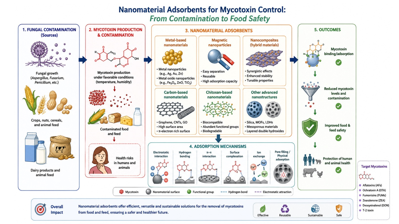
Magnetic nanoparticles, especially when incur-porated into nanocomposites, can offer high adsorption capacity and facilitate easy separation due to their mag-netic properties [43, 44]. Magnetic chitosan-based com-posites have also been applied as sorbents for contam-inants in dairy beverages [45]. This systematic review focused on major nanomaterials proposed as binders and their efficiency in reducing mycotoxin concentrations across different matrices (Figure 2).
- MATERIALS AND METHODS
2.1. Data Sources and Search Strategy
We systematically searched PubMed, Web of Science, and Scopus up to 12 August 2025 and included only English-language articles. Two investigators (AA and SZ) independently screened the titles, abstracts, and full-text articles to identify eligible studies. The keywords considered for the systematic search were: (mycotoxin OR aflatoxin OR ochratoxin A OR patulin OR fumonisins OR zearalenone OR trichothecenes) AND (nanomaterial OR nanostructure OR nanoparticle OR nano) AND (reduction OR binding OR mitigation OR removal). The full database-specific search strategies are provided in Table 1. This systematic review did not involve people, humans, or animals as participants; therefore, we did not need to register the review protocol with PROSPERO.
2.2. Study Selection
We defined inclusion and exclusion criteria using the PICOS framework (population, intervention/exposure, comparator, outcome, and study design). Published studies were considered eligible for inclusion in the present systematic review according to the following criteria: (1) evaluated the effects of nanomaterials, nano-particles, and nanocomposites and did not use other nano-structures, such as nano-emulsions, on mycotoxin reduc-tion; (2) reported the extent of mycotoxin reduction, whether resulting from toxin adsorption or degradation, or from the inhibition of fungal growth and the consequent prevention of mycotoxin production, rather than reporting adsorption capacity alone; (3) investigated the effects of nanomaterials on mycotoxin reduction in food, or culture media; (4) did not investigate nanomaterials for mycotoxin detection; (5) did not investigate the effects of nanomaterials on mycotoxin reduction in vivo or via feeding to animals and biological or cell-based gastrointestinal models. We excluded articles that did not meet the criteria (Table 2).
2.3. Data Extraction
Two authors extracted data from the included articles. The authors discussed any conflicts to reach agreement. The authors extracted data from each study based on the following characteristics: first author’s name, publication year, type of nanomaterial, type of food/medium, type of mycotoxin, nanomaterial concentration used, and myco-toxin reduction percentage. When a study tested several nanomaterials, mycotoxins, matrices, or doses, each com-bination was extracted as a separate study arm. Reduction percentages were recorded as reported (concentration units were not converted); adsorption capacity (mg/g) was not extracted. Regarding nanomaterial type, the main sub-groups included metal-based (metal and metal oxide) nanomaterials, nanocomposites, carbon-based nanomat-erials, organic nanomaterials, and other nanomaterials.
2.4. Data Analysis
Mycotoxin reduction efficiency was expressed as the percentage decrease in toxin concentration reported by each study and distinguished from adsorption capacity (mg/g). Each study arm was treated as one observation, with the midpoint used for reported ranges. Simple unweighted arithmetic means were calculated overall and by mycotoxin and nanomaterial subgroup, without adjust-tment for sample size, dose, replicates, or matrix. Given the heterogeneity of the included studies, the synthesis was descriptive rather than a meta-analysis, and mean values were interpreted cautiously rather than as a basis for ranking nanomaterials.
- RESULTS AND DISCUSSION
3.1. Systematic Search
The literature search yielded 55 records eligible for inclusion in this systematic review [2,17,19,31,32,39, 41-44, 50-94]. We excluded five full-text articles because of insufficient information, and Table 2 lists the reasons for exclusion at all stages.
3.2. Characteristics of Original Articles
Table 3 presents the characteristics of the 55 articles included in this systematic review, comprising 121 study arms. The investigated nanomaterials included metal-based nanomaterials (n = 21), nanocomposites (n = 17), organic nanomaterials (n = 9), carbon-based nano-materials (n = 5), and other nanomaterials (n = 3). The mycotoxin reductions reported in these studies may have resulted from adsorption or binding, degradation, or inhib-ition of fungal growth and the consequent prevention of mycotoxin production, or a combination of these mechanisms. Figure 3 shows the distribution of studies investigating metal-based nanomaterials for mycotoxin reduction.
Regarding the mycotoxin type, most studies (39 studies) were conducted on aflatoxins (AFB1, AFB2, AFG1, AFG2, AF total, AFM1) reduction using nano-materials; 9 studies on Ochratoxin A (OTA), 8 studies on fumonisin (FB1, FB2, FB total), 6 studies on zearalenone (ZEA), 5 studies on deoxynivalenol (DON), and 5 studies on other types of mycotoxins, including Fusaric acid, Nivalenol, Fumagillin, Gliotoxin, Neosolaniol, T2 toxin, fusarin C, moniliformin, Alternaria toxins, patulin, and trichothecenes.
Regarding the matrix used to investigate the binding capacity of mycotoxins, 21 studies were performed on food and feed products (milk, palm kernel cake, apple juice, pistachio nuts, oils, barley, cucumber, corn seeds, maize grain, water, animal feed, Romy cheese, wheat grain, rice grain, grape juice), and 34 studies were conducted on media (aqueous simulator and phosphate buffer) and culture media (Table 3).
3.3. Mycotoxin Reduction Rate
The mean mycotoxin reduction (%) considering the reduction of mycotoxins by nanomaterials was: aflatoxins (84.34%; 39 studies), ochratoxin A (79.72%; 9 studies), fumonisin (87.16%; 8 studies), zearalenone (75.71%; 6 studies), and deoxynivalenol (53.47%; 5 studies) (Figure 4). Researchers mostly detected and quantified myco-toxins using HPLC, ELISA, and ELISA/HPLC.
The overall mean mycotoxin reduction using all studied nanomaterials was 81.67%. The mean mycotoxin reduction (%) for each nanomaterial class was as follows: metal-based nanomaterials (81.07%; 21 studies), nanocomposites (83.55%; 17 studies), carbon-based nanomaterials (76.55%; 5 studies), and organic nanomaterials (72.61%; 9 studies) (Figure 5). These unweighted means are descriptive only: the classes differ in the number of studies, doses, matrices, mycotoxins, and mechanisms of reduction, and we did not statistically test differences between them
3.4. Discussion
Mycotoxins are a critical multidisciplinary issue in chemistry, food, plant, and animal sciences. However, fewer studies address nanotechnology from these disciplines. Nanoscale materials could be considered an alternative because of their unique properties and the opportunities they offer. Recently developed nanomaterials are now used commercially in medicine and other industries. Several researchers have experimentally studied the use of nanotechnology to eliminate microbial toxins, but their results have not yet been systematically reviewed. Given the high prevalence of mycotoxins in food and feed, efficient methods to remove contamination help improve product safety. This systematic review of experimental studies is to our knowledge, the first study to gather relevant data assessing the efficiency of nanomaterials in reducing fungal toxins from edible and non-edible matrices. Our findings showed that, in the included studies, nanomaterials reduced mycotoxin concentrations by up to 100%. Based on their chemical nature, the nanomaterials used included carbon-based nanomaterials, nanocomposites, metal-based nanomaterials, and organic nanomaterials. The included studies reported decreases in concentrations of different mycotoxins across various matrices. These reduction percentages are not equivalent to adsorption capacity (mg/g) and, depending on the study, may reflect adsorption, degradation, or inhibition of fungal growth or toxin production.
Beyond summarizing removal efficiency, this review adds three elements that, to our knowledge, earlier work on nano-adsorbents has not combined. First, it applies a systematic, PICOS-based search and a uniform extraction scheme to experimental data, so reduction rates for the five major mycotoxins and the four nanomaterial classes can be examined alongside each study's matrix, dose, and analytical method. Second, by explicitly separating the reported percentage reduction from adsorption capacity, it shows that part of the available evidence cannot be interpreted as binding alone, because a decrease in toxin concentration may also reflect degradation or inhibition of fungal growth and toxin production; this limits direct ranking of materials and shifts the central question from which nanomaterial removes the most toxin to under which conditions and through which mechanism it does so. Third, it quantifies reporting gaps rather than only success rates: 34 of the 55 included studies did not report the dose, and none provided toxicological data. These evidence gaps, rather than the high mean reduction rates, define the priorities for the next generation of studies.
3.4.1. Mycotoxin adsorption mechanisms
The mycotoxin adsorption by nanomaterials process is explained by several proposed mechanisms, mostly inferred from the physicochemical characteristics of the materials and from adsorption behavior rather than directly confirmed. The main proposed mechanisms include: 1) Physical Adsorption, which operates through non-covalent, weak intermolecular forces such as van der Waals interactions and pore-filling. Mesoporous silica nanoparticles containing pore channels (2-50 nm) can physically trap mycotoxin molecules of compatible size. 2) Chemical Adsorption, which involves the formation of strong, specific chemical bonds between the mycotoxin and functional groups on the nanomaterial's surface. Electrostatic Interactions: These occur between charged toxin molecules and oppositely charged nanomaterial surfaces. For example, at certain pH levels, the carboxyl groups of ochratoxin A (OTA) are negatively charged and are strongly attracted to the positively charged surfaces of chitosan nanoparticles. Hydrogen Bonding and Complexation/Chelation: These are other patterns of this mechanism. 3) π-π Stacking Interactions, which are specific and highly effective interactions where the π-electron systems of aromatic rings on the adsorbent and adsorbate overlap (Figure 6). Carbon-based nanomaterials, such as activated carbon nanotubes and graphene oxide, can physically adsorb mycotoxins because of their large surface areas, graphitic structures, and π-electron clouds. Furthermore, surface engineering through amino-functionalization or molecularly imprinted polymers can enhance adsorption capacity and selectivity [20, 37, 62, 95]. In studies of antifungal nanomaterials, a decrease in toxin concentration may also result from degradation or from inhibition of fungal growth and toxin biosynthesis rather than from adsorption.
3.4.2. The influencing factors on mycotoxin adsorption
Adsorption, the ability of a nanomaterial to bind mycotoxins, is crucial to its effectiveness. Some nanomaterials can be engineered to bind to specific mycotoxins, which is important for targeted detoxification. Besides, nanomaterials used in food or feed applications should be stable and biocompatible. The nanomaterials and developed nanostructures reduced mycotoxins, depending on major variables such as concentration, pH, contact time, matrix/media type, and mycotoxin concentration (Figure 7).
3.4.3. Effect of initial concentration of mycotoxins
Most studies found that the initial toxin concentration strongly influenced reduction rates. Adsorption experiments with varied initial AFB1 concentrations showed that removal of AFB1 using magnetic graphene composite decreased from 93.88% to 88.12% when the AFB1 content increased from 5 μg/L to 25 μg/L [43]. Because nanomaterials have a defined surface area, they can adsorb only a limited amount of mycotoxin molecules; therefore, removal efficiency may decrease at higher initial toxin concentrations.
3.4.4. Effect of nanomaterial types
As the results show, the highest mean mycotoxin reduction rate (83.55%) was related to the nanocomposite type of nanomaterials (Figure 8); however, the differences among classes were small, were not tested statistically, and are confounded by differences in dose, matrix, mycotoxin, and study design, so they do not indicate an intrinsic superiority of any class. Nanocomposites may offer a high surface area-to-volume ratio and a large interfacial area for mycotoxin binding, and, as multiphase structures containing components such as polymers, ceramics, and metals, they may perform better as adsorbent materials. Included studies mostly used chitosan nanostructures in nanocomposite mixtures with different adsorbent materials, which showed high efficiency in adsorbing mycotoxins. Furthermore, activated carbon, bentonite, hydrogels, gums, and polylysine mixed with other nano-adsorbent materials were investigated to reduce mycotoxin levels in matrices. In several cases, it was concluded that a mixture of materials synergistically increases mycotoxin reduction. However, all analyses should consider the effects of specific nanostructures in nanocomposites and the matrix effects of experimental variables. In addition, metal-based nanomaterials showed a notable mycotoxin-reducing effect across different food and media samples. The remarkable properties of metal-based nanomaterials, such as an extremely high specific surface area and abundant, highly active surface sites related to their nanoscale size and tunable surface chemistry, make them desirable for binding various agents. Carbon-based nanomaterials and organic nanomaterials also demonstrated binding efficiency in the studied research. Because nanomaterial mycotoxin-reducing performance is multi-faceted, it may be enhanced by a synergistic combination of maximized surface area, tunable surface chemistry, and unique quantum-scale properties.
3.4.5. Effect of nanomaterial dosage
Mycotoxin adsorption efficiency also depends on nanomaterial dosage. Studies reported that increasing nanoparticle (Ag-NPs, Fe-NPs, and Cu-NPs) content increased AFB1 adsorption. In addition, using nanoparticles individually provided higher AFB1 removal than using them together [54]. In a similar study, AFB1 adsorption (up to 94%) was significantly enhanced by higher nano-composite content (10 to 20 mg/mL) [43]. Obviously, higher adsorbent concentrations increase the number and availability of active sites, resulting in greater mycotoxin adsorption from matrices.
3.4.6. Effect of temperature
Temperature is another effective factor in removal efficiency, depending on the matrix or medium containing mycotoxins. In most reviewed studies, raising the temperature to a critical point increased mycotoxin removal by nanomaterials. Higher temperatures improved AFB1 removal from contaminated oil samples (81.60% to 95.64%) using a graphene composite as the temperature increased from 25°C to 60°C. However, temperatures above 60°C showed a decreasing trend in AFB1 adsorption [43]. It was reported that the AFB1 adsorption capacity of metal nanoparticles increased with rising temperature. The obtained feature indicated the endothermic nature of the adsorption process. At higher temperatures, increased toxin-molecule motility likely provides more opportunities for adsorption at nanostructure active sites [54].
3.4.7. Effect of pH
pH influences mycotoxin adsorption by altering nanomaterial surface charge and mycotoxin ionization, thereby affecting electrostatic interactions. For example, OTA adsorption onto positively charged nano-MgO is pH-dependent [17, 76]. Because these effects vary by adsorbent–mycotoxin pair, no general pH trend can be established, and each system must be optimized.
3.4.8. Effect of contact time
Most studies showed that mycotoxin adsorption with nanomaterials is time-dependent. However, the reviewed plots showed that adsorption was fast during the initial contact times and continued over time to a definite equilibrium point, at which maximum adsorption occurred. During the subsequent contact period, no significant additional adsorption occurred. This suggests that the active sites in the mycotoxin binder become occupied during the initial contact time. Sampling at different times showed that AFB1 binding to the titanium dioxide nanoparticle-based nanostructure is time-dependent, with maximum adsorption achieved after 48 hours of contact time [32, 96-99].
3.4.9. Other related factors in mycotoxin adsorption
Matrix type and composition, including solid or liquid state, may have influenced the reported mycotoxin reduction rates. Results from in vitro studies using naturally or artificially contaminated matrices may also differ from those in real-world systems. The included studies varied in nanomaterial characteristics, dose, mycotoxin type, matrix, experimental conditions, and analytical methods. These factors were not adjusted for; therefore, class means should not be used to rank nanomaterials. Dose was unreported in 34 of 55 studies. Observed reductions may have involved adsorption, chemical degradation, fungal growth inhibition, and reduced toxin production (e.g., antifungal coatings [100]). Their relative contributions could not be established. No formal risk-of-bias assessment was performed because no widely accepted tool specific to in vitro mycotoxin-binding studies was identified. Thus, reported reduction rates should be interpreted descriptively, not as definitive evidence of efficacy. Future studies should improve methodological reporting and use standardized assessment approaches where appropriate.
3.4.10. Practical applications of nanomaterial-based adsorbents for mycotoxin mitigation
Nanomaterial-based adsorption technologies offer opportunities for post-harvest management. Incorporating nanomaterials into storage platforms, packaging materials, and active preservation methods could reduce fungal growth and mycotoxin production during storage and transportation. Because nanocomposites with both antifungal and binding properties can provide dual protection against contamination, they can improve product quality and extend shelf life. Furthermore, nanomaterials can be integrated into monitoring and remediation efforts in food safety management. Multifunctional nanomaterials that can simultaneously detect and adsorb mycotoxins may facilitate rapid contamination mitigation. Such approaches could promote risk management programs and support regulatory compliance throughout the food and feed production chain. Overall, the reviewed in vitro and food-matrix studies suggest that nanomaterials may help reduce mycotoxin contamination in food and feed. However, demonstrated efficacy and applicability in food matrices do not establish gastrointestinal stability, toxicological safety, or regulatory acceptability, which still require evaluation.
Another development that single-endpoint reduction rates do not capture is the shift toward recoverable, multifunctional nanomaterials. Magnetic nanostructured agents can be separated from liquid matrices with an external magnetic field. Still, when tested in beer, milk, and distiller's dried grains with soluble, the matrix slightly decreased adsorption capacity for some toxins [62]; capacities determined in simple aqueous solutions should therefore not be transferred directly to foods. In parallel, adsorption–degradation hybrid systems immobilize photocatalytic or enzymatic modules on selective adsorbents such as metal–organic frameworks, covalent organic frameworks, and molecularly imprinted polymers, enriching trace toxins at the surface and converting them in situ, which may reduce the required dose of detoxifying agent [101]. This concept is already reflected in some of the included studies, such as nanobody-functionalized magnetic nanoparticles for selective aflatoxin capture [87] and a carbon-doped iron-based nanozyme that both adsorbs and degrades AFB1 [88]. However, degradation adds a requirement that adsorption does not: the structure and toxicity of the transformation products must be verified, because a decrease in the parent toxin does not by itself demonstrate detoxification. Future comparative studies should therefore report adsorption capacity (mg/g) with isotherm and kinetic analysis, regeneration over repeated cycles, and the toxicity of reaction products, in addition to percentage reduction.
3.4.11. Safety and environmental considerations
Safety is essential for food and feed applications, yet the included studies provided no toxicological data. Metal and metal oxide nanoparticles (e.g., Ag, ZnO, Cu, CuO, and TiO2) can migrate into food, particularly when added directly and not subsequently removed. Potential toxicity, migration, and environmental release or accumulation therefore require case-specific assessment before application, in accordance with risk-assessment guidance for nanomaterials in the food and feed chain [102].
- CONCLUSION
Overall, the included in vitro studies demonstrated that nanomaterials can reduce mycotoxin concentrations by up to 100%, with potential for multi-mycotoxin removal and enhanced selectivity through surface modification. However, substantial study heterogeneity, the absence of formal quality assessment and direct comparisons with conventional adsorbents, and the exclu-sion of in vivo and biological gastrointestinal studies limit conclusions regarding their comparative performance, safety, and practical applicability. Future research should prioritize standardized efficacy assessments, toxico-logical and environmental safety, gastrointestinal fate, and scalable, cost-effective production, supported by clear regulatory frameworks to facilitate their responsible application in food and feed systems.
- DECLARATION STATEMENTS
5.1. Acknowledgments
None.
5.2. Conflicts of Interest
The authors declare no conflict of interest.
5.3. Author Contributions
Setayesh Zamanpour and Anna Abdolshahi wrote the article and reviewed it.
5.4. Funding
The manuscript did not receive funds from external organizations.
5.5. Data Availability Statement
Dara are available by any request from corresponding author.
5.6. Ethics
Not applicable.
5.7. Use of Artificial Intelligence Chat-bots
Artificial Intelligence (AI) chatbots were used solely to improve the language, grammar, spelling, and clarity of the manuscript and to assist in the design of some figures. The authors reviewed, edited, and verified all AI-assisted text and figure elements and take full responsibility for the accuracy, originality, integrity, and final content of the manuscript.
- Adsorption
- Contamination
- Mycotoxins
- Nanomaterials,
- Reduction
ارجاع به مقاله
مراجع
1. Dwivedi M, Singh P. Novel strategies to mitigate mycotoxin contamination in raw medicinal plant materials. Def Life Sci J. 2024; 9(2): 166-175. https://doi.org/10.14429/dlsj.9.18733
2. Helmy EAM. Detoxification and anti-mycotoxigenic effect of Ochratoxin A produced by Penicillium expansum species utilizing probiotic Pichia kudriavzevii and biogenic-selenium nano-formulations. J Appl Pharm Sci. 2019; 9: 1-8. https://doi.org/10.7324/japs.2019.S101
3. Holakouie Naieni K, Ghods B, Ghorbani R, Bagheri B, Abdolshahi A. Aflatoxin contamination of pistachio and aflatoxicosis: Knowledge, attitude, and practices of people in Damghan City, Iran. J Nuts. 2020; 11(1): 91-99. https://doi.org/10.22034/jon.2020.1892535.1080
4. Neme K, Mohammed A. Mycotoxin occurrence in grains and the role of postharvest management as a mitigation strategies. A review. Food Control. 2017; 78: 412-425. https://doi.org/10.1016/j.foodcont.2017.03.012
5. Tolosa J, et al. Multi-mycotoxin occurrence in feed, metabolism and carry-over to animal-derived food products: A review. Food Chem Toxicol. 2021; 158: 112661. https://doi.org/10.1016/j.fct.2021.112661
6. Niaz W, et al. Mycotoxins: A comprehensive review of its global trends in major cereals, advancements in chromatographic detections and future prospectives. Food Chem X. 2025: 102350. https://doi.org/10.1016/j.fochx.2025.102350
7. Sdogati S, et al. Co-occurrence of aflatoxin B1, zearalenone and ochratoxin A in feed and feed materials in Central Italy from 2018 to 2022. Foods. 2024; 13(2): 313. https://doi.org/10.3390/foods13020313
8. Logrieco AF, et al. The mycotox charter: Increasing awareness of, and concerted action for, minimizing mycotoxin exposure worldwide. Toxins. 2018; 10(4): 149. https://doi.org/10.3390/toxins10040149
9. Stoev SD. Food security, underestimated hazard of joint mycotoxin exposure and management of the risk of mycotoxin contamination. Food Control. 2024; 159: 110235. https://doi.org/10.1016/j.foodcont.2023.110235
10. Casu A, et al. Changing climate, shifting mycotoxins: A comprehensive review of climate change impact on mycotoxin contamination. Compr Rev Food Sci Food Saf. 2024; 23(2): e13323. https://doi.org/10.1111/15414337.13323
11. Wang J, et al. Mycotoxin contamination in organic and conventional cereal grain and products: A systematic literature review and meta-analysis. Compr Rev Food Sci Food Saf. 2024; 23(3): e13363. https://doi.org/10.1111/1541-4337.13363
12. Sun L, et al. Current status of major mycotoxins contamination in food and feed in Asia: A review. ACS Food Sci Technol. 2023; 3(2): 231-244. https://doi.org/10.1021/acsfoodscitech.2c00331
13. Akpoghelie PO, et al. Mycotoxins in food and agriculture: From occurrence to economic consequences. Arch Microbiol. 2026; 208(1): 1-24. https://doi.org/10.1007/s00203-025-04559-x
14. Pigłowski M. The Rapid Alert System for Food and Feed (RASFF): The place in scientific research, limitations and possibilities of data use. Sci Pap Silesian Univ Technol Organ Manag. 2025; (216). https://doi.org/10.29119/16413466.2025.216.27
15. Subramanian A. Emerging trends and challenges in molecular imprinted polymer-based electrochemical and optical sensors for food safety. Food Anal Methods. 2026; 19(1): 1-19. https://doi.org/10.1007/s12161-025-02917-5
16. Zamanpour S, Noori SMA, Yancheshmeh BS, Afshari A, Hashemi M. A systematic review to introduce the most effective postbiotics derived from probiotics for aflatoxin detoxification in vitro. Ital J Food Sci. 2023; 35(4). https://doi.org/10.15586/ijfs.v35i4.2369
17. Nan M, Xin X, Xue H. Synthesis of nano-MgO positively charged inorganic ceramic membrane and electrostatic adsorption for ochratoxin A in grape juice. Food Ferment Ind. 2023; 49(24): 37-43. https://doi.org/10.13995/j.cnki.111802/ts.034919
18. Javani Jouni F, et al. Aflatoxin M1 detoxification from infected milk using Fe3O4 nanoparticles attached to specific aptamer. J Nanostruct Chem. 2018; 8(1): 13-22. https://doi.org/10.1007/s40097-017-0250-5
19. Fakhar E, et al. Core-shell nanoparticles based on ZnO/yeast cell wall: Characterization, antifungal properties and aflatoxin B1 binding. J Nanostruct Chem. 2025; 15(1). https://doi.org/10.57647/jnsc.2025.1501.01
20. Horky P, et al. Nanoparticles as a solution for eliminating the risk of mycotoxins. Nanomaterials. 2018; 8(9): 727. https://doi.org/10.3390/nano8090727
21. Sadr S, et al. Advancing gut microbiome insights: Graphene quantum dot nanobiosensors for microbial metabolite detection. Chem Eng J. 2025: 162954. https://doi.org/10.1016/j.cej.2025.162954
22. Hu W, et al. Nanomaterial-modified nanochannels electrochemical sensors for sensitive detection of zearalenone mycotoxin. Food Anal Methods. 2026; 19(1): 35. https://doi.org/10.1007/s12161-025-02958-w
23. Zhang L, et al. Development of an unlabeled aptamer-based fluorimetric method for aflatoxin B1 detection combined with nanofiber-packed solid-phase extraction. Spectrochim Acta A Mol Biomol Spectrosc. 2026; 344: 126673. https://doi.org/10.1016/j.saa.2025.126673
24. Nikravan L, Zamanpour S, Hashemi M, Marashi SMH, Noori SMA. Application of nanotechnology to develop carrageenan-based films and coatings as carriers for essential oils. Curr Pharm Biotechnol. 2025; 26(7): 972-981. https://doi.org/10.2174/0113892010287269240222054020
25. Ahmadian R, Larypoor M, Bayat M. Fabrication and evaluation of PVA-NYS-THY nanofiber scaffolds as antifungal agents against fluconazole-resistant Candida glabrata. Arch Razi Inst. 2025. https://doi.org/10.32598/ari.80.5.3399
26. Imran M, et al. Catalytic and antibacterial activities with molecular docking analysis of chitosan and polyethylene glycol-NiO2 nanostructures. J Nanostruct Chem. 2025; 15(5). https://doi.org/10.57647/jnsc.2025.1505.20
27. Azari B, Pourahmad A, Sadeghi B, Mokhtary M. Green synthesis of SiO2 from Equisetum arvense plant for synthesis of SiO2/ZIF-8 MOF nanocomposite as photocatalyst. J Coord Chem. 2023; 76(2): 219-231. https://doi.org/10.1080/00958972.2023.2166408
28. Pourjafari M, Ghane M, Kaboosi H, Sadeghi B, Rezaei A. Antibacterial properties of Ag–Cu alloy nanoparticles against multidrug-resistant Pseudomonas aeruginosa through inhibition of quorum sensing pathway and virulence-related genes. J Biomed Nanotechnol. 2022; 18(4): 1196-1204. https://doi.org/10.1166/jbn.2022.3331
29. Khodadad H, Hatamjafari F, Pourshamsian K, Sadeghi B. Microwave-assisted synthesis of novel pyrazole derivatives and their biological evaluation as anti-bacterial agents. Comb Chem High Throughput Screen. 2021; 24(5): 695-700. https://doi.org/10.2174/1386207323666201019152206
30. Karami-Osboo R, Hasantabar V, Maham M. Effective magnetic nanoadsorbent based on natural carboxymethyl cellulose/polyaniline nanotube/graphene oxide for aflatoxin B1 and B2 adsorption in rice utilizing a novel synthesis method. Int J Biol Macromol. 2024; 282: 137162. https://doi.org/10.1016/j.ijbiomac.2024.137162
31. Pirouz AA, et al. A novel adsorbent magnetic graphene oxide modified with Chitosan for the simultaneous reduction of mycotoxins. Toxins. 2018; 10(9). https://doi.org/10.3390/toxins10090361
32. Nasiri Poroj S, et al. The synergistic effect of titanium dioxide nanoparticles and yeast isolated from fermented foods in reduction of aflatoxin B1. Food Sci Nutr. 2023; 11(11): 7109-7119. https://doi.org/10.1002/fsn3.3635
33. Maham M, et al. Application of metal nanoparticle-loaded nanocomposites for extraction, removal, and detection of mycotoxins in foods. Future Foods. 2025; 11: 100672. https://doi.org/10.1016/j.fufo.2025.100672
34. Zhang X, et al. Recent advances on emerging nanomaterials for controlling the mycotoxin contamination: From detection to elimination. Food Front. 2020; 1(4): 360-381. https://doi.org/10.1002/fft2.42
35. Jafari M, et al. Nanomaterials and nano-bio-polymers characteristics for using in meat and food packaging. Food Chem Adv. 2025; 9: 101135. https://doi.org/10.1016/j.focha.2025.101135
36. Abd-Elsalam KA, et al. Nanobiotechnological strategies for toxigenic fungi and mycotoxin control. In: Food preservation. Elsevier; 2017: 337-364. https://doi.org/10.1016/B978012804303-5.00010-9
37. Lin X, et al. Application of nanomaterials for coping with mycotoxin contamination in food safety: From detection to control. Crit Rev Anal Chem. 2024; 54(2): 355-388. https://doi.org/10.1080/10408347.2022.2076063
38. Savi GD, et al. Mesoporous silica nanoparticles incorporated with zinc oxide as a novel antifungal agent against toxigenic fungi strains. J Environ Sci Health B. 2022; 57(3): 176-183. https://doi.org/10.1080/03601234.2022.2041955
39. Mekawey AAI, El-Metwally MM. Impact of nanoencapsulated natural bioactive phenolic metabolites on chitosan nanoparticles as aflatoxins inhibitor. J Basic Microbiol. 2019; 59(6): 599-608. https://doi.org/10.1002/jobm.201800481
40. Monjazeb Marvdashti L, Abdulmajid Ayatollahi S, Salehi B, Sharifi-Rad J, Abdolshahi A, Sharifi-Rad R, Maggi F. Optimization of edible Alyssum homalocarpum seed gum-chitosan coating formulation to improve the postharvest storage potential and quality of apricot (Prunus armeniaca L.). J Food Saf. 2020; 40: e12805. https://doi.org/10.1111/jfs.12805
41. Jebali A, et al. Modification of nanocellulose by poly-lysine can inhibit the effect of fumonisin B1 on mouse liver cells. Colloids Surf B Biointerfaces. 2015; 126: 437-443. https://doi.org/10.1016/j.colsurfb.2014.12.047
42. Aly S, et al. Chemical remediation of Aflatoxin B1 using encapsulated polyvinylpyrrolidone as an environmental-friendly control. Egypt J Chem. 2019; 62(10): 1933-1947. https://doi.org/10.21608/ejchem.2019.10332.1677
43. Ji J, Xie W. Detoxification of Aflatoxin B1 by magnetic graphene composite adsorbents from contaminated oils. J Hazard Mater. 2020; 381. https://doi.org/10.1016/j.jhazmat.2019.120915
44. Pirouz AA, et al. The use of innovative and efficient nanocomposite (magnetic graphene oxide) for the reduction of Fusarium mycotoxins in palm kernel cake. Sci Rep. 2017; 7(1). https://doi.org/10.1038/s41598-01712341-3
45. Marvdashti LM, Milani H, Abdolshahi A, Khodaei SM, Zad SS, Shariatifar N, et al. Evaluation of phthalate acid esters (PAEs) in kefir samples by using magnetic chitosan coated with polyaniline (Fe3O4@CHI@ PANI0/GC–MS method: A health risk assessment study. Microchem J. 2024; 207: 111801. https://doi.org/10.1016/j.microc.2024.111801
46. Mazaheri M. Effect of UV radiation on different concentrations of aflatoxin B1 in pistachio. In: I International Symposium on Mycotoxins in Nuts and Dried Fruits 963. 2011. https://doi.org/10.17660/ActaHortic.2012.963.5
47. Cheraghali A, Yazdanpanah H. Interventions to control aflatoxin contamination in pistachio nuts: Iran experience. J Food Saf. 2010; 30(2): 382-397. https://doi.org/10.1111/j.1745-4565.2010.00214.x
48. da Silva AC, et al. Microwave drying and disinfestation of Brazil nut seeds. Food Control. 2016; 70: 119-129. https://doi.org/10.1016/j.foodcont.2016.04.049
49. Balbaa M, et al. Antioxidants and radical scavenging role of Leek (Allium kurrat) against aflatoxin-contaminated peanut. Toxin Rev. 2018; 37(4): 334-343. https://doi.org/10.1080/15569543.2017.1387152
50. Abdelghany TM, Hassan MM, El-Naggar MA. GC/MS analysis of Juniperus procera extract and its activity with silver nanoparticles against Aspergillus flavus growth and aflatoxins production. Biotechnol Rep. 2020; 27. https://doi.org/10.1016/j.btre.2020.e00496
51. Al-Hamiri KAKA, Hussein HZ. Efficacy of normal and nanoscale table salt on inhibiting the growth of fungi Aspergillus flavus in the culture medium and reduction the aflatoxin B1 on the stored rice grains. Int J Agric Stat Sci. 2021; 16: 1329-1335.
52. Aljbori NM, Hussein HZ. Testing the efficiency of silver nanoparticles manufactured locally by the alga Spirogyra in inhibiting the fungus Aspergillus flavus and reduction of aflatoxin B1. IOP Conf Ser Earth Environ Sci. 2023; 1225(1): 012062. https://doi.org/10.1088/1755-1315/1225/1/012062
53. Ammar HAM, El-Desouky TA. Green synthesis of nanosilver particles by Aspergillus terreus HA1N and Penicillium expansum HA2N and its antifungal activity against mycotoxigenic fungi. J Appl Microbiol. 2016; 121(1): 89-100. https://doi.org/10.1111/jam.13140
54. Asghar MA, et al. Facile, one-pot biosynthesis and characterization of iron, copper and silver nanoparticles using Syzygium cumini leaf extract: As an effective antimicrobial and aflatoxin B1 adsorption agents. PLoS One. 2020; 15(7). https://doi.org/10.1371/journal.pone.0234964
55. Asghar MA, et al. Nano-adsorbents obtained from walnut and almond shells for the effective removal of aflatoxin B1. Biomass Convers Biorefin. 2025. https://doi.org/10.1007/s13399-025-06590-w
56. Belhedi M, et al. Silicon dioxide (SiO2) nanoparticles affect the morphology, sporulation, mycotoxin production, and patho-genicity of Fusarium brachygibbosum infecting olive trees. Front Nanotechnol. 2025; 7. https://doi.org/10.3389/fnano.2025.1569453
57. Bakri MM, et al. Efficacy of Juniperus procera constituents with silver nanoparticles against Aspergillus fumigatus and Fusarium chlamydosporum. BioNano-Science. 2020; 10(1): 62-72. https://doi.org/10.1007/s12668-019-00716-x
58. El-Desouky TA, Ammar HAM. Honey mediated silver nanoparticles and their inhibitory effect on aflatoxins and ochratoxin A. J Appl Pharm Sci. 2016; 6(6): 83-90. https://doi.org/10.7324/JAPS.2016.60615
59. Fayed A, Elsayed H, Ali T. Packaging fortified with Natamycin nanoparticles for hindering the growth of toxigenic Aspergillus flavus and aflatoxin production in Romy cheese. J Adv Vet Anim Res. 2021; 8(1): 58-63. https://doi.org/10.5455/javar.2021.h485
60. Gharieb MM, Hassan EM, Soliman AM. Nutritional conditions affecting of selenium nanoparticles synthesized by Fusarium oxysporum (CCASU-2023-F9), and their biological activities against mycotoxin-producing fungi isolated from animal feed. Braz J Microbiol. 2024; 55(4): 3465-3476. https://doi.org/10.1007/s42770-024-01494-9
61. González-Jartín JM, et al. Detoxification agents based on magnetic nanostructured particles as a novel strategy for mycotoxin mitigation in food. Food Chem. 2019; 294: 60-66. https://doi.org/10.1016/j.foodchem.2019.05.013
62. González-Jartín JM, et al. Magnetic nanostructured agents for the mitigation of mycotoxins and cyanotoxins in the food chain. Food Chem. 2024; 456. https://doi.org/10.1016/j.foodchem.2024.140004
63. Hamza Z, et al. Preparation and characterization of yeast cell wall beta-glucan encapsulated humic acid nanoparticles as an enhanced aflatoxin B1 binder. Carbohydr Polym. 2019; 203: 185-192. https://doi.org/10.1016/j.carbpol.2018.08.047
64. Hernández-Meléndez D, et al. Inhibitory effect of flower-shaped zinc oxide nanostructures on the growth and aflatoxin production of a highly toxigenic strain of Aspergillus flavus Link. Materials. 2018; 11(8). https://doi.org/10.3390/ma11081265
65. Horky P, et al. Usability of graphene oxide as a mycotoxin binder: In vitro study. PLoS One. 2020; 15(9). https://doi.org/10.1371/journal.pone.0239479
66. Hu D, et al. Biogenic Trichoderma harzianum-derived selenium nanoparticles with control functionalities originating from diverse recognition metabolites against phytopathogens and mycotoxins. Food Control. 2019; 106. https://doi.org/10.1016/j.foodcont.2019.106748
67. Hussain AH, Hussein HZ. Evaluation of Agaricus sp. and Pleurotus sp. extracts efficiency in Aspergillus flavus growth inhibition and aflatoxin B1 reduction. Syst Rev Pharm. 2020; 11(10): 564-569. https://doi.org/10.31838/srp.2020.10.84
68. Hussein AH, Hussein HZ. Effect of standard and nano-urea on the inhibition of the fungus Aspergillus flavus growth and reduction of aflatoxin B1 production in the laboratory. Arab J Plant Prot. 2024; 42(2): 184-188. https://doi.org/10.22268/AJPP-001235
69. Jabeen M, et al. Development of lignin-chitosan-iron nanocomposite for effective removal of aflatoxin B1 in food and feed systems. J Clust Sci. 2025; 36(2). https://doi.org/10.1007/s10876-025-02784-4
70. Karami-Osboo R, Maham M, Nasrollahzadeh M. Synthesised magnetic nano-zeolite as a mycotoxins binder to reduce the toxicity of aflatoxins, zearalenone, ochratoxin A, and deoxynivalenol in barley. IET Nanobiotechnol. 2020; 14(7): 623-627. https://doi.org/10.1049/iet-nbt.2020.0107
71. Karimi AR, et al. Design and characterization of tunable chitosan hydrogels cross-linked with epiclon through amide bond formation modified with multi-walled carbon nanotubes for adsorption of aflatoxin B1 from aqueous solutions. J Macromol Sci A. 2025; 62(3): 263-273. https://doi.org/10.1080/10601325.2025.2467050
72. Khalid SA, Elokle AA. Evaluation of Pumpkin seed oil and its chitosan-Arabic gum nanoparticles on the viability of Aspergillus flavus and inhibition of total aflatoxin in beef sausage. Food Control. 2025; 168. https://doi.org/10.1016/j.foodcont.2024.110854
73. Khalil NM, Abd El-Ghany MN, Rodríguez-Couto S. Antifungal and anti-mycotoxin efficacy of biogenic silver nanoparticles produced by Fusarium chlamydosporum and Penicillium chrysogenum at non-cytotoxic doses. Chemosphere. 2019; 218: 477-486. https://doi.org/10.1016/j.chemosphere.2018.11.129
74. Mohammad Nejad Khiavi N, et al. Reduction of aflatoxin M1 using mixture of Saccharomyces cerevisiae and Candida albicans cell walls immobilized on silica nanoparticles entrapped in alginate gel. J Environ Chem Eng. 2020; 8(1). https://doi.org/10.1016/j.jece.2019.103635
75. Moradian M, Faraji AR, Davood A. Removal of aflatoxin B1 from contaminated milk and water by nitrogen/carbon-enriched cobalt ferrite -chitosan nano-sphere: RSM optimization, kinetic, and thermos-dynamic perspectives. Int J Biol Macromol. 2024; 256. https://doi.org/10.1016/j.ijbiomac.2023.127863
76. Nan MN, et al. Electrostatic adsorption and removal mechanism of ochratoxin A in wine via a positively charged nano-MgO microporous ceramic membrane. Food Chem. 2022; 371. https://doi.org/10.1016/j.foodchem.2021.131157
77. Paimard G, et al. Detoxification of patulin from juice simulator and apple juice via cross-linked Se-chitosan/L-cysteine nanoparticles. LWT. 2021; 143. https://doi.org/10.1016/j.lwt.2021.111146
78. Pena GA, et al. Reduction of Fusarium proliferatum growth and fumonisin accumulation by ZnO nano-particles both on a maize based medium and irradiated maize grains. Int J Food Microbiol. 2022; 363. https://doi.org/10.1016/j.ijfoodmicro.2021.109510
79. Pérez-de León A, et al. Comparison of the in vitro antifungal and anti-fumonigenic activities of copper and silver nanoparticles against Fusarium verticillioides. J Clust Sci. 2020; 31(1): 213-220. https://doi.org/10.1007/s10876-019-01638-0
80. Rezagholizade-shirvan A, et al. Adsorptive removal of aflatoxin M1 in milk using magnetic chitosan/curcumin nanocomposite supplemented with Lactiplantibacillus plantarum postbiotic. Microchem J. 2025; 215. https://doi.org/10.1016/j.microc.2025.114436
81. Rezagholizade-shirvan A, et al. Removal of aflatoxin M1 in milk using magnetic laccase/MoS2/chitosan nanocomposite as an efficient sorbent. Chemosphere. 2024; 365. https://doi.org/10.1016/j.chemosphere.2024.143334
82. Safajoo S, et al. Synthesis of a new thiourea-polygalacturonic acid nanocomplex adsorbent for removing patulin from apple juice simulator and apple juice. J Verbrauch Lebensm. 2022; 17(3): 231-241. https://doi.org/10.1007/s00003-022-01370-4
83. Savi GD, et al. Mesoporous silica nanoparticles adsorb aflatoxin B1 and reduce mycotoxin-induced cell damage. J Environ Sci Health B. 2023; 58(1): 1-9. https://doi.org/10.1080/03601234.2022.2161251
84. Shahinfar M, et al. Simultaneous removal of four aflatoxins using magnetic nanobentonite as a green and fast sorbent: Kinetic, thermodynamic, and isotherm investigation. Environ Sci Pollut Res. 2023; 30(51): 110515-110527. https://doi.org/10.1007/s1135602329963-y
85. Silwana N, et al. Heterogeneous fenton degradation of patulin in apple juice using carbon-encapsulated nano zero-valent iron (CE-nZVI). Foods. 2020; 9(5). https://doi.org/10.3390/foods9050674
86. Vahidimehr A, et al. Saccharomyces cerevisiae and Lactobacillus rhamnosus cell walls immobilized on nano-silica entrapped in alginate as aflatoxin M1 binders. Int J Biol Macromol. 2020; 164: 1080-1086. https://doi.org/10.1016/j.ijbiomac.2020.07.089
87. Wang H, et al. Nanobody immobilization on magnetic nanoparticles via monomeric streptavidin-biotin specific interaction for aflatoxin adsorption. Adv Compos Hybrid Mater. 2024; 7(3). https://doi.org/10.1007/s42114-024-00893-8
88. Wang L, et al. Construction of carbon-doped iron-based nanozyme for efficient adsorption and degradation to synergistic removal of aflatoxin B1. Colloids Surf B Biointerfaces. 2025; 245. https://doi.org/10.1016/j.colsurfb.2024.114297
89. Yousif DA, Hussein HZ, Ali AM. Testing the efficiency of zinc oxide nanoparticles, honey, honey supported by zinc oxide nanoparticles and phylex in degradation of aflatoxin B1 in stored sea maize seeds. J Glob Pharma Technol. 2019; 11(2): 225-235.
90. Zaban MI, Azim NSA, Abd El-Aziz ARM. Antifungal and anti-aflatoxin efficacy of biogenic silver nanoparticles produced by Aspergillus species: Molecular study. Pak J Pharm Sci. 2019; 32(5): 2509-2526.
91. Zadeh MHB, Shahdadi H. Nanocellulose coated with various free fatty acids can adsorb fumonisin B1, and decrease its toxicity. Colloids Surf B Biointerfaces. 2015; 134: 26-30. https://doi.org/10.1016/j.colsurfb.2015.06.037
92. Živančev J, et al. The impact of fullerenol nanoparticles on the growth of toxigenic Aspergillus flavus and aflatoxins production in vitro and in corn flour. J Food Sci. 2024; 89(3): 1814-1827. https://doi.org/10.1111/1750-3841.16952
93. Karami-Osboo R, et al. Encapsulated Zataria multiflora's essential oil inhibited the growth of Aspergillus flavus and reduced aflatoxins levels in contaminated pistachio nut. Biocatal Agric Biotechnol. 2023; 51: 102796. https://doi.org/10.1016/j.bcab.2023.102796
94. Liu M, et al. Highly efficient and cost-effective removal of patulin from apple juice by surface engineering of diatomite with sulfur-functionalized graphene oxide. Food Chem. 2019; 300: 125111. https://doi.org/10.1016/j.foodchem.2019.125111
95. Song C, Qin J. High-performance fabricated nano-adsorbents as emerging approach for removal of mycotoxins: A review. Int J Food Sci Technol. 2022; 57(9): 5781-5789. https://doi.org/10.1111/ijfs.15953
96. Abdolshahi A, et al. Aflatoxin binding efficiency of Saccharomyces cerevisiae mannoprotein in contaminated pistachio nuts. Food Control. 2018; 87: 17-21. https://doi.org/10.1016/j.foodcont.2017.12.008
97. Emadi A, et al. In vitro strain specific reducing of aflatoxin B1 by probiotic bacteria: A systematic review and meta-analysis. Toxin Rev. 2022; 41(3): 995-1006. https://doi.org/10.1080/15569543.2021.1929323
98. Emadi A, et al. Aflatoxin reduction in nuts by roasting, irradiation and fumigation: A systematic review and meta-analysis. Crit Rev Food Sci Nutr. 2022; 62(18): 5056-5066. https://doi.org/10.1080/10408398.2021.1881436
99. Javan AJ, et al. The efficiency of lactic acid bacteria strains isolated from cottage cheese (Khiki) to aflatoxin M1 reduction in milk. J Microbiota. 2024; 1(2): e148764. https://doi.org/10.5812/jmb-148764
100. Abdolshahi A, Marvdashti LM, Salehi B, Sharifi-Rad M, Ghobakhloo S, Iriti M, et al. Antifungal activities of coating incorporated with Saccharomyces cerevisiae cell wall mannoprotein on Aspergillus flavus growth and aflatoxin production in pistachio (Pistacia vera L.). J Food Saf. 2019; 39(2): e12608. https://doi.org/10.1111/jfs.12608
101. Yang X, Yao M, Liao W, Li X. Adsorption–degradation integrated approaches to mycotoxin removal from food matrices: A comprehensive review. Toxins. 2025; 17(11): 556. https://doi.org/10.3390/toxins17110556
102. EFSA Scientific Committee, More S, Bampidis V, et al. Guidance on risk assessment of nanomaterials to be applied in the food and feed chain: Human and animal health. EFSA J. 2021; 19(8): e06768. https://doi.org/10.2903/j.efsa.2021.6768
- چکیده مشاهده شده: 97 بار
- Pdf (English) دانلود شده: 41 بار