Exploration and Molecular Characterization of Indigenous Lactic Acid Bacteria from Bada Fish (Rasbora argyrotaenia) of Lake Maninjau as Preliminary Probiotic Candidates for Traditional Functional Food Applications
بیوتکنولوژی غذایی کاربردی,
دوره 13 شماره 1 (2026),
7 آذر 2025
,
صفحه 1-27 (e34)
https://doi.org/10.22037/afb.v13i1.53488
چکیده
Background and Objective: Lactic acid bacteria (LAB) are important microorgan-isms with potential applications in food and health industries because of their probiotic and antimicrobial properties. Indigenous LAB from traditional fermented foods may provide valuable sources of functional microorganisms, including candidates for future functional food development. This study aimed to conduct preliminary phenotypic and in vitro screening of LAB isolated from fermented Bada fish (Rasbora argyrotaenia) from Lake Maninjau, West Sumatra, Indonesia, based on acid tolerance, bile tolerance, and growth-inhibitory activity, with 16S rRNA gene analysis of isolate IBD.2
Material and Methods: 20 presumptive LAB isolates were obtained from fermented Bada fish and characterized morphologically and biochemically based on Gram staining, cell morphology, catalase activity, and fermentation type. The 16 homofer-mentative isolates were then screened for probiotic potential based on tolerance to gastric acid and bile salts. Antimicrobial activity was evaluated against Escherichia coli O157, Staphylococcus aureus ATCC 25923, and Salmonella enteritidis ATCC 13076. Neutralized cell-free supernatants were further evaluated for antimicrobial activity and analyzed using SDS-PAGE for preliminary protein-size characterization. The most pro-mising isolate was identified by 16S rRNA gene sequencing and phylogenetic analysis.
Results and Conclusion: Re-evaluation using a survival criterion of ≥50% after acid exposure for 3 h and after exposure to 0.3% Oxgall for 3 h identified six preliminary probiotic candidates. Only three also retained ≥50% survival after 6 h of bile exposure. All isolates were Gram-positive, rod-shaped, catalase-negative, and homofermentative. Isolate IBD.2 produced the largest inhibition zones against the tested indicator bacteria, demonstrating growth-inhibitory activity under the assay conditions, while neutralized cell free supernatant also exhibited inhibitory activity. SDS-PAGE revealed protein bands ranging from 10–25 kDa which represents only a preliminary protein size charac-terization. Molecular identification showed that IBD.2 shared 98.94% 16S rRNA gene sequence similarity with the reference strain Lentilactobacillus buchneri (formerly Lactobacillus buchneri) strain 1955 (used for comparison), indicating close taxonomic affiliation but not confirming species- or strain-level identity. These findings identify preliminary candidates for further evaluation based on the screening endpoints measured; they do not establish comprehensive probiotic functionality, safety for consumption, or suitability for a functional food product. The study did not establish or certify the halal status of the isolates or any resulting product. No direct toxicological assessment, animal experiments, clinical studies, or gastrointestinal health outcomes were included. Potential food applications require further functional and safety characterization and evidence of relevant health effects.
- INTRODUCTION
The bada fish (Rasbora argyrotaenia) is a native fish of Lake Maninjau, inhabiting freshwater bodies with unique ecological conditions. Freshwater fish harbor diverse dig-estive tract microbiota that may provide a source of poten-tial probiotic bacteria. These bacteria may exhibit adaptive characteristics, including tolerance to low pH and bile salts and the ability to produce antimicrobial compounds [1]. Previous research has shown that bacteria derived from fer-mented fish-derived foods, particularly those of the genera Lactobacillus, Bacillus, and Pediococcus have been invest-igated for potential probiotic applications and digestive effects in specific experimental settings; such findings cannot be extrapolated to the isolates screened here [2].
Bada Salai is exclusively made from Bada Fish (Rasbora argyrotaenia). This product looks like bilaterally compr-essed raw fish with slight disintegration near the belly and tail, preserved through traditional smoking techniques, resulting in a distinctive aroma, savory flavor, and a longer shelf life. Bada Salai is traditionally made by fermenting dried fish under microaerophilic conditions in tightly closed banana leaf containers for about 3-4 weeks in a semi-open hut. The most common dominant micro-organisms in many fermented fish products have been reported to be Lactic Acid Bacteria (LAB), Bacillus, and Staphylococcus [3]. Using 16S rRNA sequencing, LAB in Bada Fish (Rasbora argyrotaenia) can be identified. Fermented fish products may serve as sources of lactic acid bacteria (LAB) with potential probiotic-associated charac-teristics. However, the presence of LAB in fermented fish does not necessarily establish the probiotic status or safety of the microorganisms. These properties require strain-specific functional and safety assessments [4].
Interest in probiotic products, including products intended to meet halal requirements, provides context for exploring microorganisms from local fermented foods. Reported digestive, immune, or nutritional effects of particular probiotics depend on the strain, product, and supporting evidence and cannot be attributed to the isolates in this study. The present investigation did not evaluate immunity, nutrient absorption, gastrointestinal health, or stunting prevention. Its contribution is preliminary screening of local isolates for further investigation [5].
The use of indigenous lactic acid bacteria (LAB) from bada fish (Rasbora argyrotaenia) provides a food-derived source of preliminary probiotic candidates. Their potential use in products intended to meet halal requirements would depend on separate verification of the organisms, culture media, processing materials, and production process. Recovery from a traditional food identifies the ecological source of the isolates but does not demonstrate safety for consumption, physiological benefits, or performance in a formulated food. These potential applications require separate evaluation. Previous studies have shown that the dominant microorganisms in fermented fish products generally consist of LAB, Bacillus, and Staphylococcus, which contribute to the fermentation characteristics and functional potential of the product, especially when identified through molecular approaches such as 16S rRNA gene sequencing [6].
However, to date, comprehensive scientific studies on the molecular characterization, screening of probiotic properties, and potential applications of fermented fish from Lake Maninjau as functional foods remain very limited. Therefore, molecular identification based on genetic analysis is needed to establish the taxonomic identity of the LAB isolates obtained. Safety (e.g., GRAS or QPS status), genetic stability, and the absence of transferable antibiotic-resistance genes cannot be established by 16S rRNA sequencing alone and require separate assessment,
Soaking with salt (±30% of fish weight)
such as whole-genome analysis and antibiotic susceptibility testing, in future studies. Through 16S rRNA-based molecular identification, researchers can determine the taxonomic affiliation of isolates (species-level identification only when supported by sufficient sequence evidence) and analyze phylogenetic relationships. Probiotic potential, however, must be assessed separately through functional and safety assays. Molecular identification does not establish halal status. Any future halal designation would require separate assessment of the microorganisms, raw materials, culture media, processing materials, and production process, together with the applicable certification of the final product. Thus, this research is preliminary step in the development of local probiotics based on Indonesian biological resources that may support future functional food innovation [7]. This study combines phenotypic screening, acid- and bile-tolerance assays, growth-inhibitory testing, and 16S rRNA gene analysis of IBD.2 to identify candidates from fermented Bada fish for subsequent investigation [8].
Studies on the microbiology of fermented fish products from Lake Maninjau have been conducted, but the genotypic identity and functional properties of their lactic acid bacteria (LAB) remain insufficiently characterized. Therefore, this study combines gene-based molecular identification with in vitro assessment of LAB isolated from fermented Bada fish (Rasbora argyrotaenia) to identify isolates with potential probiotic properties [9]. Sequence analysis provides evidence of taxonomic affiliation for IBD.2, while the screening assays measure survival under defined in vitro acid and bile conditions and inhibition of indicator-bacterium growth. The scope is preliminary screening; the principal functional and safety limitations are discussed below [10]. In this manuscript, the current valid name Lentilactobacillus buchneri (formerly Lactobacillus buchneri, reclassified in 2020), is used throughout.
- MATERIALS AND METHODS
2.1 Fermentation of Bada Fish in Lake Maninjau
The fermented Bada fish (Rasbora argyrotaenia) analyzed in this study were prepared using the same traditional procedure for all four sample groups (IBA–D). Freshly caught fish were placed in separately labelled laboratory sample containers and transported from the collection sites to the laboratory in a cooler box. The fish were washed with clean water without removing the viscera or scales, then salted at approximately 30% of their weight (w/w) and mixed with chili, turmeric, onion, and grated coconut. The mixture was enclosed in banana or taro leaves and fermented at ambient room temperature for 2–4 days; the duration for each sample group is reported in Table 1. Ambient relative humidity, oxygen concentration, and redox potential were not measured. After fermentation, the fish were removed from the leaf wrapping and smoked. Samples for lactic acid bacteria (LAB) isolation were collected after smoking. This preparation reflects a traditional approach to fermented fish production [11], and the process is illustrated in Figure 1.
2.2 Sampling location of Bada Fish in Lake Maninjau
Sampling of Bada fish (Rasbora argyrotaenia) was conducted at four locations in the Lake Maninjau area between 10 and 12 June 2026. The locations were selected to encompass differences in habitat characteristics and proximity to human activities. The Sungai Batang site was located near a traditional market, whereas the Tanjung Raya site was situated in a tourism area. Activities associated with these areas may influence local water conditions and, potentially, fish growth; however, their effects on growth were not directly assessed in this study. Other human activities in the surrounding area included floating net cage aquaculture, fishing, and household waste disposal. A total of 53 fish were captured directly at an approximate collection depth of 20 m: 10 from Tanjung Raya (IBA; 0.266100° S, 100.210649° E), 14 from Sungai Batang (IBB; 0.327146° S, 100.223601° E), 13 from Tanjung Sani (IBC; 0.350838° S, 100.187854° E), and 16 from Sungai Batang Antokan (IBD; 0.291012° S, 100.152593° E). Fish measuring at least 10 cm in length were selected; their average weight was approximately 30 g. The collected fish were processed into fermented Bada fish, a traditional local food product. Samples were kept separate and labelled according to their collection sites for subsequent laboratory analysis [12].
The four sampling locations represented different areas around Lake Maninjau, West Sumatra, Indonesia. Sample 1 was collected from Sungai Batang, near the traditional market. Sample 2 was collected from the central area of Lake Maninjau at an estimated depth of approximately 20 m. Sample 3 was collected from the upstream section of the Batang Antokan River, near the Lake Maninjau Hydroelectric Power Plant, while Sample 4 was collected from the tourist area of Tanjung Raya, near Tapian Kualo Beach. The estimated sampling depth ranged from approximately 20 to 50 m, although the depth varied depending on the sampling location and fish-catching method. Fish were captured using fishing hooks and nets. Immediately after collection, the fish were placed in a cooler box and transported to the laboratory for subsequent processing. LAB isolation was conducted in the Microbiology Laboratory of the University of West Sumatra, while molecular characterization was performed at the Biotechnology Laboratory of Andalas University, Indonesia. The sampling locations are illustrated in Figure 2, and the corresponding sample information, including fermentation duration, is provided in Table 1.
2.3 Isolation of LAB from Bada Fish in Lake Maninjau
The LAB isolation process begins with the enrichment process. A total of 25 grams of each sample was put into 225 mL of physiological saline solution (0.85% NaCl) to obtain a dilution of 10-1, then homogenized [13]. The solution was pipetted 1 mL, then put into a tube containing 9 mL of sterile physiological solution to obtain a dilution of 10-2, and continued until a dilution of 10-6. Each dilution was pipetted 1 mL and transferred into a sterile Petri dish. Each dilution was transferred into 2 sterile Petri dishes (duplo). Then, 15 mL of MRS agar + CaCO3 3% media. After the MRS agar medium solidifies, the Petri dish is stored upside down in microaerophilic conditions by overlaying (pouring MRS Agar on the Petri dish that has been previously inoculated with each dilution) at 37°C for 48 hours [14]. Counting is carried out on a plate containing 25-250 colonies according to [15]. LAB isolation is carried out by taking 20 colonies from the plate that can be counted and then streaking them on MRS Agar + CaCO3 to obtain a pure LAB culture.
Purification begins by selecting distinct colonies with a clear zone surrounding them. The purification stage can be repeated 2-3 times to ensure the purity of the colonies. Representative colonies are sampled from the plate and streaked onto plates containing MRS Agar, incubated at room temperature for 48 hours. Colonies with different morphologies are purified by sampling and streaking again onto new plates containing MRS Agar until colonies with uniform shape and size are obtained. A single colony is sampled and streaked onto an agar slant. To preserve the culture, a 1:1 glycerol stock is prepared. Each isolate is inoculated into MRS broth containing 30% glycerol and then stored frozen at -80°C/-20°C. The stock culture is renewed after 6 months of storage. For long-term storage, lyophilization of the selected isolates is performed [16].
2.4 Characterization of LAB from Bada Fish of Lake Maninjau
2.4.1. Identification of LAB Morphology
Morphological identification was performed macroscopically by inoculating LAB cultures onto MRS Agar to observe the shape, color, and diameter of the LAB isolates. Morphological characterization was then continued microscopically using Gram staining to determine cell shape and color. Meanwhile, catalase and fermentation type tests were used to characterize biochemical properties [17].
2.4.2. Selection of LAB from Bada Fish of Lake Maninjau as a Probiotic Candidate
- Resistance of LAB Isolates of Bada Fish (Rasbora argyrotaenia) from Lake Maninjau to Stomach pH
The resistance assessment for gastric pH was carried out based on [18], method with modifications. This assay used two types of media, including MRS broth with addition of 37% HCl to achieve a pH of 2.5 and MRS broth without the addition of HCl to maintain a pH of 6.8 (as a control). The medium was sterilized at 121 °C for 15 min using autoclave. Moreover, 5 ml of MRS broth-HCl were added to 0.5 mL of the bacterial isolate and incubated at 37oC for 3 and 6 h. Then, absorbance was read at 600 nm. This was carried out three times. Resistance was expressed as a percentage. The percentage of the resistance of LAB isolates can be calculated using the Eq. 1 [19]:
Eq. 1
- Resistance of LAB Isolates from Bada Fish of Lake Maninjau to Bile Salts
The resistance assessment of bile salt was carried out based on a method from [19] with modifications. Various concentrations of bile salt of 0, 0.3, and 0.5% were added to the MRS broth media. The media were autoclaved at 121 °C for 15 min. The bacterial isolate (0.5 ml) was transferred into 5 ml of MRS broth containing 0, 0.3, and 0.5% ox-gall and incubated at 37 oC for 5 h. The MRS broth containing no bile salt was set as the control and compared with the treatments. The LAB growth was assessed using UV spectrophotometry based on the absorbance at 600 nm. All assessments were carried out in three replications. Isolate resistance was expressed as a percentage. According to [13], the percentage of the resistance of LAB isolates can be calculated using the Eq. 2:
Eq. 2
2.5. Screening of LAB Bada Fish in Lake Maninjau for Antimicrobial Potential
2.5.1. Antimicrobial activity test of LAB
Three pathogens of Salmonella enteritidis ATCC 13076, Staphylococcus aureus and Escherichia coli, were assessed for antimicrobial activity of LAB using the modified paper diffusion method production [1]. Briefly, 1 ml of LAB culture enriched for 48 h was collected using a micropipette and transferred into a sterile microtube, and the supernatant was collected for antimicrobial resistance assessment. Then, 0.4 g of NA media was homogenized by heating using a hot plate and sterilized using an autoclave. Then, 40 µl of the bacterial isolate enriched for 24 h was poured into a Petri dish containing 20 ml of NA medium and cooled down until the media hardened. The paper disk was soaked in the LAB isolate suspension for approximately 10 min. After the agar solidified, the paper disk was set on NA medium, which contained isolates of pathogenic bacteria. Then, a positive control was set by dropping LAB supernatant (20 µl) onto sterile test papers, including 10 g of penicillin, 30 g of kanamycin, and 10 g of ampicillin. This was incubated at 37 °C for 24 h anaerobically. After 24 h, the diameter of the inhibition zone was measured using caliper [18].
2.5.2. Antimicrobial activity test of cell-free supernatant
Briefly, 1 mL was cultured in 9 ml of MRS solution at 37 oC and incubated for 2 d. This was centrifuged at 14,000 rpm for 5 min. Then, 0.22-µl membrane filter was used to filter the supernatant. To eliminate the barrier effect because of the presence of organic acids, 1 N NaOH solution was added to the cell-free supernatant to maintain pH 6.5 [1]. The bacterial pathogens were grown aerobically at 37 °C for 24 h. Then, 0.2% pathogenic bacteria were transferred onto 20 mL of MHA solution at 50 °C. After the gelatin was solid, a 6-mm well was made in the media using a cork borer. Furthermore, 50 µl of supernatant was transferred into the wells and set 10–15 min. The incubation was carried out at 37°C for 24 h under aerobic conditions. Antimicrobial activity of bacteriocins in the supernatant was verified by varying the time intervals of 15, 30, and 60 min at 100 ºC. The supernatant of LAB was investigated for its ability to inhibit bacteria, including Escherichia coli O157, Staphylococcus aureus ATCC 25923 and Salmonella enteritidis ATCC 13076, via similar methods. The inhibition zone was associated with the presence of bacteriocin compounds, and its dimension was recorded using a caliper [20].
2.6 Isolation and characterization of 16S rRNA LAB
Genomic DNA was extracted using the PureLink™ Genomic DNA Mini Kit (Invitrogen) according to the manufacturer’s instructions, with lysozyme (20 mg/mL) added to facilitate lysis of the bacterial cell wall. The extracted DNA was used as the template for amplification of the 16S rRNA gene with primers 63F (5′-CAGGCCTAACACATGCAAGTC-3′) and 1387R (5′-GGGCGGWGTGTACAAGGC-3′; W = A or T). The 50-µL PCR contained 25 µL of GoTaq Green Master Mix (2×), 5 µL of each primer, 2 µL of genomic DNA template, and 13 µL of nuclease-free water . Amplification consisted of initial denaturation at 95 °C for 5 min; 35 cycles of denaturation at 95 °C for 30 s, annealing at 58 °C for 30 s, and extension at 72 °C for 1 min; followed by final extension at 72 °C for 5 min. PCR products were separated on a 1.5% agarose gel in TBE buffer at 100 V for 60 min, stained with GelRed, and visualized using a gel documentation system. A 100-bp DNA ladder was used as the size marker, and the expected amplicon for primers 63F and 1387R was approximately 1,300 bp. For purification, the PCR product was mixed with 0.1 volume of 3 M sodium acetate (pH 5.2) and 2.5 volumes of absolute ethanol, incubated at −80 °C for 30 min, and centrifuged at 15,000 × g for 20 min at 4 °C. The DNA pellet was washed with 70% ethanol, centrifuged at 15,000 × g for 5 min at 4 °C, air-dried, and dissolved in ultrapure water. The PCR products were subsequently purified by sodium acetate–absolute ethanol precipitation [21].
Purified PCR products were sequenced by First BASE Laboratories (Malaysia). Sequence data were analyzed using BioEdit 7.2.5 and MEGA 11 software. Molecular identification was performed by comparing the 16S rRNA gene sequences with GenBank reference sequences using BLASTn (https://blast.ncbi.nlm.nih.gov/Blast.cgi). Query coverage and percentage identity were recorded for each isolate. A species-level identification was considered provisional and assigned only when the sequence showed ≥98.7% identity and ≥98% query coverage with a reference type strain, with no comparably close match to another species. Isolates that did not meet these criteria or could not be distinguished from closely related species were identified at the genus level. Furthermore, alignment was performed using the MUSCLE algorithm (MEGA 11 software). The 16S rRNA gene sequence of isolate IBD.2 was analyzed together with 15 reference sequences retrieved from GenBank. Phylogenetic analysis was performed in MEGA 11 using the Neighbor-Joining method with the p-distance model and 1,000 bootstrap replicates. No outgroup was designated; therefore, the tree was presented as an unrooted tree to determine the phylogenetic relationships between isolates [22] .
2.7 Data Analysis
Data were presented as mean ± standard deviation (SD). Each sample was measured in triplicate as technical replicates. The mean and standard deviation (SD) were calculated from the three measurements using SPSS software version 26 (IBM, USA). These technical replicates represented repeated measurements of the same sample and were not considered independent biological replicates. Statistical significance was assessed using one-way analysis of variance (ANOVA), followed by Tukey’s post-hoc test to evaluate differences between group means. A p-value < 0.05 was considered statistically significant [8].
- RESULTS AND DISCUSSION
3.1 Isolation of LAB from Bada Fish of Lake Maninjau
A total of 53 bada fish (Rasbora argyrotaenia) weighing more than 20 g were collected directly from four sampling locations around Lake Maninjau. At each location, eligible fish were assigned unique numbers, which were then drawn at random without replacement. This simple random sampling procedure yielded 10 fish from Tanjung Raya (IBA), 14 from Sungai Batang (IBB), 13 from Tanjung Sani (IBC), and 16 from Sungai Batang Antokan (IBD). Lactic acid bacteria (LAB) were isolated from the fish samples by serial dilution followed by plating on de Man, Rogosa, and Sharpe (MRS) agar. These MRS broth and agar were used to favor the recovery and cultivation of putative lactic acid bacteria (LAB). Because unsupplemented MRS medium is not exclusive to LAB and may also support the growth of other microorganisms, growth on MRS alone was not considered sufficient for LAB identification. The serial dilution process aims to reduce the density of microbes in the sample, so that the formed colonies can grow separately and are easily observed [23].
Bada fish (Rasbora argyrotaenia) samples that have been inoculated into MRS Broth and diluted in stages are then grown on MRS Agar. Following incubation on MRS agar, well-isolated colonies were examined for preliminary morphological characteristics, including a circular shape, convex elevation, yellowish-white coloration, a shiny surface, and variation in colony diameter [18]. Representative colonies were selected and restreaked on fresh MRS agar to obtain pure cultures. At this stage, the purified isolates were regarded as presumptive lactic acid bacteria (LAB), because colony morphology alone is insufficient to confirm LAB identity. This purification process is important to ensure that the isolates obtained come from one type of bacteria, so that they can be used for further analysis both morphologically, physiologically, and molecularly. A total of 20 LAB isolates have been isolated and purified with details; IBA, five isolates (IBA.1–IBA.5); IBB, six isolates (IBB.1–IBB.6); IBC, seven isolates (IBC.1–IBC.7); and IBD, two isolates (IBD.1 and IBD.2). The 20 purified isolates were retained as presumptive lactic acid bacteria for subsequent phenotypic characterization, with molecular identification performed on the selected isolate (Figure 3).
The number of presumptive LAB isolates recovered differed among sampling locations. Although Sungai Batang was located near a traditional market and Tanjung Raya in a tourism area, this study did not measure environmental conditions at either site. Therefore, any contribution of local human activities to the observed differences in isolate recovery remains speculative. The IBA sample came from Maninjau Village, which has a relatively high population density and intensive traditional market activity, while IBC.3 was taken from Duo Koto Village, a tourist area with relatively high human mobility. These conditions have the potential to increase anthropogenic pressure on the aquatic ecosystem, particularly through the entry of domestic and market waste into Lake Maninjau due to poorly managed waste disposal practices. This increased organic load can affect the composition and dynamics of microbial communities, including LAB, through changes in nutrient availability and environmental quality [24].
In contrast, IBC and IBD samples were taken from the central area of the lake, around Tanjung Sani Village and Sungai Batang Antokan Village, respectively, which are relatively far from dense residential areas. These locations are characterized by deeper waters (more than 100 m) and serve as upstream areas, thus tending to experience lower levels of anthropogenic disturbance. The more stable and minimally contaminated environment allows for the formation of distinct microbial communities compared to coastal areas that are more exposed to human activity. Therefore, the distribution and survival of microorganisms, including LAB, are strongly influenced by environmental factors such as nutrient availability, water quality, depth, and pollution levels, which collectively determine the microbial community structure at each location [23].
3.2 Characterization of LAB from Bada Fish of Lake Maninjau
The morphological characteristics of all isolates were observed microscopically using the Gram staining technique to determine the cell shape and properties of the bacterial cell wall (Table 2). LAB are included in the group of Gram-positive bacteria, namely bacteria that are able to bind the crystal violet-iodine complex and maintain the purple color in their cells. The Gram staining process involves several stages, namely the use of crystal violet as the main dye, Lugol's solution (KI-I₂) as a mordant, 96% alcohol as a decolorizing agent, and safranin as a balancing dye. Crystal violet will dissociate into CV⁺ ions that bind to negatively charged components in the cell, then be strengthened by the formation of an insoluble crystal violet-iodine (CV-I) complex [25].
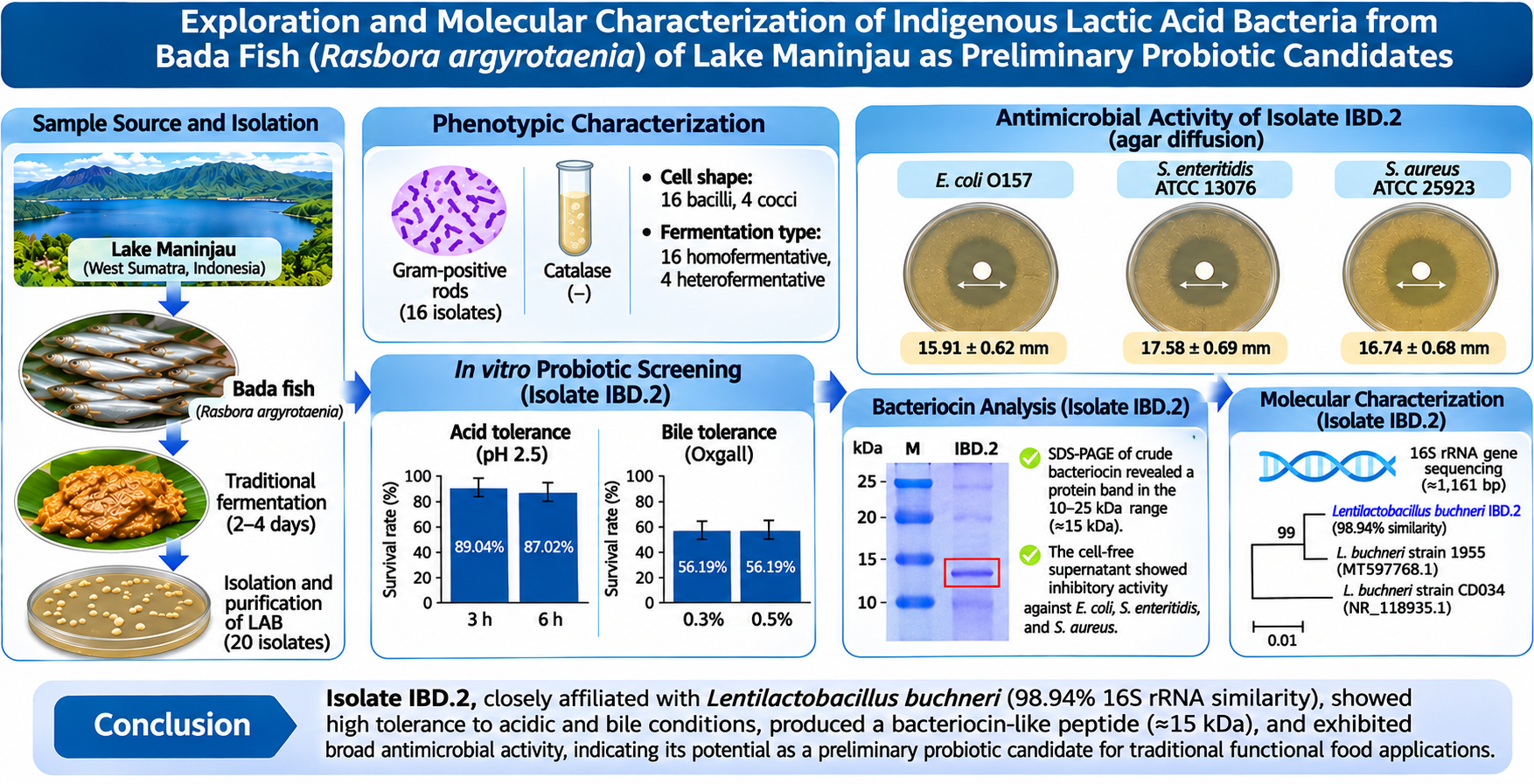
The difference in staining response between Gram-positive and Gram-negative bacteria is determined by the structure of their cell walls. Gram-positive bacteria have a thick peptidoglycan layer (approximately 50–90%), so during the decolorization stage, dehydration occurs, causing the pores to narrow and the CV I complex to remain trapped inside the cell. The Gram staining results in this study showed that all 20 LAB isolates were Gram-positive (+), indicated by a purple color that was maintained after the decolorization process. Of these, 16 isolates were rod-shaped (bacilli), and 4 isolates were spherical (cocci) (Table 2 and Figure 4), corresponding to 80% (16/20) and 20% (4/20) of the isolates, respectively. Gram staining and cell shape are presumptive phenotypic criteria that indicate a Gram-positive cell-wall architecture and cellular morphology; the terms bacilli and cocci describe cell shape only and do not denote a bacterial genus. All four coccoid isolates (IBB.1, IBB.2, IBC.1, and IBC.5) were also heterofermentative (Table 2), whereas all rod-shaped isolates were homofermentative, indicating phenotypic heterogeneity within the LAB collection. Because morphological and physiological traits alone cannot resolve LAB at the genus or species level, taxonomic assignment of the isolates requires molecular identification, and only the selected isolate IBD.2 was identified by 16S rRNA gene sequencing (Section 3.4). This Gram-positive nature not only confirms the basic characteristics of LAB but is also related to resistance to conditions (Figure 4). The results of this study are not much different from the results of the study [9] on tilapia, which found 20 isolates classified as Gram-positive bacteria and rod-shaped (bacilli), while [5] found 15 isolates classified as Gram-positive bacteria with rod-shaped (bacilli) in Bada fish, which live in densely populated areas in Lake Maninjau.
This is in accordance with the statement [25], which states that Gram-positive bacteria will take on a purple crystal violet color even though they have been washed with alcohol, and when given red safranin, the bacteria will remain purple, while the red color indicates Gram-negative bacteria.
Meanwhile, biochemical characterization was carried out through catalase tests and LAB fermentation types (Table 2). The catalase test aims to determine the ability of a microorganism to produce the catalase enzyme, an enzyme that converts H2O2 into water and O2 gas [9]. This test is carried out by dropping a 3% H2O2 solution on the test microorganism. A positive reaction in the catalase test is indicated by the formation of O2 gas bubbles. LAB is catalase-negative, which means it cannot produce the catalase enzyme, so it cannot convert H2O2 into water and O2 gas [26]. This is in accordance with the results obtained; no O2 gas bubbles were formed in the 20 Bada fish LAB isolates tested, so all LAB isolates obtained were catalase-negative. Further biochemical characterization was performed through LAB fermentation type testing (Table 2). Lactic acid bacteria are classified into two main groups, homofermentative and heterofermentative, based on the end product of carbohydrate fermentation. Homoferm-entative LAB produce lactic acid as the main product, while heterofermentative LAB produce various products such as lactic acid, ethanol, carbon dioxide (CO₂), and acetic acid. Determination of fermentation type was carried out by observing gas formation in a Durham tube positioned upside down in a liquid culture medium after 48 hours of incubation. Isolates were categorized as heterofermentative if gas formed in the Durham tube, while the absence of gas indicated homofermentative behavior. In this study, 16 LAB isolates from Bada fish (Rasbora argyrotaenia) showed homofermentative behavior, while 4 other isolates were heterofermentative, consisting of 2 isolates from sample IBB and 2 isolates from sample IBC.
Differences in fermentation characteristics between isolates are thought to be influenced by environmental factors, particularly the conditions of the sampling locations. Samples IBB and IBC originated from areas with high human activity, such as densely populated residential areas, traditional markets, and tourist destinations. Anthropogenic activities in these areas have the potential to increase pollution loads, particularly due to the direct discharge of domestic and market waste into Lake Maninjau. This condition can disrupt the balance of the aquatic ecosystem and affect the dynamics of the microbial community, including variations in the physiological and metabolic characteristics of the isolated LAB. Thus, environmental factors play an important role in determining the diversity and functional properties of microorganisms that thrive in these habitats [19].
3.3. LAB Selection of Bada Fish from Lake Maninjau, West Sumatra as a Probiotic Candidate
Of the 20 isolates, the four heterofermentative isolates (IBB.1, IBB.2, IBC.1, and IBC.5), all of which were coccoid and produced gas in the Durham tube, were excluded from probiotic screening. LAB selection as probiotic candidates was therefore conducted on the remaining 16 isolates, which were Gram-positive, catalase-negative, and homofermentative (no gas production in the fermentation test).
3.3.1 Resistance of LAB Isolates of Bada Fish from Lake Maninjau, West Sumatra to Stomach pH.
Testing the resistance of LAB to gastric pH was carried out. Testing the resistance of LAB to gastric pH was carried out at pH 2.5 because the pH in the proventriculus and gizzard was 2.5 - 3.5; the feed transit time was 70 minutes, and tested for 3 hours and 6 hours. The LAB isolates tested for resistance to pH 2.5 were evaluated from viable plate counts (CFU/mL) on MRS agar, as described in Section 2.4.2 [24], The results were obtained in Table 3.
As shown in Table 3, all 16 presumptive LAB isolates remained culturable after exposure to pH 2.5 for 3 and 6 h. Survival rates, calculated from viable counts (CFU/mL) relative to the initial counts at 0 h, ranged from 72.34% to 89.04% after 3 h and from 58.34% to 87.02% after 6 h. The variation among isolates suggests differences in their ability to withstand the acidic conditions used in this assay. Acid tolerance was used as one of the screening endpoints [8]. Sixteen LAB isolates were tested for their resistance to pH 2.5 using the viable plate-count method described in the The isolates with the highest resistance were IBD.2, IBB.5, IBA.2, IBB.3, IBC.7, IBC.4, IBA.4 and IBA.3, which showed the highest level of resistance to acidic conditions. All isolates were derived from fermented bada fish obtained from Lake Maninjau, West Sumatra. The survival rates after 3 hours of incubation were 89.04%, 88.76%, 88.12%, 86.61%, 86.27%, 84.91%, 83.25% and 81.06%, respectively. After 6 hours of incubation, there was a slight decrease in viability, with values of 87.02%, 86.42%, 85.18%, 84.01%, 83.05%, 81.19%, 80.73%, and 78.65%, respectively. These results indicate sustained acid tolerance in the eight isolates highlighted here. Acid tolerance alone did not determine final candidate selection; eligibility also required ≥50% mean survival in the 3-h bile-salt assay. The survival rate differences of isolates IBD.2, IBB.5, IBA.2, IBB.3, IBC.7, IBC.4, IBA.4, and IBA.3 were 2.02, 2.34, 2.94, 2.60, 3.22, 3.72, 2.52, and 2.41, respectively. These relatively small survival rates indicate high cell viability stability under acidic conditions. This finding is consistent with previous reports, which stated that LAB isolates with low viability decline rates have better probiotic potential. In addition, the results of this study showed superior performance compared to previous studies that reported a survival rate of LAB from okara of 78.34% after 2 hours of incubation at pH 2.5 [5]. Compared with other studies, strains such as Lactobacillus brevis, Lactobacillus plantarum, and Pediococcus ethanol-indurans isolated from traditional fermented products showed survival rates at pH 2.5 for 4 hours of 33–64%, 35–85%, and 40–76%, respectively [27], Meanwhile, Lentilactobacillus buchneri strains isolated from fermented flour were reported to have a survival rate of ≥80% after 4 hours of incubation at the same pH [26]. Thus, LAB isolates from bada fish (Rasbora argyrotaenia) in this study showed competitive acid resistance, even tending to be higher than some other isolate sources. Physiologically, probiotic candidates must be able to survive extreme conditions throughout the digestive tract, from the mouth to the intestines. The acidic environment of the stomach acts as a first barrier that selects microorganisms before they reach the intestines, making the ability to survive at low pH a critical parameter in probiotic evaluation [28].
3.3.2. Resistance of Lab Isolates of Bada Fish from Lake Maninjau, West Sumatra to Bile Salts
Bile-salt tolerance of the presumptive LAB isolates from bada fish was assessed in MRS broth containing 0.3% (w/v) Oxgall. Viable counts (CFU/mL) were determined immediately after inoculation (0 h) and after 3 and 6 h of incubation at 35 °C. Survival percentages at 3 and 6 h were calculated relative to the corresponding 0-h viable count for each isolate, and the results are presented in Table 4.
Following exposure to 0.3% Oxgall, all 16 presumptive LAB isolates remained culturable, with survival rates ranging from 39.86% to 58.66% at 3 h and from 30.15% to 56.19% at 6 h (Table 4). Applying the ≥50% criterion to the mean survival values at 3 h identified six isolates that met the bile-salt selection threshold: IBD.2 (58.66%), IBB.5 (57.35%), IBA.2 (55.41%), IBB.3 (53.49%), IBC.7 (52.18%), and IBC.4 (50.73%). These six isolates also met the acid-tolerance criterion and were therefore retained as preliminary probiotic candidates. IBA.4 (49.57%) and IBA.3 (48.95%) did not meet the 3-h bile-salt threshold and were excluded from this candidate count, although their antimicrobial screening results remain reported. At the separate 6-h bile-exposure endpoint, only IBD.2 (56.19%), IBB.5 (54.11%), and IBA.2 (52.14%) retained ≥50% survival; IBB.3, IBC.7, and IBC.4 fell below the threshold. The resulting candidate count was six at the 3-h bile endpoint and three at 6 h [29].
The decline in survival between 3 and 6 h indicates that bile tolerance depended on exposure duration. IBB.3, IBC.7, and IBC.4 met the 3-h selection criterion but did not retain ≥50% survival at 6 h. Candidate selection was based on the stated 3-h endpoint[30].
3.3.3. Antimicrobial Activity Test of LAB Isolates of Bada Fish from Lake Maninjau, West Sumatra,
Eight acid-tolerant LAB isolates from Bada fish (Rasbora argyrotaenia) were screened for antimicrobial activity, including the six isolates meeting the 3-h bile-salt criterion and two additional isolates, IBA.3 and IBA.4, that did not meet this criterion. The eight tested isolates showed inhibitory activity against the growth and development of pathogenic bacteria, namely Escherichia coli O157, Staphylococcus aureus ATCC 25923, and Salmonella enteritidis ATCC 13076. This inhibitory activity was shown through the formation of inhibition zones as presented in Table 5.
The results obtained from the study can be seen in Table 5, showing that the area of the clear zone/inhibition zone of each isolate varies greatly. This is due to the different abilities of each bacterium from each isolate. The formation of a clear zone is due to the metabolic results of LAB producing lactic acid, hydrogen peroxide, and possibly other antimicrobial metabolites, such as bacteriocin-like substances (not characterized in this study), which have antimicrobial properties [31]. The LAB isolate with the largest inhibition zone on Escherichia coli O157 was IBD.2 isolate with a diameter of 15.91 mm, while the lowest was IBA.4, namely 12.37 mm. The largest diameter of the inhibition zone against Staphylococcus aureus ATCC 25923 bacteria in IBD.2 isolate was 16.74 mm, while the lowest was IBA.3, namely 13.85 mm. The largest diameter of the inhibition zone against Salmonella enteritidis ATCC 13076 bacteria which had the largest inhibition zone was IBD.2 isolate with a diameter of 17.58 mm, while the lowest was IBA.2 isolate at 15.23 mm.
Based on the results of previous tests, the LAB IBD.2 isolate was selected to continue the antimicrobial activity test against pathogenic bacteria using penicillin, kanamycin, and ampicillin antibiotics as positive controls to compare the diameter of the inhibition zone formed in the test bacteria. The use of these antibiotics is based on their ability to inhibit the growth of pathogenic bacteria, especially Gram-positive bacteria used in this study. According to [5], penicillin antibiotics are β-lactam antibiotics that work by inhibiting bacterial cell wall synthesis so they are effective against Gram-positive bacteria. In addition, ampicillin as a penicillin derivative has a broader spectrum of action because it is able to inhibit the growth of Gram-positive and Gram-negative bacteria through the same mechanism, namely inhibiting bacterial cell wall synthesis. Meanwhile, kanamycin is an aminoglycoside antibiotic that works by inhibiting bacterial protein synthesis through interference with ribosomal subunits, so it can provide antimicrobial activity against various pathogenic bacteria [32].
Antibiotics were administered using paper discs containing the specified concentrations of (µg) penicillin 10, ampicillin 10, and kanamycin 30. To determine the re-sistance and sensitivity of the pathogenic bacteria, a positive control antibiotic was used. The inhibition-zone diameters observed for the antimicrobial preparation der-ived from isolate IBD.2 against the tested indicator bacteria are presented in Table 6. These results demonstrate in vitro inhibitory activity under the assay conditions used. Based on Table 6, the LAB IBD.2 isolate was able to inhibit the growth of all pathogenic test bacteria used. The highest inhibitory activity was shown against Escherichia coli O157:H7 with a clear zone diameter of 24.57 mm, followed by Staphylococcus aureus ATCC 25923 at 18.82 mm, and Salmonella enteritidis ATCC 13076 at 10.32 mm. The results showed that the LAB IBD.2 isolate had higher antimicrobial activity against Escherichia coli O157:H7 compared to the control antibiotics 10 μg penicillin, 30 μg kanamycin, and 10 μg ampicillin (Figure 5). Overall, Figure 5 shows that the stained LAB IBD.2 isolate has antimicrobial activity against all tested pathogenic bacteria.
This activity is better than the control antibiotic, where penicillin was unable to inhibit the growth of S. aureus ATCC 25923 and S. enteritidis ATCC 13076, while kanamycin did not show antimicrobial activity against all tested pathogenic bacteria. These results are in line with research [28], which reported that penicillin was unable to inhibit the growth of Escherichia coli O157, so the bacteria were categorized as resistant to penicillin antibiotics. The high antimicrobial activity of LAB IBD.2 isolate is indicated by the large diameter of the inhibition zone formed against pathogenic bacteria. According to [29], antimicrobial activity based on the diameter of the inhibition zone is classified into four categories, namely weak (<5 mm), moderate (5–10 mm), strong (>10–20 mm), and very strong (>20–30 mm). Based on this classification, Among the LAB isolates evaluated, IBD.2 exhibited the strongest antimicrobial activity against the tested pathogenic bacteria. In addition, this isolate demonstrated promising probiotic characteristics based on its acid and bile salt tolerance, Gram-staining character-ristics, catalase reaction, and fermentation type. Nevertheless, the inhibition-zone results should be interpreted as evidence of in vitro antimicrobial activity under the experimental con-ditions and not as evidence of superiority over conventional antibiotics or clinical effectiveness. The LAB IBD.2 isolate from fermented Bada fish (Rasbora argy-rotaenia) is classified as having high activity against all three pathogenic test bacteria. Inhibition of indicator bac-teria provides an in vitro screening endpoint for candidate selection. It does not demonstrate suppression of micro-organisms in the gastrointestinal tract or a health benefit to the host; such effects require separate evaluation [25].
3.3.4. Antimicrobial Activity of the Crude Bacteriocin Supernatant
After pH neutralization of the LAB supernatant, the antimicrobial activity of the neutralized cell-free supernatant from LAB isolate IBD.2 was tested against pathogenic bacteria. Neutralization was carried out to reduce the influence of organic acids, especially lactic acid, which is the main antimicrobial component of LAB [33]. The test results in Table 7 show that the neutralized supernatant still showed residual antimicrobial activity against Escherichia coli O157:H7 and Staphylococcus aureus ATCC 25923 after pH neutralization, whereas no activity was observed against Salmonella enteritidis ATCC 13076. However, pH neutralization only reduces the contribution of organic acids and does not confirm the presence or activity of bacteriocins, because other antimicrobial compounds may also contribute to the observed inhibition. Because confirmatory assays such as protease sensitivity or partial purification were not performed, the inhibition is interpreted only as residual antimicrobial activity of the neutralized supernatant.
Results of the study can be seen in Table 7 which show the antimicrobial activity after the pH of the lactic acid bacteria supernatant was neutralized against Escherichia coli O157: H7 of 17.23 ± 0.31 mm and S. aureus ATCC 25923 of 21.42 ± 0.28 mm mm, but antimicrobial activity was not shown against Salmonella enteritidis ATCC 13076. The results of the study obtained were higher when compared with the results of the study conducted by [6], in his study, finding LAB isolates from Bilih fish from Lake Singkarak had antimicrobial activity in the cell-free supernatant against Staphylococcus aureus ATCC 25923 (13.1 mm) and Escherichia coli O157: H7 (12.7 mm). Meanwhile, the study [26] did not find antimicrobial activity in LAB isolates from raw beef after the pH of the supernatant was neutralized, this is thought to be because LAB produces organic acids that affect antimicrobial activity. Previous research reported that Lactobacillus plantarum NS isolated from various fermented freshwater fish-based food products showed that organic acids have an important role in antimicrobial activity [15]. Inhibitory activity began to be detected in the final phase of exponential growth, namely at an incubation time of 12–15 hours, especially against the pathogenic bacteria Bacillus cereus, Escherichia coli, and Listeria monocytogenes. Meanwhile, the highest antibacterial activity against Staphylococcus aureus and Salmonella enteritidis ATCC 13076murium ATCC 14028 was reported to occur at an incubation time of 21–24 hours. Other research also showed that Lactobacillus plantarum and Lactobacillus brevis isolated from fermented bekasam food from South Sumatra have antimicrobial activity through cell-free culture supernatant and can inhibit the growth of E. coli and L. monocytogenes [5]. The antimicrobial activity of LAB supernatants may involve organic acids and other metabolites and, in some strains, bacteriocins (not confirmed in the present study). Bacteriocins are antimicrobial peptides or proteins that can act through several mechanisms, such as inhibiting the formation of lipid II as a cell wall precursor, forming pores in the target cell membrane, and inhibiting cell wall biosynthesis [28]. This mechanism begins with the interaction of bacteriocin peptides with the phospholipid layer of the target cell membrane through electrostatic bonds between the positively charged groups on the peptides and membrane fatty acids. This interaction causes the formation of pores in the membrane, increasing cell permeability, and ultimately resulting in bacterial cell death [23]. Bactericidal effects of bacteriocins have been described in the literature [34] . These mechanisms and effects were not demonstrated for IBD.2 in the present study; the inhibition-zone assay supports only growth-inhibitory activity under the tested conditions.
The neutralized cell-free supernatant of isolate IBD.2 retained antimicrobial activity against Escherichia coli O157 and Staphylococcus aureus ATCC 25923, whereas no inhibitory activity was detected against Salmonella enteritidis ATCC 13076. These findings suggest that factors other than the direct effect of organic acid-mediated acidity may contribute to the observed inhibition. Bacteriocins or other proteinaceous compounds are possible candidates; however, their involvement remains hypothetical because protease-sensitivity assays, activity-guided purification, and direct identification of the active compound were not performed. Therefore, the observed inhibition is interpreted as antimicrobial activity of the neutralized cell-free supernatant rather than confirmed bacteriocin activity.
3.3.5. Preliminary Protein-Size Charac-terization by SDS-PAGE of Isolate IBD.2 from Bada Fish of Lake Maninjau, West Sumatra, Indonesia
Testing using SDS Page is intended to determine the size of the protein using the SDS-PAGE method which is determined through the formation of protein bands to determine the molecular weight of protein bands present in the analyzed sample (Figure 6). Based on the results of SDS-PAGE analysis, protein bands with molecular sizes ranging from 10–25 kDa were detected. This result should be regarded only as a preliminary protein-size charac-terization. SDS-PAGE estimates the apparent molecular mass of proteins, but it does not by itself demonstrate that a detected band is responsible for the observed antimic-robial activity or that it belongs to a particular bacteriocin class. In addition, class III bacteriocins are conventionally defined as large (>30 kDa), heat-labile proteins, so the 10–25 kDa bands detected here cannot be used to classify the antimicrobial compound as a class III bacteriocin.
Confirming the identity and class of the active compound would require additional biochemical evidence, such as protease sensitivity assays, partial purification with activity-guided identification of the active band (e.g., gel overlay assay), and/or mass spectrometry. Previous studies reported that Lactobacillus pentosus SJ65 was able to produce bacteriocins with molecular weights of 16 and 39 kDa after being purified using acetone extraction, cross-linked glucosan chromatography, and reversed-phase high-performance liquid chromatography [16]. Meanwhile, It has been reported that [14] Lactobacillus pentosus can also produce bacteriocins with a lower molecular weight, approximately 2.9 kDa, through purification using macroporous resin, gel filtration chromatography, and reversed-phase high-performance liquid chromatography. These differences in bacteriocin molecular weight indicate that the characteristics of bacteriocins are strongly influenced by the bacterial strain and the purification method used.
3.4. Identification of Selected Isolates of IBD.2 Bada Fish Using 16S rRNA
The electrophoresis results showed that the PCR process successfully amplified the 16S rRNA gene of LAB isolated from Bada fish (Rasbora argyrotaenia). The success of amplification was indicated by the formation of a DNA fragment measuring ±1,500 bp using the universal primer 27F (AGAGTTTGATCCTGGCTGAG) as the forward primer and 1429R (GTTTACCTTACGACTT) as the reverse primer. Amplification of the 16S rRNA gene was carried out to ensure the successful isolation of genomic DNA as well as the basis for molecular identification of LAB isolates [35]. The PCR products obtained were then used for nucleotide sequencing analysis to determine the identity of the bacteria more specifically. The electrophoresis results of the PCR products of LAB isolates are shown in Figure 7. A molecular approach through 16S rRNA gene analysis was used to evaluate the genetic relationships of LAB by comparing ribosomal RNA sequences between isolates [36]. The 16S rRNA gene is a commonly used molecular marker in bacterial identification because it has conserved and variable regions that are able to differentiate bacteria down to the species and strain levels [37]. In addition, the relatively small size of the 16S rRNA molecule allows for direct sequencing without the need for PCR amplicon cloning. Figure 8 shows the results of DNA amplification with a fragment size of approximately 1.514 bp on an agarose gel, indicating that the specific primers used in this study were able to optimally amplify the target gene and support the molecular identification process of LAB isolates down to the strain level. The chromatograms of the sequencing results from the forward and reverse primer readings of the 16S rRNA gene in the IBD.2 isolate bacteria were aligned using the SeqMan program. Amplification of the 16S rRNA gene using primers 16SrRNA_27F and 16SrRNA_1525R produced an amplicon of 1.514 bp, with the chromatogram readings of the forward primer 16SrRNA_27F being 726 bp long and the reverse primer 16SrRNA_1525R being 1,131 bp long. As a limitation of the Sanger sequencing method, some bases at the ends of the chromatogram showed overlapping peaks, were less distinct, and had low signal intensity, which could affect the accuracy of the nucleotide base reading.
Therefore, a trimming process was carried out on the low-quality base sections before further assembly and sequence analysis [38]. The chromatogram of the 16SrRNA_27F forward primer reading results showed a decrease in signal quality at the end of the sequence, so trimming was performed along the 5' end of 40 bp. At a position of approximately 400 bp towards the 3' end, the chromatogram peak appears increasingly sloping and even tends to be flat, indicating low quality base reading. Therefore, the editing process was carried out by adjusting the base sequence based on the results of the reverse primer reading to improve sequence accuracy. Meanwhile, the chromatogram of the 16SrRNA_1525R reverse primer reading results also underwent trimming along the 3' end of 81 bp and 5' end of 30 bp to remove bases with low signal quality.
After the trimming, editing, and assembly stages were carried out, the sequence of bacterial 16S rRNA gene frag-ments was obtained. isolate IBD.2 with a final length of 1.161 bp. The visualization of the edited chromatogram is as follows [39]. The base sequence of the 16S rRNA gene fragment of the IBD.2 isolate bacteria obtained from the re-sults of reading the primers 16SrRNA_27F to 1525R with a length of 1,161 bp is as follows >Contig_IBD.2 (1,1160) The base sequence of the 16S rRNA gene of the IBD.2 bacterial isolate from the primary reading results 16S rRNA_27F to 1525Rthe above was then BLASTed. The BLAST results show information based on the taxonomy, from 100 BLAST sequences of Isolate IBD.2 bacteria, a query cover value of 100% was obtained. The sequence identity to the NCBI reference strain 1955 (MT597768.1) was 98.94%, as reported in the editable BLAST table. The BLASTn analysis showed that the 16S rRNA gene sequence of isolate IBD.2 shared 98.94% sequence identity with the reference sequence of Lactobacillus buchneri strain 1955 (GenBank accession number MT597768.1), with a query coverage of 100%. This result indicates a high degree of sequence similarity between isolate IBD.2 and the reference sequence. However, this similarity alone does not establish definitive species-level identification. The taxonomic interpretation should also consider the align-ment length, the reliability of the reference sequence, com-parisons with closely related taxa, and the phylo-genetic analysis. A total of 15 sequences of Lentilac-tobacillus buchneri bacteria from Gene Bank were selected for usage in genetic kinship analysis. The information characteris-tics of 15 bacterial and sequences and accession numbers are as Table 8.
Based on Table 8, isolate IBD.2 showed a similarity level of 98.94% to the 16S rRNA gene sequence of the reference strain Lentilactobacillus buchneri strain 1955 (MT597768.1), which is used here only as a reference/comparison strain. This similarity level indicates a close taxonomic affiliation of isolate IBD.2 with L. buchneri, but it does not confirm species- or strain-level identity. This is in accordance with the statement that a sequence can be said to be homologous if it has a similarity value of more than 70% [1]. Homologous analysis was performed using the Basic Local Alignment Search Tool (BLAST) method; then the results were visualized in the form of a phylogenetic tree using the MEGA application after previously carrying out the sequence alignment process using the MUSCLE algorithm in MEGA 11 [40].
Phylogenetic visualization results indicate that the lactic acid bacteria isolate from fermented Bada fish (Rasbora argyrotaenia) is apomorphically related to Lentilactobacillus buchneri. Apomorphic characters are derived characters that have undergone evolutionary changes and are shared by ingroups, while synapomorphic characters are derived characters shared by a monophyletic group [36]. The close phylogenetic relationship indicates that isolate IBD.2 shares strong evolutionary similarities with Lentilactobacillus buchneri, indicating close taxonomic affiliation but not confirming that it is the same species. The phylogenetic tree above was constructed by selecting 15 species from the BLAST results based on their query cover values. The top 15 species were then downloaded in FASTA format for use in constructing the phylogenetic tree.
The 16S rRNA gene sequence of isolate IBD.2 was analyzed using the NCBI BLAST tool and compared with sequences available in the GenBank database for molecular identification. Accession number MT597768.1 was used as a reference sequence for comparison and does not represent an accession number assigned to isolate IBD.2. The sequence of isolate IBD.2 has not been deposited in GenBank; therefore, no isolate-specific GenBank accession number is currently available.
3.5 Phylogenetic tree
Based on the BLAST results, a phylogenetic tree was constructed to determine the level of kinship between the isolate and other species in NCBI. The phylogenetic tree was constructed using the MEGA 11 by opening the FASTA file downloaded during the BLAST run [41]. The downloaded sequences, together with the IBD.2 sequence, were first aligned using the MUSCLE algorithm in MEGA 11, and then the phylogenetic tree was constructed. The phylogenetic tree was constructed using the Neighbor-Joining method with 1,000 bootstrap replicates to test the confidence level of the phylogenetic branches. Evolutionary distance analysis was performed using the p-distance model. No outgroup was designated, so the tree is interpreted as unrooted. The phylogenetic tree construction illustrates the kinship relationship between the IBD.2 isolate and 15 comparison bacterial sequences obtained from GenBank based on 16S rRNA gene fragments.
The BLAST analysis showed that the 16S rRNA gene sequence of isolate IBD.2 shared 98.94% sequence identity with Lentilactobacillus buchneri strain 1955 (GenBank accession no. MT597768.1).Molecular identification using 16S rRNA gene sequencing was performed only for isolate IBD.2. The other isolates were characterized using preliminary phenotypic methods and were not subjected to molecular sequencing in this study. Therefore, the molecular identification results reported here apply only to isolate IBD.2, whereas the identities of the remaining isolates have not been confirmed at the species level. This limitation should be considered when interpreting the identification results, and further molecular characterization is required to establish the taxonomic identities of the other isolates . Phylogenetic analysis placed IBD.2 alongside this reference strain, which is included for comparison and does not by itself represent the confirmed identity of isolate IBD.2, as shown in Figure 9. The close phylogenetic relationship indicates that the IBD.2 isolate likely belongs to the same genus group and has the potential to produce antimicrobial compounds. In addition, the position of the IBD.2 isolate on the phylogenetic branch adjacent to the Lentilactobacillus buchneri strain 1955 reference (comparison) strain indicates a strong evolutionary relationship based on 16S rRNA gene sequence analysis [13]. The phylogenetic tree showing the kinship relationship between the IBD.2 isolate and 15 comparison bacteria from GenBank is presented in Figure 9.
Phylogenetic trees illustrate evolutionary relationships between organisms based on genetic sequence similarities. The neighbor-joining method is known to be effective in reconstructing evolutionary history and grouping taxonomically close organisms into the same cluster, supported by high bootstrap values [42]. In this study, phylogenetic analysis was performed using MEGA software version 11 using the neighbor-joining method based on the p-distance model, as previously reported by [26]. We used the 16S rRNA gene sequence of isolate IBD.2 and 15 GenBank comparison sequences (16 sequences in total) to evaluate the evolutionary relationships among the bacterial isolates. The resulting phylogenetic tree showed the formation of two main branches encompassing all the bacteria analyzed. The low variation between sequences caused most isolates to cluster very closely. This is due to the high level of similarity of 16S rRNA gene fragment sequences, which ranged from 99.21% to 98.94%, as supported by BLAST analysis, multiple sequence alignment, and genetic distance calculations [3]. This very high level of sequence conservation means that the IBD.2 isolate cannot be clearly identified at the species level based solely on 16S rRNA gene markers. Therefore, the isolate is more accurately identified at the genus level, namely as a member of the genus Lentilactobacillus (closest GenBank reference: L. buchneri strain 1955, used for comparison only; species-level identity was not confirmed).
The homofermentative phenotype observed in this study describes fermentation behavior under the conditions tested. LAB have been reported in fermented foods, and particular strains have been investigated for probiotic applications [32]. The functional and safety limitations of applying these observations to IBD.2 are summarized below. Isolate IBD.2 was obtained from fermented Bada fish (Rasbora argyrotaenia), a traditional food source. This origin describes the source of isolation and does not demonstrate the halal status of the microorganism, culture components, processing materials, or any final product. The present study did not assess or obtain halal certification for these components. The ASUH concept (safe, healthy, whole, and halal) provides contextual background for animal-derived foods [43] , but it is not an experimentally established property of the isolates. Use of these isolates in functional food products intended to meet halal requirements remains a possible future application, contingent on separate verification of the complete production chain and applicable certification [42]. Published observations on particular Lentilactobacillus buchneri strains provide background for investigating related isolates, but they do not establish the safety or functional properties of IBD.2 [12]. The present study is limited to preliminary phenotypic characterization, acid- and bile-tolerance screening, growth-inhibitory assays, preliminary protein-size characterization, and 16S rRNA gene analysis of IBD.2. It did not include direct toxicological safety assessment, animal experiments, clinical studies, or direct evaluation of gastrointestinal or other health outcomes. These data are insufficient for comprehensive probiotic characterization. Further strain-specific functional and safety assessment, together with evidence of relevant host benefits, is required before food or health applications can be substantiated [28].
- CONCLUSION
This study provided preliminary phenotypic and in vitro screening of indigenous lactic acid bacteria (LAB) isolated from fermented Bada fish (Rasbora argyrotaenia) originating from Lake Maninjau, West Sumatra, Indonesia. A total of 20 LAB isolates were obtained and identified as Gram-positive and catalase-negative bacteria, with most of the isolates showing homofermentative characteristics. Re-evaluation of the reported mean survival values identified six preliminary probiotic candidates meeting the ≥50% criterion in both the 3-h acid assay and the 3-h assay with 0.3% Oxgall. These were IBA.2, IBB.3, IBB.5, IBC.4, IBC.7, and IBD.2, with 3-h acid survival of 84.91–89.04% and 3-h bile survival of 50.73–58.66%. At 6 h of bile exposure, only IBA.2, IBB.5, and IBD.2 retained ≥50% survival. Eight acid-tolerant isolates were evaluated for growth-inhibitory activity using inhibition-zone measurements, including IBA.3 and IBA.4, which were excluded from the final candidate count because their 3-h bile survival was below 50%, and isolate IBD.2 produced the largest inhibition zones against Escherichia coli O157 (15.91 mm), Staphylococcus aureus ATCC 25923 (16.74 mm) and Salmonella enteritidis ATCC 13076 (17.58 mm), whereas the inhibition zones produced by the cell-free supernatant against Escherichia coli O157 were 24.57 mm, Staphylococcus aureus ATCC 25923 were 18.82 mm, and Salmonella enteritidis ATCC 13076 were 10.32 mm. These inhibition-zone results demonstrate growth inhibition under the tested conditions and do not establish bactericidal activity or bacterial killing. SDS-PAGE analysis revealed protein bands with molecular weights ranging from 10–25 kDa, which represents only a preliminary protein-size characterization. The identity of the active compound and its bacteriocin class could not be determined because protease sensitivity, activity-guided purification, and mass spectrometry were not performed; the inhibition is therefore reported as antimicrobial activity of the cell-free supernatant. Molecular characterization based on 16S rRNA gene sequencing revealed 98.94% sequence similarity between isolate IBD.2 and the reference sequence of Lentilactobacillus buchneri strain 1955 deposited in NCBI GenBank (accession no. MT597768.1), supporting close taxonomic affiliation with L. buchneri; strain 1955 is a reference/comparison strain and does not represent the confirmed identity of isolate IBD.2, and species- and strain-level identification was not established. These screening results support further investigation of the candidates; safety, host benefits, and suitability for food applications remain to be established through strain-specific functional and safety assessment.
- DECLARATION STATEMENTS
5.1. Funding
This research was funded by the Institute for Research and Community Service (LPPM), Universitas Negeri Padang for the financial support provided through the Beginner Research Grant Scheme (Penelitian pemula) under Grant Contract No. 1953/ UN35.15/LT/2025 and Universitas Negeri Medan
5.2. Ethics and Consent to Participate
Not applicable. This article is a review of previously published literature and involved no human participants, animals, or identifiable personal data.
5.3. Data Availability
datasets were generated or analysed. The literature sources supporting this review are cited in the reference list.
5.4. Competing Interest
The authors declare that they have no competing interests.
5.5. Authors’ Contribution
All authors have made substantial contributions to the conception and design of the study, laboratory experiments, data collection, analysis, interpretation of results, and manuscript preparation. Heppy Setya Prima contributed to the experimental work, data analysis, and manuscript drafting; Rusfidra contributed to supervision, methodology development, and critical revision of the manuscript; Selvia Dewi Pohan contributed to microbiological analysis and manuscript editing; Fajri Maulana and Fadhli Fajri contributed to molecular identification and 16S rRNA analysis; and Malikil Kudus Susalam contributed to study conceptualization, supervision, antimicrobial analysis, and final manuscript revision. All authors have read and approved the final manuscript and agree to be accountable for the integrity and originality of the work.
5.6. Declaration of Generative AI
Artificial Intelligence (AI) chatbots were used solely to improve the language, grammar, spelling, and clarity of the manuscript. The authors reviewed, edited, and verified all AI-assisted text and take full responsibility for the accuracy, originality, and final content of the manuscript.
5.7. Acknowledgements
The authors would like to thank the Biotechnology Study Program, Universitas Andalas, the Department of Biology, Faculty of Mathematics and Natural Sciences, Universitas Negeri Medan (UNIMED), and the Faculty of Animal Science, Universitas Negeri Padang (UNP) for providing research facilities and technical support during this study, as well as all colleagues and laboratory staff who assisted in sample collection, laboratory analysis, and manuscript preparation. The authors also appreciate the valuable suggestions and scientific discussions contributed throughout the research process.
- antimicrobial activity
- fermented fish
- Lentilactobacillus buchneri
- Lactic acid bacteria
- probiotic candidates
ارجاع به مقاله
مراجع
1. Ameen, F. A., Soliman, M. E., Hamdan, A. M., & Hammad, S. F. (2026). Extraction, Purification, and Characterization of a Bacteriocin from Marine Lactococcus lactis NAN6399: Evaluating Antioxidant and Antimicrobial Activities. Microorganisms, 14(5), 1030. https://doi.org/10.3390/microorganisms14051030
2. Ardani, L. R., Marlida, Y., Zain, M., Jamsari, J., & Fassah, D. M. (2023). Lactic acid bacteria and yeast strains isolated from fermented fish (Budu) identified as candidate ruminant probiotics based on in vitro rumen fermentation characteristics. Veterinary World, 16(2), 395. https://doi.org/10.14202/VETWORLD.2023.395-402
3. Borthakur, D., Sharma, B. K., & Upadhye, V. J. (2025). Probiotic and aroma-producing Bacillus sp. isolates from teles, a traditional fermented fish from Assam, India: functional characterisation and potential application in fermented foods. BMC Microbiology, 25(1), 579-. https://doi.org/10.1186/S12866-025-04312
4. Brunet, S., Grankvist, A., Jaen-Luchoro, D., Bergdahl, M., Tison, J. L., Wester, A., Elfving, K., Brandenburg, J., Gullsby, K., Lindsten, C., Arvidsson, L. O., Larsson, H., Eilers, H., Strand, A. S., Lannefors, M., Keskitalo, J., Rylander, F., Welander, J., Jungestrom, M. B., … Hallbäck, E. T. (2025). Nationwide multicentre study of Nanopore long-read sequencing for 16S rRNA-species identification. European Journal of Clinical Microbiology and Infectious Diseases, 44(8), 1907–1916. https://doi.org/10.1007/S10096-025-05158
5. Chan, C. H., Chen, L. H., Chen, K. Y., Chen, I. H., Lee, K. T., Lai, L. C., Tsai, M. H., Chuang, E. Y., Lin, M. T., & Yan, T. R. (2024). Single-strain probiotics enhance growth, anti-pathogen immunity, and resistance to Nocardia seriolae in grey mullet (Mugil cephalus) via gut microbiota modulation. Animal Microbiome, 6(1), 67. https://doi.org/10.1186/S42523-024-00353-0/FIGURES/6
6. Colombo, M., Nero, L. A., & Todorov, S. D. (2020). Safety profiles of beneficial lactic acid bacteria isolated from dairy systems. Brazilian Journal of Microbiology, 51(2), 787–795. https://doi.org/10.1007/S42770-020-00227-Y/METRICS
7. de Souza, P. A., Ramos, J. N., Vasconcellos, L., Costa, L. V., Forsythe, S. J., & Brandão, M. L. L. (2025). Application and Limitations of 16S rRNA Gene Sequencing for Identifying WHO Priority Pathogenic Gram-Negative Bacilli. Infection and Drug Resistance, 18, 6353–6375. https://doi.org/10.2147/IDR.S550704
8. Dissasa, G., Lemma, B., & Mamo, H. (2022). Isolation and identification of major bacteria from three Ethiopian rift valley lakes live and processed fish, and water samples: implications in sanitary system of fish products. BMC Veterinary Research, 18(1), 439 . https://doi.org/10.1186/S12917-022-03508-W/TABLES/5
9. Elsaadany, K., EL-Sayed, A. I. M., & Awad, S. (2024). Identification, Safety Assessment, and Antimicrobial Characteristics of Cocci Lactic Acid Bacteria Isolated from Traditional Egyptian Dairy Products. Foods, 13(19), 3059. https://doi.org/10.3390/FOODS13193059
10. Fotso Techeu, U. D., Kaktcham, P. M., Momo, H. K., Foko Kouam, E. M., Tchamani Piame, L., Ngouenam, R. J., & Zambou Ngoufack, F. (2022). Isolation, Characterization, and Effect on Biofilm Formation of Bacteriocin Produced by Lactococcus lactis F01 Isolated from Cyprinus carpio and Application for Biopreservation of Fish Sausage. BioMed Research International, 2022(1), 8437926. https://doi.org/10.1155/2022/8437926
11. Hamdaoui, N., Benkirane, C., Bouaamali, H., Azghar, A., Mouncif, M., Maleb, A., Hammouti, B., Al-Anazi, K. M., Kumar, P., Yadav, K. K., Choi, J. R., & Meziane, M. (2024). Investigating lactic acid bacteria genus Lactococcus lactis properties: Antioxidant activity, antibiotic resistance, and antibacterial activity against multidrug-resistant bacteria Staphylococcus aureus. Heliyon, 10(11), e31957. https://doi.org/10.1016/j.heliyon.2024.e31957
12. Harnentis, H., Marlida, Y., Nur, Y. S., Wizna, W., Santi, M. A., Septiani, N., Adzitey, F., & Huda, N. (2020). Novel probiotic lactic acid bacteria isolated from indigenous fermented foods from West Sumatera, Indonesia. Veterinary World, 13(9), 1922. https://doi.org/10.14202/vetworld.2020.1922-1927
13. Harnentis, H., Nurmiati, N., Marlida, Y., Adzitey, F., & Huda, N. (2019). γ-Aminobutyric acid production by selected lactic acid bacteria isolate of an Indonesian indigenous fermented buffalo milk (dadih) origin. Veterinary World, 12(8), 1352. https://doi.org/10.14202/VETWORLD.2019.1352-1357
14. Harun, H., Wirasti, Y., Purwanto, B., & Purwati, E. (2020). Characterization of Lactic Acid Bacteria and Determination of Antimicrobial Activity in Dadih from Air Dingin Alahan Panjang District, Solok Regency-West Sumatera. Systematic Reviews in Pharmacy, 11(3). https://doi.org/10.31838/srp.2020.3.76
15. Javadi, K., Emadzadeh, M. R., Mohammadzadeh Hosseini Moghri, S. A. H., Halaji, M., Parsian, H., Rajabnia, M., & Pournajaf, A. (2025). Anti-biofilm and antibacterial effect of bacteriocin derived from Lactobacillus plantarum on the multidrug-resistant Acinetobacter baumannii. Protein Expression and Purification, 226, 106610. https://doi.org/10.1016/J.PEP.2024.106610
16. Kahraman-Ilıkkan, Ö. (2024). Comparative genomics of four lactic acid bacteria identified with Vitek MS (MALDI-TOF) and whole-genome sequencing. Molecular Genetics and Genomics, 299(1), 31. https://doi.org/10.1007/S00438-024-02129-2
17. Konno, N., Maeno, S., Tanizawa, Y., Arita, M., Endo, A., & Iwasaki, W. (2024). Evolutionary paths toward multi-level convergence of lactic acid bacteria in fructose-rich environments. Communications Biology 2024 7:1, 7(1), 902. https://doi.org/10.1038/s42003-024-06580-0
18. Kudaibergenova, A., Begdildayeva, N., Amirkhanova, A., Nurgazina, A., Ospanova, A., Kondybayev, A., Jumagaziyeva, A., Iskakbayeva, Z., Jumabayeva, S., & Akhmetsadykova, S. (2025). Probiotic Characterization of Lactic Acid Bacteria Isolated from Traditional Fermented Camel and Mare’s Milk Products. Https://Www.Espublisher.Com/, 21(0), 1763. https://doi.org/10.30919/FAF1763
19. Li, J., Wang, Y., Xu, X., Zheng, X., Wang, Z., Chen, X., Wang, L., Mao, L., Ge, W., Liu, H., Gao, W., Wang, T., & Wang, Z. (2025). Safety and Probiotics Evaluation of Bifidobacterial Genomes Isolated from Probiotic Products. Current Microbiology, 82(6), 246. https://doi.org/10.1007/S00284-025-04139-8
20. Li, X., Zhu, L., Wang, X., Li, J., & Tang, B. (2022). Evaluation of IR Biotyper for Lactiplantibacillus plantarum Typing and Its Application Potential in Probiotic Preliminary Screening. Frontiers in Microbiology, 13, 823120. https://doi.org/10.3389/FMICB.2022.823120
21. Madoromae, H., Atipairin, A., Tayeh, M., & Lertcanawanichakul, M. (2025). Investigating the production and synergistic antibacterial activity of bacteriocin-like substance from Brevibacillus laterosporus SA-14 (TISTR 2453) for enhanced wound healing. Heliyon, 11(4). https://doi.org/10.1016/j.heliyon.2025.e42510
22. Malakian, A., Dehghan, S., Abbasi Montazeri, E., Aramesh, M. R., Dehdashtian, M., & Aletayeb, S. M. H. (2025). 16 S rRNA–based molecular identification of coagulase-negative Staphylococcus species in neonates with sepsis and their antibiotic resistance patterns in Ahvaz, Iran. BMC Microbiology, 25(1), 114. https://doi.org/10.1186/S12866-025-03809-8
23. Marlida, Y., Harnentis, H., Nur, Y. S., & Ardani, L. R. (2023). New probiotics (Lactobacillus plantarum and Saccharomyces cerevisiae) supplemented to fermented rice straw-based rations on digestibility and rumen characteristics in vitro. Journal of Advanced Veterinary and Animal Research, 10(1), 96. https://doi.org/10.5455/JAVAR.2023.J657
24. Mazlumi, A., Panahi, B., Hejazi, M. A., & Nami, Y. (2022). Probiotic potential characterization and clustering using unsupervised algorithms of lactic acid bacteria from saltwater fish samples. Scientific Reports 2022 12:1, 12(1), 11952. https://doi.org/10.1038/s41598-022-16322-z
25. Nofiani, R., Ardiningsih, P., Adhitiyawarman, & Sarwiyati. (2022). Characteristics of Lactic Acid Bacteria isolated from traditional fermented fish. Biodiversitas: Journal of Biological Diversity, 23(11), 5662. https://doi.org/10.13057/BIODIV/D231116
26. Nurhayati, A. P. D., Zulaika, E., Amin, M., Setiawan, E., & Wijaya, Z. M. (2023). Isolation and screening of lactic acid bacteria producing anti-Edwardsiella from the gastrointestinal tract of wild catfish (Clarias gariepinus) for probiotic candidates. Open Agriculture, 8(1). https://doi.org/10.1515/opag-2022-0212
27. Pereira, W. A., Piazentin, A. C. M., de Oliveira, R. C., Mendonça, C. M. N., Tabata, Y. A., Mendes, M. A., Fock, R. A., Makiyama, E. N., Corrêa, B., Vallejo, M., Villalobos, E. F., & Ricardo, R. P. (2022). Bacteriocinogenic probiotic bacteria isolated from an aquatic environment inhibit the growth of food and fish pathogens. Scientific Reports 2022 12:1, 12(1), 5530. https://doi.org/10.1038/s41598-022-09263-0
28. Prima, H. S., Maulana, F., Rusfidra, Agasi, S. Y., Susalam, M. K., & Agusti, M. A. (2026). Effect of Feeding a Combination of Fish Waste and Indigofera zollingeriana Leaves as Diet for Laying Quails (Coturnix coturnix japonica): Impact on Production Performance. Journal of Animal Health and Production, 14(1), 38–45. https://doi.org/10.17582/JOURNAL.JAHP/2026/14.1.38.45
29. Prima, H. S., Rusfidra, R., Yansen, F., Maulana, F., & Gusti, M. A. (2025). Characterization, molecular identification and antimicrobial activity of lactic acid bacteria with potentials as halal probiotics isolated from Rinuak fish (Psilopsis sp.) in Lake Maninjau, West Sumatra, Indonesia. Applied Food Biotechnology, 12(1), 1-16. https://doi.org/10.22037/afb.v12i1.48364
30. Prima, H. S., Rusfidra, R., Yansen, F., & Satrianto, A. (2023). Characterization of Gamma Aminobutyric Acid-producing Lactic Acid Bacteria Isolated from Budu Fish in Padang Pariaman West Sumatra Indonesia and their Potentials as Probiotics. Applied Food Biotechnology, 10(4), 257-270. https://doi.org/10.22037/AFB.V10I4.42951
31. Prima, H. S., Satrianto, A., & Amar, S. (2022). Antimicrobial Potential of Limosilactobacillus Fermentum Isolated from Bilih Fish (Mystacoleucus padangensis) of Singkarak Lake, West Sumatera, Indonesia. Applied Food Biotechnology, 9(4), 297-309. https://doi.org/10.22037/AFB.V9I4.38487
32. Raimi, A., & Adeleke, R. (2023). 16S rRNA gene-based identification and plant growth-promoting potential of cultivable endophytic bacteria. Agronomy Journal, 115(3), 1447-1462. https://doi.org/10.1002/AGJ2.21241
33. Reuben, R. C., Roy, P. C., Sarkar, S. L., Rubayet Ul Alam, A. S. M., & Jahid, I. K. (2020). Characterization and evaluation of lactic acid bacteria from indigenous raw milk for potential probiotic properties. Journal of Dairy Science, 103(2), 1223-1237. https://doi.org/10.3168/JDS.2019-17092
34. Saad, M. F., Elsayed, M. M., Khder, M., Abdelaziz, A. S., & El-Demerdash, A. S. (2024). Biocontrol of multidrug resistant pathogens isolated from fish farms using silver nanoparticles combined with hydrogen peroxide insight to its modulatory effect. Scientific Reports 2024 14:1, 14(1), 7971. https://doi.org/10.1038/s41598-024-58349-4
35. Sadeghi, M., Panahi, B., Mazlumi, A., Hejazi, M. A., & Nami, Y. (2022). Screening of potential probiotic lactic acid bacteria with antimicrobial properties and selection of superior bacteria for application as biocontrol using machine learning models. LWT, 162, 113471. https://doi.org/10.1016/J.LWT.2022.113471
36. Sharma, A., Lee, S., & Park, Y. S. (2020). Molecular typing tools for identifying and characterizing lactic acid bacteria: a review. In Food Science and Biotechnology (Vol. 29, Number 10, pp. 1301–1318). The Korean Society of Food Science and Technology. https://doi.org/10.1007/s10068-020-00802-x
37. Singh, J. K., Devi, P. B., Rani, P. U., Rajapuram, D. R., Kavitake, D., Reddy, G. B., & Shetty, P. H. (2025). Purification and characterization of a bacteriocin produced by the probiotic Lactiplantibacillus plantarum subsp. argentoratensis strain I1B. 3 Biotech, 15(5), 137. https://doi.org/10.1007/S13205-025-04279-5
38. Susalam, M. K., Harnentis, Jamsari, & Marlida, Y. (2023). New Growth Medium for Culturing Lactic Acid Bacteria As Probiotic Consortium Isolated From Fermented Fish (Budu). Online Journal of Animal and Feed Research, 13(3), 199-203. https://doi.org/10.51227/OJAFR.2023.30
39. Syah, S. P., Mukhlisah, A. N., Irfan, M., Ningtyas, W. D., Marsanda, Armayanti, Yutia, R., Bulan, R. E., Andika, Aswin, & Wahid, A. (2026). Probiotic Potential of Lactic Acid Bacteria Isolated from Dangke: In Vitro Antimicrobial Activity, Hemolytic Activity, and Antibiotic Susceptibility Profile. IOP Conference Series: Earth and Environmental Science, 1603(1), 012017. https://doi.org/10.1088/1755-1315/1603/1/012017
40. Taghipour, T., Rasti, S., Saba, M., Delavari, M., Moosavi, G. A., Hooshyar, H., & Eslamirad, Z. (2022). Molecular detection and genotype identification of Acanthamoeba species from bronchoalveolar lavage of patients with pulmonary symptoms suspected of cancer. Journal of Parasitic Diseases, 46(4), 1028-1035. https://doi.org/10.1007/S12639-022-01524-Z
41. Uddin, M. S., Chamonara, K., Nayem, M. R., Siddiqua, A., Chowdhury, S., Hossain, I., Ahasan, A. S. M. L., & Masum, M. H. U. (2026). Comparative gut microbiome analysis of Rohu fish from Halda River and Kaptai Lake using 16S rRNA sequencing. Scientific Reports 2025 16:1, 16(1), 8811. https://doi.org/10.1038/s41598-025-33754-5
42. Vargas, C., Botero, L., Rodriguez, E., Dávila, L., Racedo, C., Barrera, C., Bernal, A. J., Jiménez, H., Zuluaga, P., & González, C. (2025). Genome mining to elucidate antimicrobial and plant growth promoting potentials in Lactic acid bacteria. European Journal of Plant Pathology, 172(1), 7-22. https://doi.org/10.1007/S10658-024-02984-0
43. Yusra, Y., Syandri, H., Efendi, Y., & Huda, N. (2022). Characterization of lactic acid bacteria isolated from budu, a West Sumatra fish fermentation product, and their ability to produce exopolysaccharides. F1000Research, 11, 1139. https://doi.org/10.12688/f1000research.109401.1
- چکیده مشاهده شده: 119 بار
- Pdf (English) دانلود شده: 26 بار