Development and Characterization of Polysaccharide-plant Protein Composite Hydrogels as Potential Scaffolds for Cultivated Meat
بیوتکنولوژی غذایی کاربردی,
دوره 13 شماره 1 (2026),
7 دسامبر 2025
,
صفحه 1-18 (e23)
https://doi.org/10.22037/afb.v13i1.52705
چکیده
Background and Aim: Food-grade scaffold materials with appropriate physicochemical characteristics and cell compatibility are important for cultivated meat production. This study aimed to develop and characterize composite hydrogels based on food-grade polysaccharides and plant protein isolates and assess their swelling, structural stability and preliminary cytocompatibility.
Material and Methods: Composite hydrogels were prepared using agarose, κ-carrageenan, gellan gum, or xanthan gum with pea or corn protein isolates at 0, 0.5, 1 and 2% (w/v). Protein isolates were characterized for selected compositional and functional characteristics. Hydrogel swelling and weight changes were assessed during incubation in cell culture media for 8 d. Selected formulations were assessed using live/dead staining and apoptosis/necrosis analysis using C2C12 murine myoblasts. Bovine satellite cells isolated from semimem branosus muscle were characterized using Pax7 immunofluorescence following postmortem tissue storage intervals.
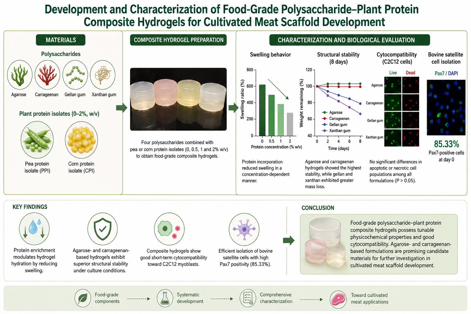
Results and Conclusion: Pea and corn protein isolates varied in protein content, apparent solubility, water and oil absorption, emulsifying characteristics and protein subunit profiles. Protein incorporation generally decreased hydrogel swelling. Agarose and κ-carrageenan-based hydrogels showed minimal relative weight changes (< 3%) within 8 d, whereas gellan hydrogels showed approximately 10% weight loss after 1 d and 15% on Day 8. Xanthan-based hydrogels demonstrated the greatest early mass loss (approximately 30–40% after 1 d) with significant dry-matter loss. Apoptotic and necrotic rates in C2C12 myoblasts exposed to hydrogels showed no significant differences from the controls. Freshly isolated bovine satellite cells showed 85.33% Pax7 positivity, whereas a few viable cells were recovered after 8 d of post-mortem tissue storage. The results demonstrate that plant protein incorporation can effectively tailor the swelling and structural stability of food-grade polysaccharide hydrogels. Agarose and κ-carrageenan-based hydrogels containing up to 1% (w/v) plant protein showed favorable short-term stability and preliminary C2C12 cytocompatibility, highlighting their potential as food-grade scaffold matrices for cultivated meat uses. These findings with the recovery of viable Pax7-positive bovine satellite cells after short-term tissue storage provided a promising foundation for further development of these composite hydrogels for three-dimensional cell culture and cultivated meat production.
- INTRODUCTION
The growing global population and increasing demand for sustainable protein sources have intensified interests in alternative meat production systems with decreased environmental effects. Conventional livestock production needs extensive land, water and energy resources and contributes significantly to greenhouse gas emissions [1]. Additionally, animal-derived meat products can serve as vehicles for zoonotic pathogens, posing challenges to food safety and public health [2]. Therefore, cultivated meat produced through in vitro cultivation of animal cells has emerged as a promising approach to complement conven-tional meat production while improving resource efficiency and production control [3, 4]. However, one major challenge in cultivated meat production is the development of edible materials capable of providing an appropriate three-dimensional (3-D) microenvironment for cell culture [5]. Unlike scaffolds developed for biomedical tissue engineering, materials intended for cultivated meat must not only support cellular functions but also satisfy food-specific requirements, including edibility, food safety, sustainability, scalability, cost-effectiveness and acceptable sensory characteristics [4, 6, 7]. Therefore, considerable attentions have been paid to food-grade hydrogel systems as candidate matrices for cultivated meat uses.
Polysaccharide-based hydrogels are from the most promising food-grade materials because of their biocompatibility, availability, gel-forming ability and tunable physicochemical characteristics. Hydrogel charac-teristics such as swelling behavior, structural stability, water-holding capacity and network architecture can affect the physicochemical environment surrounding cultured cells and are therefore important considerations in scaffold design [8]. Agarose, carrageenan, gellan gum and xanthan are examples of food-grade polysaccharides capable of forming hydrated polymer networks with distinct structural and functional characteristics [8, 9]. Beyond hydrogel and cell-culture uses, polysaccharide-based biopolymers have also been investigated extensively for food uses, including film fabrication and functional food-packaging systems, highlighting the broader versatility of these materials as food-compatible polymeric matrices [10]. Nevertheless, hydrogels prepared alone from polysaccharides may show limited structural stability or functionality depending on their composition, encouraging the development of compo-site systems with improved and tunable performance. In this context, the incorporation of plant-derived proteins into polysaccharide hydrogels has attracted increasing interest because protein–polysacch-aride interactions can affect water retention, gel stability and network organization [8, 11]. From a food perspective, plant proteins can also contribute nutritional value while supporting the animal-free and sustainability-oriented nature of cultivated meat production. Such composite systems may therefore provide a useful basis for the development of food-grade scaffold materials for cultivated meat uses [9].
Pea and corn proteins are attractive plant-derived ingredients because of their availability and functional characteristics relevant to food formulations. Pea proteins show useful water and oil-holding and emulsifying characteristics and have increasingly been investigated for food structuring and gel-forming uses [12]. Corn proteins, particularly zein-rich fractions, possess distinctive hydrophobic and film-forming characteristics that can affect their interactions with other food polymers [13]. Incorporating these proteins into polysaccharide matrices may therefore modify hydrogel hydration, structural stability and overall functionality while preserving a food-compatible composition. Importantly, differences in the intrinsic physicochemical characteristics of plant proteins may affect their interactions with polysaccharide networks and therefore the characteristics of the resulting composite hydrogels.
Although protein-enriched polysaccharide hydrogels have been investigated for cultivated-meat-associated uses, the available literature is still limited for systematic comparisons within various plant proteins and polysaccharide matrices. For example, Wollschlaeger et al. [9] assessed pea and soy-protein-enriched agarose and gellan hydrogels as well as a xanthan-locust bean gum system, while Kamel et al. [14] investigated protein and starch-enriched alginate hydrogels for bovine myoblast culture. These studies demonstrate the potential of protein-polysaccharide combinations, but they do not provide a direct comparison of various plant proteins across a similar broader set of food-grade polysaccharides. In addition, the relationship between the intrinsic physicochemical characteristics of the protein isolates and the resulting hydrogel behavior has received comparatively limited attention. Thus, an important unresolved issue is how various plant proteins affect swelling and structural stability when incorporated into distinct polysaccharide networks. Addressing this gap is important for the rational selection of food-grade hydrogel formulations and for decreasing the need to assess individual protein-polysaccharide combinations alone through empirical screening.
The novelty of the present study included its comparative assessment of pea and corn protein isolates incorporated into four food-grade polysaccharide systems (agarose, carrageenan, gellan gum and xanthan) at various protein concentrations with independent characterization of the two protein isolates. By combining protein-level physicochemical characterization with assessments of hydrogel swelling and structural stability, the study provides a basis for relating differences in protein functionality to the behavior of the resulting composite materials. In addition, preliminary cell compatibility was assessed using C2C12 murine myoblasts, while bovine satellite cells were isolated and characterized by Pax7 immunofluorescence as a relevant muscle-cell source for subsequent scaffold studies. Accordingly, the objectives of the present study were to develop and characterize polysaccharide-plant protein composite hydrogels and assess the effects of protein incorporation on their swelling behavior and structural stability with preliminary in-vitro cell compatibility. The findings identified promising food-grade formulations for further investigation as scaffold materials in cultivated meat research.
- MATERIALS AND METHODS
Unless otherwise indicated, all polysaccharide and protein concentrations were expressed as weight/volume percentages (% w/v; g per 100 ml). Sunflower oil concentration in the emulsifying activity assay was expressed as volume/volume percentage (% v/v). No molar concentrations were used.
2.1. Materials
Agarose, κ-carrageenan, gellan gum, xanthan gum, pea protein isolate, corn protein isolate, dimethyl sulfoxide (DMSO), paraformaldehyde, Triton X-100, bovine serum albumin (BSA) and Tween-20 were purchased from Merck, Germany. High-glucose Dulbecco's modified eagle media (DMEM-hg), fetal bovine serum (FBS), horse serum, stable L-glutamine, Primocin, sodium pyruvate, calcium and magnesium-free phosphate-buffered saline (PBS), Dulbecco's phosphate-buffered saline (DPBS), 0.05% trypsin-EDTA, collagenase type II, DNase I, D-glucose, gentamicin, penicillin, streptomycin and amphotericin B were purchased from Gibco, Thermo Fisher Scientific, USA. The murine myoblast cell line C2C12 was purchased from CLS Cell Lines Service, Germany (no. 400476). Matrigel and 8-well Ibidi µ-slides were purchased from Corning, USA, and Ibidi, Germany, respectively. Anti-Pax7 primary antibody, Alexa Fluor 488-conjugated secondary antibody, DAPI and ProLong Gold antifade mountant were purchased from Thermo Fisher Scientific, USA. Commercial sunflower oil was purchased from a local market in Amol, Iran.
2.2. Preparation of Composite Hydrogels
Four polysaccharides (agarose, κ-carrageenan, gellan gum and xanthan gum) and two plant protein isolates (pea and corn protein isolates) were used to prepare the composite hydrogels. Stock solutions of agarose, κ-carrageenan and gellan gum were prepared at 2% (w/v), whereas xanthan gum was prepared at 1% (w/v). Complete dissolution was achieved using microwave heating followed by equilibration at the appropriate working temperature. The lower xanthan concentration was selected based on its distinct viscosity and handling characteristics relative to the other polysaccharides and the previously reported compositional approach [9].
Pea and corn protein stock solutions were prepared at concentrations corresponding to final hydrogel protein concentrations of 0, 0.5, 1 and 2% (w/v) after 1:1 (v/v) mixing with the polysaccharide solutions. All solutions were prepared aseptically using class-II biological safety cabinet. Before mixing, the polysaccharide solutions were set at 37 °C for agarose, κ-carrageenan and gellan gum and at 65 °C for xanthan gum. Equal volumes of polysaccharide and protein solutions were gently mixed to achieve homogeneous formulations. The final polysaccharide concentrations were 1% (w/v) for agarose, κ-carrageenan and gellan gum and 0.5% (w/v) for xanthan gum [9, 15]. The lower xanthan concentration was selected based on its distinct gel-forming and handling characteristics relative to the other polysaccharides, following the reported approaches of Kumar et al. [15] and Wollschlaeger et al. [9].
Aliquots of 100 µl were transferred into cylindrical polytetrafluoroethylene (PTFE) molds (7 mm diameter × 7 mm height) placed in 24-well plates and set to gel at room temperature (RT) (22°C ±2) for 15 min. Gellan-based hydrogels were immersed in 1 ml of DMEM-hg for 15 min to promote ionic cross-linking through divalent cations in the culture media [9, 16]. All formulations were visually investigated after gelation for shape retention, structural integrity, dimensional stability and handling characteristics. Formulations containing 0, 0.5 and 1% (w/v) proteins were selected for cell-based experiments because they preserved sufficient structural integrity for handling. In contrast, single-protein formulations containing 2% (w/v) protein were preserved for physic-chemical characterization but excluded from cell-based experiments because they were visibly softer and further fragile after gelation with samples showing partial collapse or edge tearing during transfer from the mould. These observations were qualitative and were not derived from mechanical assessments. In addition, a combined formul-ation containing 1% (w/v) pea protein and 1% (w/v) corn protein (HP and HC; total protein concentration of 2% w/v) was included in the cytocompatibility experiments to assess the combined effects of the two proteins.
2.3. Characterization of Pea and Corn Protein Isolates
2.3.1. Proximate composition
The moisture, dry matter, fat, ash and protein contents of the pea and corn protein isolates were assessed using standard AOAC methods [17]. Protein content was calculated from nitrogen content using conversion factor of 6.25. Protein, fat and ash contents are expressed on a dry-matter basis, whereas moisture and dry matter are expressed on an as-is basis. All assessments were carried out in triplicate.
2.3.2. Protein solubility
Protein solubility was assessed at pH 7 using standardized food-protein solubility procedure [18]. Protein dispersions were prepared in phosphate buffer, mixed and centrifuged. Protein content in the supernatant was assessed and solubility was expressed as the percentage of soluble protein relative to the total protein content. Assessments were carried out in triplicate.
2.3.3. Water and oil absorption capacity
Water absorption capacity (WAC) was assessed by mixing approximately 1 g of protein isolate with 10 ml of deionized water, followed by continuous stirring for 30 min at RT and centrifugation at 2000× g for 20 min. The WAC was expressed as grams of water absorbed per gram of sample. Oil absorption capacity (OAC) was assessed using a similar procedure with commercial sunflower oil, followed by centrifugation at 4000 × g for 20 min. WAC and OAC were assessed based on Beuchat [19] in triplicate.
2.3.4. Emulsifying activity
The emulsifying characteristics of the pea and corn protein isolates were assessed at pH 7 using turbidimetric method of Pearce and Kinsella [20]. A 0.5% (w/v) protein dispersion was homogenized with commercial sunflower oil at 25% (v/v) of the total volume at 15000 rpm for 1 min. Aliquots of the emulsion were collected immediately after homogenization and 10 min of standing, diluted 100-fold (DF = 100) with 0.1% (w/v) sodium dodecyl sulfate (SDS) solution and then absorbance was assessed at 500 nm. The emulsifying activity index (EAI-Eq. 1) and emulsion stability index (ESI-Eq.2) were calculated as follows:
EAI (m²/g) = (2 × 2.303 × A₀ × DF) / (C × φ × 10⁴)
Eq. 1
ESI (min) = (A₀ × Δt) / (A₀ − A₁₀)
Eq. 2
where, A₀ and A₁₀ were the absorbances of the diluted emulsion at 0 and 10 min, respectively; DF was the dilution factor (100); C was the protein concentration in the aqueous phase before emulsification (0.005 g ml-1); φ was the oil volume fraction (0.25); and Δt was the time interval (10 min). Assessments were carried out in triplicate.
2.3.5. Color measurement
The color of compressed protein-isolate powder samples was assessed using Konica Minolta CR-400 colorimeter (Konica Minolta, Japan) under the CIE D65 standard illuminant. Moreover, L*, a* and b* values were recorded and chroma (C*) and hue angle (h°) were calculated as C* = (a*² + b*²)^½ and h° = tan⁻¹(b*/a*). Assessments were carried out in triplicate.
2.3.6. Sodium dodecyl sulfate–polyacryla-mide gel electrophoresis
Sodium dodecyl sulfate–polyacrylamide gel electrophoresis (SDS-PAGE) was carried out under decreasing conditions based on the Laemmli method [21] using 12% (w/v) separating gel and a 4% (w/v) stacking gel. Protein samples were dispersed at 2 mg ml-1, mixed with SDS-containing loading buffer supplemented with 5% (v/v) β-mercaptoethanol and heated at 95 °C for 5 min. Twenty micrograms of the protein were loaded per lane with a molecular-weight standard. Following electro-phoresis, protein bands were visualized using Coomassie brilliant blue staining and compared with the molecular-weight marker to estimate the apparent molecular-weight profile of the protein isolates.
2.4. Swelling Characteristics and Structural Stability of Hydrogels
Four independent hydrogel samples were prepared for each formulation containing 0, 0.5, 1 or 2% (w/v) pea or corn protein combined with agarose, κ-carrageenan, gellan gum or xanthan gum. Hydrogel preparation and weighing were carried out under aseptic conditions. Immediately after gelation, each hydrogel was immersed in 1 ml of growth media consisting of DMEM-hg supplemented with 10% FBS, 1% stable L-glutamine and 0.2% Primocin. After 15-min equilibration, excess surface moisture was removed and the initial wet weight (W₀) was recorded. Hydrogels were incubated under standard cell-culture conditions and wet weights were recorded after 1, 3 and 8 d. The culture media were completely replaced on Day 3.
For dry-weight assessment, a separate set of four hydrogels for each formulation was dried at 60 °C until constant weight (7 d). This protocol was selected to set comparability with a previously reported procedure used for associated polysaccharide-protein hydrogels [9]. Because drying could alter hydrogel structure and might induce thermal or structural changes, the resulting dry weights were interpreted as method-dependent values rather than completely structure-neutral measure of the original hydrogel state. The swelling ratio was calculated using Eq. 3:
Swelling ratio (%) = [(W₀ − Wd) / Wd] × 100
Eq. 3
where, W₀ was the initial wet weight and Wd was the dry weight. Relative hydrogel weight was calculated from the wet weight assessed on Days 1, 3 and 8 relative to the initial Day 0 weight and used as an indicator of mass retention during incubation.
2.5. Xanthan Hydrogel Dry-matter Loss
Because xanthan-based hydrogels demonstrated significant wet-weight loss during the first 24 h, a separate gravimetric assessment was carried out to assess dry-matter loss. Xanthan hydrogels without protein and those containing 0.5 or 1% (w/v) pea protein isolate were incubated in similar culture media at 37 °C for 24 h. After incubation, samples were recovered, dried using similar 60 °C drying protocol for dry-weight assessment and weighed after reaching constant weight. Dry-matter loss was calculated relative to the corresponding initial dry weight. This gravimetric approach was used to assess loss of solid material but did not distinguish within dissolution, release of uncross-linked polymer or protein, fragmentation and other compositional changes.
2.6. The C2C12 Cell Culture and Cytocompatibility Assessment
The C2C12 murine myoblast cell line was cultured in DMEM-hg supplemented with 10% FBS, 1% stable L-glutamine and 0.2% Primocin at 37 °C under humidified atmosphere containing 5% CO₂. Cells were routinely set at an initial density of 5 × 10³ cells per cm² and passaged at approximately 70% confluence using 0.05% trypsin-0.53 mM EDTA following washing with calcium and magnesium-free PBS [9, 22].
For cytocompatibility experiments, cells were seeded at 1.5 × 10⁴ cells per well in 48-well plates containing preformed cylindrical hydrogels (7-mm diameter × 7-mm height). Before cell seeding, hydrogels were equilibrated using complete culture media under sterile conditions. Cells were seeded directly onto the hydrogel surface rather than encapsulated with the hydrogel matrix. Phenol red-free DMEM-hg supplemented with 10% FBS, 1% stable L-glutamine and 0.2% Primocin was used during the cytocompatibility experiments. Cell viability was assessed using live/dead fluorescence staining and apoptosis/necrosis was quantified after 24 and 48 h of culture [9, 23].
2.7. Bovine Satellite Cell Isolation
Bovine satellite cells were isolated from semimem-branosus muscle from Holstein dairy cattle in a commercial abattoir. Muscle samples were transported under refrigerated conditions and either processed within 2 h post-mortem or stored at 4 °C for 2, 5 or 8 d in DMEM-hg containing gentamicin, penicillin, streptomycin and amphotericin B. Approximately 5 g of the muscle tissue were finely minced and enzymatically digested in 20 ml of Ca²⁺/Mg²⁺-free DPBS containing 0.25% trypsin, 427.5 U ml-1 collagenase type II, 0.01% DNase I and 1% (w/v) D-glucose for 1 h at 37 °C with gentle agitation. After digestion, released cells were transferred into chilled growth media consisting of DMEM-hg supplemented with 10% FBS, 10% horse serum, 1 mM sodium pyruvate and antibiotics. The rest of tissue was subjected to a second digestion cycle and the resulting cell fractions were mixed.
The cell suspension was then passed through 100 and 40-µm cell strainers, centrifuged at 1000× g for 10 min at 4 °C and resuspended in cryopreservation media consisting of 90% FBS and 10% (v/v) DMSO. Cell suspensions were aliquoted into cryovials and cryopreserved until further use. The isolation and storage procedures were based on a previously reported bovine satellite-cell protocol [24], with the post-mortem storage intervals adapted for the present study.
2.8. Immunofluorescence Analysis
For immunofluorescence analysis, isolated bovine satellite cells were seeded at 5,000 cells per well onto Matrigel-coated 8-well Ibidi µ-slides and cultured for 3 d in proliferative media consisting of DMEM-hg supplemented with 20% FBS, 10% horse serum, 1% stable L-glutamine and 0.2% Primocin. Where applicable, preformed hydrogel discs were introduced after cell attachment using surface-contact approach without encapsulation.
Cells were fixed with 3.7% paraformaldehyde for 10 min and permeabilized with 0.1% Triton X-100 for 15 min. Samples were blocked for 1 h at RT in DPBS containing 1% (w/v) BSA and 0.1% Tween-20. Cells were incubated with anti-Pax7 primary antibody (1:200) at 4 °C overnight followed by Alexa Fluor 488-conjugated secondary antibody (1:400) at RT for 2 h. The DAPI (1 µg ml-1) was added to the mixture during the final 30 min to counterstain nuclei. Samples were mounted using ProLong Gold antifade mountant and investigated using Nikon Eclipse confocal microscope (Nikon, Japan). The proportion of Pax7-positive cells was assessed by counting Pax7-positive nuclei relative to the total number of DAPI-stained nuclei using ImageJ software [25].
2.9. Statistical Analysis
Unless otherwise stated, experiments were carried out using four independent replicates (n = 4). Data were analyzed using one-way analysis of variance (ANOVA) followed by Tukey's post hoc test. Statistical analyses were carried out using GraphPad Prism 9 (GraphPad Software, USA) and differences were recorded as statistically significant at P < 0.05.
- RESULTS AND DISCUSSION
3.1. Characterization of Pea and Corn Protein Isolates
To characterize the protein ingredients used for hydrogel enrichment, the corn and pea protein isolates were assessed for proximate composition, apparent solubility, water and oil absorption capacity, emulsifying character-istics, color characteristics and protein subunit profiles (Table 1). The corn and pea protein isolates contained 92.85% ±0.46 and 81.94% ±0.68 protein (dry basis), respectively. The pea protein isolate showed higher fat and ash contents and higher moisture content (7.29 against 1.60%) than that the corn protein isolate did (Table 1). These differences indicated that the two protein ingredients varied in composition and therefore provided various starting materials for incorporation into the polysaccharide networks.
At pH 7, the apparent solubility of the corn protein isolate was 62.00%, compared with 40.74% for the pea protein isolate. These values represented the protein recovered in the supernatant under the conditions of the solubility assay and should therefore be interpreted as apparent solubility or dispersion behavior rather than as direct measure of molecular solvation. This distinction was particularly relevant when comparing proteins with various molecular structures and aggregation behavior [12].
The two isolates varied in their water/oil absorption and emulsifying characteristics. The WAC was 4.03 ±0.03 and 4.64 ±0.22 g g-1 for corn and pea protein isolates, respectively, while the corresponding OAC values were 1.46 ±0.55 and 1.79 ±0.10 g g-1. The EAI was significantly higher for pea protein (21.4 ±1.8 m² g-1) than for corn protein (7.8 ± 1.2 m² g-1). In contrast, the ESI was slightly higher for corn protein (14.3 ±1.1 min) than pea protein (12.6 ±0.7 min). These differences demonstrated that the two isolates included distinct functional characteristics under the experimental conditions and might therefore interact differently with the polysaccharide matrices.
The color parameters revealed clear differences between the two protein isolates (Table 1). The pea protein isolate showed significantly higher lightness (L* = 87.23) than that the corn protein isolate did (L* = 67.82) with higher redness (a* = 5.36 against 1.91) and yellowness (b* = 21.68 against 15.97). Accordingly, the pea isolate demonstrated higher chroma (C* = 22.33) than that the corn isolate did (16.08), whereas the lower hue angle of pea protein (h° = 76.11 against 83.17) reflected a relatively further reddish-yellow appearance. These differences were relevant to the development of cultivated-meat scaffold materials because the intrinsic color of plant proteins might affect the visual appearance of the resulting hydrogel and, ultimately, the appearance of a scaffold intended for food uses. The observed color differences also provided additional evidence of the distinct physicochemical characteristics of the two protein isolates used for hydrogel enrichment.
The SDS-PAGE analysis under decreasing conditions further demonstrated differences in the apparent subunit profiles of the two protein isolates (Figure 1). The corn protein isolate demonstrated prominent bands at approximately 20 and 23 kDa and a weaker band near 45 kDa, consistent with the predominant presence of low-molecular-weight zein subunits [13]. The pea protein isolate showed several prominent bands majorly in the approximately 35–45 kDa region, with no prominent bands greater than 100 or 60 kDa, consistent with the dissociation of legumin and vicilin into their constituent subunits under decreasing conditions [12]. These patterns provided qualitative information on the apparent protein subunit profiles of the two isolates and supported the compositional differences in their functional characterization. However, the bands should not interpreted as complete molecular-weight distribution of the isolates.
Overall, the characterization results demonstrated that the pea and corn protein isolates varied in protein content, hydration behavior, emulsifying activity and apparent subunit profile. These differences provided useful context for interpreting their effects on hydrogel swelling and stability, although direct causal relationships between individual protein characteristics and hydrogel performance could not be established from the present data alone.
3.2. Hydrogel Swelling and Structural Stability
The swelling behavior of the hydrogels was assessed to investigate how incorporation of plant proteins affected water uptake by the four polysaccharide matrices (Figure 2). From all four polysaccharides, protein-free formulations demonstrated significantly higher swelling ratios than those the corresponding protein-enriched hydrogels did under the conditions. For pea and corn-protein formulations, increasing protein concentration from 0 to 2% (w/v) generally decreased the swelling ratio, particularly in the pea-protein-containing systems.
The decrease in swelling following protein incorporation was consistent with changes in the interactions within the composite network. Protein-polysaccharide interactions might decrease the quantity of water-accessible space within the matrix and thereby decrease water uptake. Similar effects have been reported for protein-enriched polysaccharide hydrogel systems [9, 11, 14]. The present swelling assessments therefore demonstrated that plant protein incorporation could be used to modulate the hydration behavior of these polysaccharide hydrogels. An associated concentration-dependent relationship between protein–polysaccharide interactions and gel network characteristics has been reported for mung bean protein isolate-gellan gum composites; in which, protein concentration and pH affected the balance between the gel structure and water retention [26]. This provided a relevant comparison with the present findings; although the protein source and experimental conditions varied. In the present study, the concentration-dependent decrease in swelling similarly indicated that protein incorporation altered the hydration characteristics of the composite network, without setting a specific molecular mechanism to be established.
The effect of protein incorporation was evident in the dry weights used for calculation of the swelling ratios. For each polysaccharide, increasing protein concentration occurred by an increase in assessed dry weight, consistent with the greater quantity of solid material incorporated into the hydrogel formulation. Because the swelling ratio was calculated using dry weights after drying at 60 °C to constant weight, the potential effects of the drying procedure should be considered. Drying can alter the structure of hydrogel matrices, particularly for protein-containing systems and polysaccharides, whose network morphology is sensitive to dehydration. Previous studies have shown that drying at approximately 60 °C can induce structural collapse, shrinkage and changes in rehydration behavior in polysaccharide-based gels [27, 28]. In the present study, all formulations were subjected to similar drying procedures and the resulting dry weights were used consistently for the swelling-ratio calculation. Neverthe-less, drying-induced structural changes could be complete-ly excluded as a potential source of systematic variation. The reported swelling ratios should therefore be interpreted as comparative assessments achieved under the specified drying conditions rather than absolute descriptors of the hydrated network structure.
The structural stability of the hydrogels during incubation in cell culture media varied significantly within the polysaccharide systems (Figure 3). Agarose and carrageenan-based hydrogels showed minimal changes in wet weight, resulting in approximately 3% of their initial values throughout the 8-d incubation. In contrast, gellan-based hydrogels showed an approximately 10% decrease in wet weight on Day 1, followed by a further decrease to approximately 15% on Day 8.
Xanthan-based hydrogels demonstrated the greatest decrease, with approximately 30–40% loss of initial wet weight within the first 24 h, followed by a slower decline during the rest incubation time. The differences within the polysaccharides were similar to differences in their network formation and water-retention behavior. Importantly, the wet-weight assessments alone could not distinguish between loss of water, release of soluble or weakly associated components, fragmentation of the hydrogel or other changes occurring during incubation. The wet-weight data were therefore interpreted as indicator of the relative mass stability of the hydrogels.
To further investigate the pronounced mass decrease for xanthan-based hydrogels, dry-matter loss was additionally assessed after 24 h of incubation (Table 2). Xanthan hydrogels without added protein showed approximately 20% dry-weight loss, whereas formulations containing 0.5 and 1% pea proteins showed approximately 25 and 27% losses, respectively. Thus, a significant proportion of the initial dry matter was no longer recovered from the hydrogel after 24 h. These results supported the interpretation that the large wet-weight decrease for xanthan hydrogels was associated partly with loss of solid materials.
The stability patterns observed in the present study were broadly consistent with previous reports on protein-enriched polysaccharide systems. Wollschlaeger et al. [9] investigated pea and soy-protein-enriched agarose and gellan systems and reported that agarose and gellan-based formulations could form stable cell-laden gels, whereas xanthan–locust bean gum systems did not produce stable cell-laden constructs. Although the formulations and experimental conditions were not identical, the pronounced instability observed for xanthan in the present study was generally similar.
The present results also indicated that protein concentrations around 1% (w/v) provided a practical upper range for formulations that preserved satisfactory handling and short-term structural stability. This observation was similar to the maximum protein concentration reported for the systems by Wollschlaeger et al. [9]. However, this concentration should not be considered a universal protein-loading threshold, as the practical upper limit was likely to depend on the specific protein, polysaccharide, concentration, processing conditions and network-forming mechanism.
The present findings were broadly similar to those of Kamel et al. [14], who reported formulation-dependent mass loss in alginate-based systems containing xanthan and plant-derived protein/starch components. In their study, mass losses of up to 57.39% for the alginate-xanthan formulation and 36.03% for the tapioca starch-alginate-xanthan formulation were reported within 7 d. However, differences in the base polysaccharide, formulation composition and experimental conditions prevented direct quantitative comparison. Collectively, these findings emphasized the importance of assessing structural stability for each specific polysaccharide-protein combination rather than assuming that performance could be concluded within formulations.
Collectively, the stability results indicated that agarose and carrageenan-based hydrogels preserved their mass most effectively within a 8-d experiment, whereas gellan showed moderate loss and xanthan showed pronounced early instability. Under the conditions, the xanthan formulations therefore did not provide adequate compositional stability for uses needing prolonged aqueous incubation. Further formulation development such as blending with further stable polysaccharide or modifying the cross-linking strategy is needed before xanthan-based systems are recommended for extended cell-culture uses.
3.3. Characterization of Isolated Bovine Satellite Cells
Bovine satellite cells were isolated from semimembranosus muscle and characterized using Pax7 immunofluorescence to assess the recovery of a predominantly myogenic cell population after various post-mortem storage intervals (Figure 4). Freshly processed tissue yielded a Pax7-positive fraction of 85.33%. After 2 and 5 d of storage at 4 °C, the corresponding Pax7-positive fractions were 78.19 and 80.11%, respectively.
The Pax7-positive fraction therefore was relatively high during the first 5 d of post-mortem storage, although some variation was observed between individual storage intervals. The increase from 78.19% on Day 2 to 80.11% on Day 5 should not be interpreted as evidence of improved cell recovery with storage, but rather as biological and isolation-associated variation. After 8 d of storage, only a few viable cells were recovered and these isolates were not appropriate for further experiments.
The present observations were similar to those by Skrivergaard et al. [24], who demonstrated that bovine satellite cells could be recovered from semimembranosus muscle after 2 and 5 d of post-mortem storage while preserving characteristics relevant to cell culture. Skrivergaard et al. [24] also assessed 8 d of storage and reported poor cell recovery at this time point. Thus, the present observations were broadly similar to those within 2 to 8-d storage intervals. The relatively high Pax7-positive fraction achieve from freshly processed tissue provided evidence that the isolation procedure generated a predominantly satellite-cell population. These findings included practical relevance for cultivated-meat research because the availability and quality of the starting cell population are important considerations alongside scaffold selection. Under the isolation conditions, refrigerated post-mortem storage for up to 5 d did not prevent recovery of a predominantly Pax7-positive cell population, whereas recovery after 8 d was severely compromised.
The storage tolerance in the present bovine satellite-cell system varied from those in other species and tissue types. Latil et al. [29] reported recovery of satellite cells from mouse and human skeletal muscles after up to 14 and 17 d of post-mortem storage, respectively, although recovery decreased with prolonged storage. In contrast, Aoued and Singh [30] reported recovery of fibroblast-like cells from goat skin after 160 d of refrigerated storage. These differences indicated that post-mortem storage tolerance strongly depended on species, tissue type and cell type; therefore, the storage window established should not be generalized over the bovine muscle satellite-cell system investigated in the present study.
3.4. Cytocompatibility of Composite Hydro-gels
The cytocompatibility of selected hydrogel formulations was assessed using C2C12 murine myoblasts through live/dead staining and quantitative analysis of apoptotic and necrotic cells (Figures 5 and 6). Cells were seeded directly onto the surface of the hydrogels. The live/dead images showed predominantly viable cells under the conditions, with relatively few dead cells. Quantitative analysis showed modest increase of approximately one percentage point in the combined apoptotic and necrotic fraction in some protein-containing groups, compared with the protein-free control. However, none of the differences within the experimental groups reached statistical significance (P > 0.05). Thus, the investigated hydrogel formulations did not produce a detectable increase in apoptosis or necrosis in C2C12 myoblasts under these conditions.
The lack of significant differences in C2C12 apoptosis and necrosis was generally similar to that of significant cytotoxicity reported for protein-enriched polysaccharide hydrogel systems. For example, Wollschlaeger et al. [9] reported no cytotoxic effects of material leachates on C2C12 cells, while Kamel et al. [14] reported no significant toxicity of their formulations to bovine myoblasts and maintenance of PAX7 and desmin expression. However, such comparisons should be interpreted cautiously because cell type, assay format, hydrogel composition and exposure conditions vary within various studies. The present findings therefore provided evidence of preliminary short-term cytocompatibility under the specific experimental conditions rather than definitive evidence of biocompatibility.
The results from the hydrogel stability and C2C12 assessments indicated that agarose and carrageenan-based formulations warranted priority for further investigation. Their relatively stable behavior during the 8-d incubation with the absence of a detectable increase in C2C12 apoptosis or necrosis provided a useful basis for further formulation optimization. In contrast, the significant early mass loss for xanthan indicated that its formulation needed further modification before the uses involving prolonged aqueous cell culture. These findings supported further investigations of agarose and carrageenan-based formulations, particularly at protein concentrations up to 1% (w/v), as candidate food-grade materials for cultivated-meat scaffold development.
- CONCLUSION
This study assessed food-grade composite hydrogels prepared from agarose, κ-carrageenan, gellan gum and xanthan gum combined with pea or corn protein isolates as candidate materials for cultivated-meat scaffold development. Incorporation of plant proteins generally decreased hydrogel swelling, while the extent of the effect depended on the polysaccharide and protein formulation. Within the highlighted systems, agarose and carrageenan-based hydrogels showed the greatest structural stability during the 8-d incubation, whereas gellan demonstrated moderate mass loss and xanthan showed pronounced early instability with significant dry-matter loss.
The pea and corn protein isolates also demonstrated distinct compositional and functional characteristics, including differences in apparent solubility, water and oil absorption, emulsifying characteristics and apparent protein subunit profiles. These differences provided a useful basis for further optimization of protein-polysaccharide composite formulations. Preliminary cytocompatibility assessment using surface-seeded C2C12 myoblasts showed no significant increases in apoptosis or necrosis under the conditions. Moreover, bovine satellite-cell characterization demonstrated a relatively high Pax7-positive fraction after fresh isolation and after 2 and 5 d of post-mortem tissue storage, whereas cell recovery was severely compromised after 8 d.
The results identified agarose and κ-carrageenan-based formulations containing up to 1% (w/v) plant protein as promising candidates for further scaffold development, based on their short-term structural stability and preliminary C2C12 cytocompatibility. These findings represented an initial material-screening stage in the development of a functional cultivated-meat scaffold.
Further studies should assess 3-D cell encapsulation, long-term stability, mechanical and rheological character-istics, cell proliferation and myogenic differentiation, as well as food-associated characteristics, scalability and processing feasibility. Food-safety functionality warrants investigation, as polysaccharide-based carriers containing antimicrobial plant-derived essential oils have shown potentials for extending ground-meat shelf life [31]. Alternative processing strategies, including ultrasound and enzyme-assisted extraction, may provide new routes for achieving functional polysaccharide components [32]. Fermentation-derived polysaccharides represent a potential source of functional scaffold materials, with solid-state fermentation reported to produce bioactive polysaccharides with distinct structural and immunomodulatory characteristics [33]. Their appropriate-ness for cultivated-meat uses must be established.
- Polysaccharide hydrogels
- Plant protein isolates
- Edible scaffolds
- Cytocompatibility
- Cultivated meat
ارجاع به مقاله
مراجع
[1]. Mace JL, Knight A, Vieira F, Tatemoto P, Gameiro M. The missing target: Why industrialized animal farming must be at the core of the climate agenda. Animals. 2025; 15(22): 3256. https://doi.org/10.3390/ani15223256
[2]. Heredia N, García S. Animals as sources of food-borne pathogens: A review. Anim Nutr. 2018; 4(3): 250-255. https://doi.org/10.1016/j.aninu.2018.04.006
[3]. Melzener L, Verzijden KE, Buijs AJ, Post MJ, Flack JE. Cultured beef: From small biopsy to substantial quantity. J Sci Food Agric. 2021; 101(1): 7-14. https://doi.org/10.1002/jsfa.10663
[4]. Seah JSH, Singh S, Tan LP, Choudhury D. Scaffolds for the manufacture of cultured meat. Crit Rev Biotechnol. 2022; 42(2): 311-323. https://doi.org/10.1080/07388551.2021.1931803
[5]. Baker BM, Chen CS. Deconstructing the third dimension: How 3D culture microenvironments alter cellular cues. J Cell Sci. 2012; 125(13): 3015-3024. https://doi.org/10.1242/jcs.079509
[6]. O'Neill EN, Cosenza ZA, Baar K, Block DE. Considerations for the development of cost-effective cell culture media for cultivated meat production. Compr Rev Food Sci Food Saf. 2021; 20(1): 686-709. https://doi.org/10.1111/1541-4337.12678
[7]. Chng VJY, Wan ACA. The scaffold concept for alternative proteins. J Food Eng. 2023; 357: 111622. https://doi.org/10.1016/j.jfoodeng.2023.111622
[8]. Fan Z, Cheng P, Zhang P, Zhang G, Han J. Rheological insight of polysaccharide/protein based hydrogels in recent food and biomedical fields: A review. Int J Biol Macromol. 2022; 222: 1642-1664. https://doi.org/10.1016/j.ijbiomac.2022.10.082
[9]. Wollschlaeger JO, Maatz R, Albrecht FB, Klatt A, Heine S, Blaeser A, et al. Scaffolds for cultured meat on the basis of polysaccharide hydrogels enriched with plant-based proteins. Gels. 2022; 8(2): 94. https://doi.org/10.3390/gels8020094
[10]. Junaid PM, Saikumar A, Islam RU, Patel MK, Eranda DHU, Islam M, Zaidi S, Badwaik LS. Polysaccharide-based biopolymers: exploring film fabrication techniques, molecular interactions and their potential food applications. Quality Assurance and Safety of Crops & Foods. 2025; 17(4): 257-283. https://doi.org/10.15586/qas.v17i4.1581
[11]. Albano KM, Cavallieri ÂLF, Nicoletti VR. Electrostatic interaction between proteins and polysaccharides: Physicochemical aspects and applications in emulsion stabilization. Food Rev Int. 2019; 35(1): 54-89. https://doi.org/10.1080/87559129.2018.146744
[12]. Lam ACY, Can Karaca A, Tyler RT, Nickerson MT. Pea protein isolates: Structure, extraction and functionality. Food Rev Int. 2018; 34(2): 126-147. https://doi.org/10.1080/87559129.2016.1242135
[13]. Shukla R, Cheryan M. Zein: The industrial protein from corn. Ind Crops Prod. 2001; 13(3): 171-192. https://doi.org/10.1016/S0926-6690(00)00064-9
[14]. Kamel J, Lee JY, Yadav U, Afrin S, Yadav CJ, Zo SM, et al. Development of polysaccharide hydrogels enriched with protein and starch for bovine myoblast cell culture. J Food Sci. 2025; 90: e70350. https://doi.org/10.1111/1750-3841.70350
[15]. Kumar A, Rao KM, Han SS. Application of xanthan gum as polysaccharide in tissue engineering: A review. Carbohydrate Polymers. 2018; 180: 128-144. https://doi.org/10.1016/j.carbpol.2017.10.009
[16]. Morris ER, Nishinari K, Rinaudo M. Gelation of gellan – A review. Food Hydrocoll. 2012; 28(2): 373-411. https://www.aoac.org/10.1016/j.foodhyd.2012.01.004
[17]. AOAC International. (2019). Official Methods of Analysis of AOAC International (21st ed.). AOAC International. Retrieved from https://www.aoac.org/official-methods-of-analysis-21st-edition-2019/
[18]. Morr CV, German B, Kinsella JE, Regenstein JM, Van Buren JP, Kilara A, et al. A collaborative study to develop a standardized food protein solubility procedure. J Food Sci. 1985; 50(6): 1715-1718. https://doi.org/10.1111/j.1365-2621.1985.tb10572.x
[19]. Beuchat LR. Functional and electrophoretic characteristics of succinylated peanut flour protein. J Agric Food Chem. 1977; 25(2): 258-261. https://doi.org/10.1021/jf60210a044
[20]. Pearce KN, Kinsella JE. Emulsifying properties of proteins: Evaluation of a turbidimetric technique. J Agric Food Chem. 1978; 26(3): 716-723. https://doi.org/10.1021/jf60217a041
[21]. Laemmli UK. Cleavage of structural proteins during the assembly of the head of bacteriophage T4. Nature. 1970; 227(5259): 680-685. https://doi.org/10.1038/227680a0
[22]. Yaffe D, Saxel O. Serial passaging and differentiation of myogenic cells isolated from dystrophic mouse muscle. Nature. 1977; 270(5639): 725-727. https://doi.org/10.1038/270725a0
[23]. Vermes I, Haanen C, Steffens-Nakken H, Reutelingsperger C. A novel assay for apoptosis. Flow cytometric detection of phosphatidylserine expression on early apoptotic cells using fluorescein labelled Annexin V. J Immunol Methods. 1995; 184(1): 39-51. https://doi.org/10.1016/0022-1759(95)00072-I
[24]. Skrivergaard S, Rasmussen MK, Therkildsen M, Young JF. Bovine satellite cells isolated after 2 and 5 days of tissue storage maintain the proliferative and myogenic capacity needed for cultured meat production. Int J Mol Sci. 2021; 22(16): 8376. https://doi.org/10.3390/ijms22168376
[25]. Schneider CA, Rasband WS, Eliceiri KW. NIH Image to ImageJ: 25 years of image analysis. Nat Methods. 2012; 9(7): 671-675. https://doi.org/10.1038/nmeth.2089
[26]. Lergchinnaboot P, Saengsuk N, Sonklin C, Phumsombat P. Influence of mung bean protein isolate concentration and pH on gel formation and physicochemical properties of mung bean protein–gellan gum composites. Quality Assurance and Safety of Crops & Foods. 2026; 18(SP2): 1-17. https://doi.org/10.15586/qas.v18sp2.1637
[27]. Cassanelli M, Prosapio V, Norton I, Mills T. Role of the drying technique on the low-acyl gellan gum gel structure: Molecular and macroscopic investigations. Food Bioprocess Technol. 2019; 12(2): 313-324. https://doi.org/10.1007/s11947-018-2210-6
[28]. da Silva Júnior MAV, Feltre G, Dacanal GC. Mathematical modelling of drying of hydrogels via finite element method and texture analysis. Processes. 2024; 12(8): 1564. https://doi.org/10.3390/pr12081564
[29]. Latil M, Rocheteau P, Châtre L, Sanulli S, Mémet S, Ricchetti M, et al. Skeletal muscle stem cells adopt a dormant cell state post mortem and retain regenerative capacity. Nat Commun. 2012; 3: 903. https://doi.org/10.1038/ncomms1890
[30]. Aoued HS, Singh M. Recovery of fibroblast-like cells after 160 days of postmortem storage of goat skin tissues in refrigerated media. J Vet Sci Technol. 2015; 6(4): 236. http://dx.doi.org/10.4172/2157-7579.1000236
[31]. Mirmoeini SS, Tajik H, Almasi H, Moradi M. Development of antimicrobial bioaerogel based on Zataria multifloraessential oil to extending ground meat shelf life. Quality Assurance and Safety of Crops & Foods. 2025; 17(1): 42-56. https://doi.org/10.15586/qas.v17i1.1518
[32]. Mezhoudi M, Abdelhedi O, Elfalleh W, Bendif H, Fakhfakh N, Zouari N, Jridi M. Ultrasound- and enzyme-assisted extraction of Moringa oleifera polysaccharides: bioactivity evaluation. Quality Assurance and Safety of Crops & Foods. 2025; 17(4): 1-16. https://doi.org/10.15586/qas.v17i4.157438
[33]. Kaewkumsan P, Gavahian M, Tseng WT, Guo JH. Structural and immunomodulatory properties of bioactive polysaccharide from solid-state fermented brown rice with Antrodia cinnamomea mycelia. Quality Assurance and Safety of Crops & Foods. 2025; 17(3). https://doi.org/10.15586/qas.v17i3.1561
- چکیده مشاهده شده: 14 بار
- Pdf (English) دانلود شده: 8 بار