Bioactive Composition and Ex Vivo Cutaneous Effects of Lactiplanti-bacillus plantarum T5-derived Cell-free Supernatants from Tarkhineh
Applied Food Biotechnology,
Vol. 13 No. 1 (2026),
7 Azar 2025
,
Page 1-20 (e20)
https://doi.org/10.22037/afb.v13i1.52738
Abstract
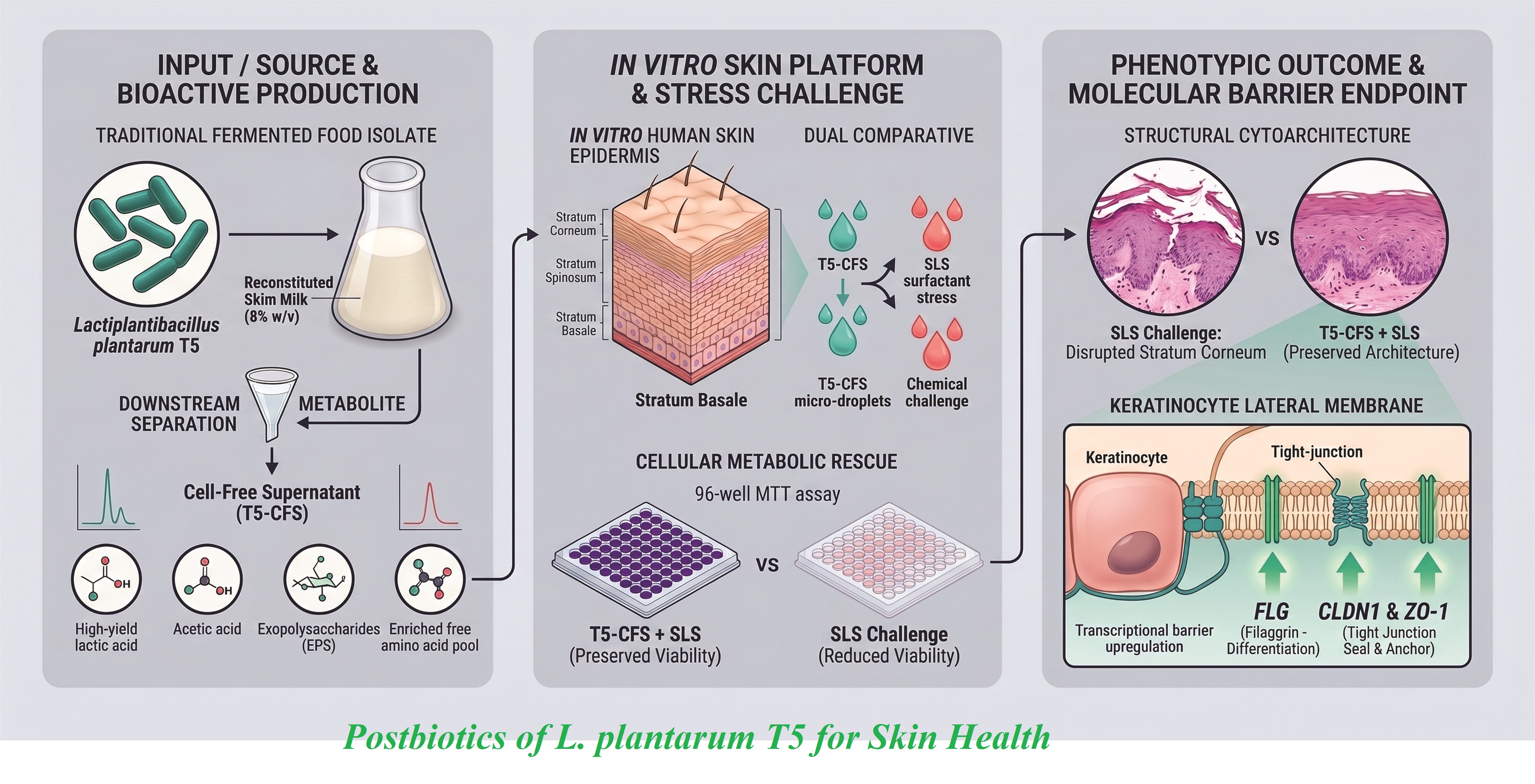
Background and Objective: The skin microbiome preserves cutaneous health and barrier function and its dysbiosis contributes to diverse dermatological disorders. Although probiotics are widely investigated, the composition and cutaneous effects of strain-specific cell-free preparations from traditional fermented foods are poorly characterized. This study characterized selected fermentation components in cell-free supernatant derived from Lactiplantibacillus plantarum T5 (T5-CFS) isolated from Iranian traditional food and assessed its protective effects using ex vivo human skin model.
Materials and Methods: Lactiplantibacillus plantarum T5 was cultured in reconstituted skim milk. The resulting cell-free supernatant was analysed using high-performance liquid chromatography to quantify organic and free amino acids and exopolysaccharides were assessed via the phenol-sulphuric acid method. Ex vivo human foreskin explants treated with various T5-CFS concentrations were assessed for viability and sodium lauryl sulphate-induced damages using 3-(4,5-di methyl thiazol-2-yl)-2,5-diphenyltetrazolium bromide assays. Morphology was investigated using hematoxylin-eosin staining and barrier-associated gene expression was quantified using quantitative real-time polymerase chain reaction.
Results and Conclusion: The T5-derived cell free supernatant contained lactic acid (2.44 mg/ml), acetic acid (0.97 mg/ml) and exopolysaccharide content (2705 mg/l). In an exploratory amino-acid screening, total free amino acids increased from 2340 to 13130 mg/l after fermentation. Under the ex vivo conditions, T5-derived CFS treatment preserved tissue viability, preserved epidermal morphology, decreased sodium lauryl sulphate-associated tissue damages and upregulated the mRNA expression of key barrier-associated genes, including ZO1, CLDN1 and FLG. The Lactiplantibacillus plantarum T5-derived CFS contained a complex fermentation-associated composition and was associated with preserved epidermal morphology and increased expression of barrier-associated transcripts in ex vivo human skin. Collectively, these findings indicate that the preparation contains a diverse pool of fermentation-derived components that may contribute to the observed barrier-associated responses; however, component-specific causality and functional barrier restoration were not established. These findings support further investigation of T5-derived CFS as a candidate preparation for future topical formulation development.
- INTRODUCTION
The skin microbiota contributes to cutaneous home-ostasis, immune response regulation and epidermal integrity preservation [1,2]. Disturbance of this microbial balance by environmental, chemical or immunological stressors increases skin permeability and contributes to inflammatory conditions such as atopic dermatitis, acne and psoriasis [3]. Accordingly, strategies aimed at pre-serving or restoring epidermal barrier homeostasis are important in dermatological research and topical product development. Recent microbiome research has increased interests in probiotic and postbiotic preparations as potential tools for supporting intestinal and cutaneous health. Although the skin and gastrointestinal tract differ substantially in their anatomy and microbial ecology, the two rely on interactions within microbial, epithelial and immune components to respond to environmental chal-lenges [4,5]. Fermented foods represent valuable sources for isolating novel probiotic strains and evidence shows that the origin and strain-specific characteristics of probiotics greatly affect their functional and therapeutic characteristics [6]. Probiotic strains belonging to genera such as Lactiplantibacillus and Bifidobacterium have been reported to affect skin barrier-associated outcomes, inflammatory skin conditions [4,7] and wound healing processes [8]. However, the topical use of live probiotics presents formulation challenges linked to preserving viability, ensuring product stability and controlling potential safety risks [9].
Based on the International Scientific Association for Pro-biotics and Prebiotics consensus, a postbiotic is a pre-paration of inanimate microorganisms and/or their com-ponents that confers a health benefit on the host [10]. Such preparations may offer practical advantages over live microorganisms, including improved formulation stability and decreased concerns linked to microbial viability; however, their composition and biological effects depend on the processing method and the components in the final preparation [11,10]. Depending on their composition and biological context, fermentation-associated components e.g. organic acids, EPSs and free amino acids (FAA) have been reported to exhibit antioxidant, anti-inflamm-atory or immunomodulatory activities in previous studies [8,9, 12,13]. These reports provide a rationale for investigating their potential relevance to cutaneous biology but such activities were not directly assessed in the present study.
Previous studies suggest that selected non-viable microbial preparations or their components may affect epithelial and dermal cell responses, including the expression of genes or proteins involved in tissue hom-eostasis [14,15]. Within probiotic candidates, Lactiplanti-bacillus (L.) plantarum has shown strain-dependent functional characteristics in diverse biological systems, including reported antimic-robial and immunomodulatory activities [14,16,17]. For example, the strain L. plantarum GMNL6 has been reported to affect collagen-linked responses, melanin production and skin microbiome-associated outcomes in previous studies [15]. In particular, L. plantarum T5, an indigenous strain isolated from a traditional Iranian fermented foods [18], was selected because of its complementary strain-specific bioactive and bioprocessing attributes. Previous studies have demon-strated antimuta-genic activity in its cellular fraction and cell-free super-natant, supporting the presence of bio-logically active extracellular fermentation products [19]. In addition, T5 has been reported to promote cutaneous wound healing in an animal model, accompanied by decreased inflammation and accelerated tissue repair, thereby providing a relevant rationale for its investigation in skin-linked uses [20]. The strain has shown EPS-pro-duction capacity [18] and robust biomass production under optimized laboratory-scale fermentation conditions [21], making it a promising candidate for generating a bioactive, metabolite-enriched cell-free preparation for cutaneous assessment. So, isolated or characterized EPS preparations from L. plantarum have been reported to promote human dermal fibroblast prolif-eration and migration in previous studies [22,23]. These findings provide a literature-based rationale for considering EPS as one possible contributor to the biological characteristics of complex fermentation-derived preparations; however, EPS-specific activity was not assessed in the present study. Despite these reports, the fermentation-associated composition and cutaneous effects of L. plantarum strains isolated from traditional Iranian fermented foods are insufficiently characterized.
This study characterized selected fermentation-associa-ted components of the T5-derived CFS and assessed its effects using ex vivo human skin model. The authors hypothesized that T5-derived CFS was well tolerated by ex vivo human skin explants and attenuated sodium lauryl sulphate (SLS)-associated tissue damages while preserving epidermal morphology and influencing the expression of barrier-associated genes. To assess this hypothesis, the authors characterized selected components of T5-derived CFS, assessed tissue viability under baseline conditions, investigated its effects on SLS-associated tissue damages and epidermal morphology in ex vivo human skin explants and assessed changes in the expression of selected barrier-associated genes.
- MATERIALS AND METHODS
2.1. Bacterial Strain and Culture Conditions
Briefly, L. plantarum T5, previously isolated from Tarkhineh (a traditional Iranian fermented food product) described by Noori et al. [21], was used. The partial 16S rRNA gene sequence is deposited in GenBank under accession number of JQ301796. Cryopreserved stocks were preserved at -80 °C in 25% (v/v) glycerol. Working cultures were prepared by inoculating 1% (v/v) of the thawed suspension into 10 ml of de Man, Rogosaand Sharpe (MRS) broth (Sigma‑Aldrich, USA) supplemented with 0.15% (w/v) L‑cysteine hydrochloride, followed by incubation at 37 °C for 24 h.
2.2. Preparation of Lactiplantibacillus plantarum T5-derived Cell-free Supernatant
The T5-derived CFS was prepared by dissolving skimmed milk powder (Scharlau, Spain) in distilled water (DW) to a final concentration of 8% (w/v) with 0.15% (w/v) L-cysteine hydrochloride and inoculating with 1% (v/v) L. plantarum T5. The cultures were incubated at 37 °C with shaking at 100 rpm for up to 24 h. Bacterial growth was monitored at 0, 4, 8, 12, 16, 20 and 24 h using viable plate counting. At the stationary phase (20 h), cultures were centrifuged at 10,000× g for 20 min and the supernatant was filtered through 0.22-µm syringe filters (Sartorius, Germany). The T5-derived CFS was assessed in its native, non-neutralized state to preserve the complete physico-chemical composition of the preparation, including its nat-urally occurring organic acids. Unfermented reconstituted skim milk (RSM) (pH 6.50 ±0.14), prepared using the identical media composition, served as the vehicle-matrix control. The sterile T5-derived CFS was aliquoted and stored at -20 °C until compositional analysis and ex vivo tissue assessments [24].
2.3. Assessment of Growth Kinetics and Acidification Dynamics
Viable cell counts (VCC) were assessed using standard serial dilution and plating on MRS agar supplemented with 0.15% (w/v) L‑cysteine hydrochloride [16]. Briefly, samples were collected at the designated time points, serially diluted in a sterile 0.9% (w/v) NaCl solution and 100-µl aliquots were spread-plated in triplicate. The plates were incubated at 37 °C for 24–48 h and then colony-forming units (CFUs) were enumerated. Parallel pH values were assessed at each sampling point using calibrated pH meter (Model PHS 550, INESA, China), standardized with buffer solutions at pH 4.0 and 7.0 (Merck, Germany).
2.4. Analysis of Organic Acids, Exopoly-saccharides and Free Amino Acids in T5-derived CFS
2.4.1. Organic acid quantification via high-performance liquid chromatography
The organic acids, specifically lactic acid and acetic acid, were quantified using high-performance liquid chromatography (HPLC) system (Shimadzu, Japan). Concentrations were assessed by comparing retention times with those of standard solutions of acetic acid and lactic acid (Sigma-Aldrich, Germany). The HPLC system was equipped with a UV absorbance detector set at 220 nm, a C18 column (250 × 4.6 mm, 5 µm particle size; V.D.S., Germany), a mobile phase consisting of 25 mM (pH 2.5) with a flow rate of 0.6 ml/min under isocratic elution conditions with a sample injection volume of 20 µl and a column temperature of 45 °C [16,17].
2.4.2. Exopolysaccharides isolation and quantification
The EPS were isolated and quantified from L. plantarum T5 fermentation broth using modified version of the method described by Kim et al. [25]. Briefly, proteins were precipitated by adding trichloroacetic acid (TCA) to a final concentration of 10% (w/v), followed by incubation at 4 °C for 30 min and centrifugation at 12,000× g for 15 min at 4 °C. The EPS-containing supernatant was mixed with two volumes of ice-cold absolute ethanol and incubated at 4 °C for 24 h to precipitate the EPS fraction. The resulting pellet was harvested by centrifugation (6,000× g, 20 min) and washed twice with 70% (v/v) ethanol. To remove residual milk sugars, the pellet was dissolved in DW and dialyzed against running DW for 48 h using 12–14 kDa molecular weight cut-off membrane (Sigma-Aldrich, USA). Total EPS content was quantified using phenol-sulphuric acid colorimetric method [26] and D-glucose as the standard calibration reference.
2.4.3. Amino acid profile
The FAA profile of the T5-derived CFS was analysed using HPLC system (Agilent Technologies, USA) equipped with a diode array detector (DAD). Chrom-atographic separation was achieved using Agilent Zorbax Eclipse AAA reversed-phase column (150 46 mm, 3.5 µm) incubated at 40 °C. Online pre-column derivatization was automatically carried out via automated reaction of primary amino acids with o-phthalaldehyde (OPA) and secondary amino acids with 9-fluorenylmethyl chloroformate (FMOC) (Merck, Germany) prior to injection. Sarcosine and norvaline (Sigma-Aldrich, USA) were used as internal standards. Derivatized amino acids were detected at 338 nm for o-phthalaldehyde derivatives and 262 nm for FMOC derivatives. Absolute concen-trations were calculated using linear regression calibration curves generated from certified amino acid standards (Sigma‑Aldrich, USA) [27]. The FAA profiling was carried out as an exploratory quantitative screening to characterize the free amino acid composition of the fermented skim milk-derived preparation after 20 h of culture. This analysis was carried out using a pooled fermentation sample and was therefore reported as a descriptive n = 1 dataset. Accordingly, the results are presented as absolute concentrations of individual FAAs (mg/L), and no variance estimates [mean ± standard deviation (SD)] or inferential statistical analyses were applied.
2.5. Ex Vivo Foreskin Explant Culture and Treatment
Foreskin tissue segments (approximately 1 × 2 ) were initially rinsed three times with a 1% (v/v) penicillin/streptomycin solution (Gibco, USA). Subcuta-neous fat was meticulously excised from the lower dermal layer using surgical scalpel and stereomicroscope. The prepared samples were cultured in DMEM (Gibco BRL, France). This media was supplemented with 100 U/ml penicillin, 100 μg/ml streptomycin (Gibco BRL, France), 200 μg/ml L-glutamine (Gibco BRL, France) and 2% foetal bovine serum (FBS). Samples were cultured at 37 °C for 20–24 h under 96% humidity and 5% using incubator (Memmert, Germany). After the pre-incubation, each tissue sample was treated with 100 µl/ of the T5-derived CFS. The explants were harvested for downstream analysis after short-term (24 and 48 h) or long-term (8 d) exposures. Control wells received an equal volume of sterile phosphate-buffered saline (PBS). The basal culture media were refreshed every 48 h throughout the expe-riment [14]. Human foreskin tissue explants were achieved as discarded surgical tissue after routine, elective paediatric circumcisions in accordance with the ethical principles of the Declaration of Helsinki. Prior to sample collection, written informed consents were collected from the legal guardians for the scientific use of discarded surgical materials. All tissues were rendered fully anonymous and de-identified at the source. The procurement and handling of discarded biological waste complied with standard institutional safety and bioethics guidelines for anony-mized clinical materials.
2.6. in vitro Cytotoxicity and Biocompatibil-ity Assessment
Tissue viability and metabolic activity were assessed using 3-(4,5-di methyl thiazol-2-yl)-2,5-diphenyltetra-zolium bromide (MTT) assay, a colorimetric method for assessing cellular metabolic activity [28]. To assess the cytocompatibility of the T5-derived CFS, ex vivo skin explants were exposed topically to various concentrations of the T5-derived CFS (25, 50, 75 and 100% v/v) for 24 h. Each explant was topically treated with 100 µl/cm² of the assigned treatment solution. Explants receiving an equivalent volume of sterile PBS (100 µl/cm²) under similar culture conditions served as the PBS vehicle control. After the incubation, the treatment solutions were removed and the skin explants were washed three times with sterile PBS to remove residual treatment solution. The explants were then submerged in culture media containing 0.5 mg/ml of MTT reagent (Sigma-Aldrich, USA) and incubated at 37 °C for 3 h under humidified atmosphere containing 5% setting mitochondrial dehydrogenases of viable cells to reduce the tetrazolium salt to insoluble formazan crystals. Then, the tissue pieces were transferred into microcentrifuge tubes containing 1 ml of dimethyl sulfoxide (DMSO) (Sigma, Germany) and incubated under constant agitation at room temperature (RT) for 4 h to extract the intracellular formazan crystals from the tissue matrix. The optical density (OD) of the resulting solutions was assessed using enzyme-linked immunosorbent assay (ELISA) microplate reader (BioTek Instruments, USA) at a primary wavelength of 570 nm, with background turbidity corrected by subtracting the absorbance at a reference wavelength of 630 nm. Relative tissue viability was calculated as a percentage relative to the untreated control group based on the Eq. 1:
Eq. 1
2.7. Cytoprotective Efficacy against Sod-ium Lauryl Sulphate-induced Damages
To assess whether pretreatment with T5-derived CFS affected tissue responses to surfactant-induced stress, an ex vivo SLS-challenge assay was carried out. The ex vivo skin explants were topically pretreated with T5-derived CFS at 50 or 100% (v/v) for 8 d, with the basal culture media refreshed every 48 h. Control explants were similarly treated and received a similar volume of PBS as the vehicle control. After 8-d pretreatment time, the explants were exposed to SLS (0, 0.1, 0.3, 0.5 and 1% w/v; 99% purity; Sigma-Aldrich, Germany) for 3 h at 37 °C. After SLS exposure, the explants were rinsed with PBS to remove residual chemical traces. The rest tissue metabolic activity and relative cell rescue rates were immediately quantified via the solid-tissue MTT reduction protocol and chemical formazan extraction method as explicitly detailed in Section 2.6. Explants receiving PBS pretreatment and exposed to 0% SLS served as the reference group for normalization of relative tissue metabolic activity.
2.8. Histological Assessment of Epidermal Architecture
After 8-d pretreatment and then 3-h exposure to 0.3% (w/v) SLS (with corresponding control and vehicle-treated groups), foreskin explants were collected for morpho-logical analysis. Samples were fixed in 4% (w/v) neutral buffered formalin (NBF) for 24 h at RT, dehydrated in graded ethanol, cleared in xylene and embedded in paraffin. Tissues were sectioned into 4–5 µm slices using microtome (Leica Biosystems, Germany). Sections were mounted on glass slides and stained with hematoxylin and eosin (H&E) (Sigma-Aldrich, Germany). After staining, sections were dehydrated, cleared and mounted with Entellan (Merck, Germany) under glass coverslips. Morphological characteristics of the epidermis and dermo-epidermal junction were assessed using light microscope equipped with a digital imaging system (LABOMED, USA) by an observer blinded to the treatment group [29].
2.9. Total RNA Extraction, cDNA Synthesis and Quantitative Real-time PCR
Total RNA was extracted from harvested skin explants using Qiazol lysis reagent (Qiagen, Germany) following the manufacturer’s protocol. To eliminate potential genomic DNA contamination, isolated RNA was treated with RNase-free DNase I (Sinaclone, Iran) at 37 °C for 30 min, followed by thermal inactivation with EDTA at 65 °C for 10 min. Purified RNA concentration and purity were assessed spectrophotometrically using Nanodrop spectro-photometer (Thermo Fisher Scientific, USA). Similar quantities of purified RNA (1µg) were reverse-transcribed into complementary DNA )cDNA( using RevertAid first strand cDNA synthesis kit (Thermo Scientific, USA) based on the thermal profile of 25 °C for 5 min (priming) and 42 °C for 60 min (reverse transcription), followed by termination at 70 °C for 5 min. Quantitative real-time PCR (qRT-PCR) was carried out using ABI StepOne real-time PCR system (Applied Biosystems, USA) and RealQ Plus 2× master mix green (HIGH ROX; Amplicon, Denmark). Target-specific primers were synthesized by Sinaclone, Iran (Table 1). The reaction contained Master Mix, cDNA, of each primer (10 pmol) and nuclease-free water. The cycling conditions included initial activation and denaturation at 95 °C for 15 min and then 40 cycles of 95 °C for 15 s and 60 °C for 30 s. A final melting curve analysis (60 to 95 °C) verified amplicon specificity. All reactions were run in technical duplicates in independent biological replicates ( ). Glyceraldehyde-3-phosphate dehydrogenaseserved as the endogenous reference gene, for normalization and relative fold changes in gene expression were calculated using comparative method.
2.10. Statistical Analysis
All biological assays (growth kinetics, organic acids /EPS quantification, MTT viability, cytoprotection assay, histological analysis and RT-qPCR gene expression) were carried out using three independent biological replicates ( ). Data analysis was carried out using GraphPad Prism v.8.0.2 (GraphPad, USA). Results were expressed as mean ±SD (standard deviation). To evaluate differences across multiple experimental cohorts, data were analyzed using one-way analysis of variance (ANOVA) followed by Tukey’s post-hoc test pairwise multiple comparisons. For two-group comparisons between vehicle-control and T5-derived CFS-treated explants in the barrier gene expression analyses, statistical significance was determined using a two-tailed, unpaired Student’s t-test. Differences were considered statistically significant at (*P < 0.05, **P < 0.01, ***P < 0.001). The FAA profiling was carried out as an exploratory single-run analysis using one pooled fermented sample (n=1), as described in Section 2.4.3. Accordingly, these data were reported as individual absolute concentrations without calculation of SD, inferential statistical testing or between-replicate comparisons.
- RESULTS AND DISCUSSION
3.1. Growth Kinetics and Acidification Dynamics of Lactiplantibacillus plantarum T5 During Fermentation
As shown in Figure 1, viable cell counts demonstrated a typical bacterial growth curve for L. plantarum T5 during fermentation in 8% RSM. The viable cell counts increased from 7.4 ± 0.01 log₁₀ CFU/ml at inoculation to a peak density of 8.4 ± 0.02 log₁₀ CFU/ml at 20 h, indicating attainment of the stationary phase under the conditions used in this study. By 24 h, the count decreased slightly to 7.6 ± 0.04 log₁₀ CFU/ml, suggesting the onset of an early decline phase. The culture media pH decreased from 6.5 ± 0.14 at the start to 4.7 ± 0.03 over the 24-h fermentation time. This acidification occurred concurrently with bacterial growth and was consistent with the expected production of organic acids during lactic fermentation. Although the culture was investigated for 24 h, the T5-derived CFS was collected at 20 h because the culture reached the stationary phase at that time point and the desired fermentation conditions were achieved. Previous studies have reported that the transition from the late-exponential to the stationary phase in lactic acid bacteria is commonly accompanied by the accumulation of organic acids and other extracellular fermentation-associa-ted components [30,31]. The acidification pattern in the present study was broadly consistent with fermentation profiles reported for L. plantarum strains grown in dairy matrices [32]. However, the rate and extent of pH decrease may vary based on strain-specific metabolic activity, milk composition, inoculum size and fermentation conditions [32]. Overall, the present growth and acidification data supported the ability of strain T5 to grow and acidify the RSM media under the current conditions. The resulting T5-derived CFS represented an unfractionated, soluble fermentation-derived preparation. Therefore, the biological responses observed in the ex vivo experiments should be interpreted as effects of the complete CFS matrix and should not be attributed to single metabolite or component in isolation.
3.2. Composition of T5-derived CFS
3.2.1. Quantification of organic acids and exopolysaccharides in T5-derived CFS
The HPLC analysis verified lactic acid as the predominant organic acids in the T5-derived CFS, reaching a concentration of , accompanied by acetic acid at (Table 2).
Table 2. Bioactive component concentrations in Lactiplantibacillus plantarum T5 fermentation
Bioactive Component
concentration
Lactic acid (mg/mL)
2.44 ± 0.02
Acetic Acid (mg/mL)
0.97 ± 0.04
Exopolysaccharides (mg/L)
2705 ± 0.2
Data are presented as mean ± SD (n = 3).
These concentrations were in the range reported for dairy-adapted L. plantarum strains [16], reflecting the active primary metabolic capacity of strain T5 under the RSM fermentation conditions. The resulting organic acids composition, with the final supernatant pH of , established an acidic physicochemical environment. In literature, lactic acid and acetic acid have been reported to affect skin acidification and contribute to antimicrobial regulation [13,33,34]. While skin surface pH under physiological conditions typically ranges 4.1-5.8 [35], the assessed organic acids content in the present preparation provided a descriptive biochemical rationale for its assessment in cutaneous models. In addition to organic acids, fermentation yielded 2705 ± 0.2 mg/l of crude EPS in skim milk matrix. This yield was higher than that documented for several other dairy-fermenting L. plantarum isolates such as strains HY7714 and RO30 [23,36]. However, direct quantitative comparisons in studies must be interpreted with caution, as assessed yields are affected by differences in milk composition, fermentation parameters, extraction protocols and analytical quantification methods. Previous literature has documented diverse, strain-specific cutaneous activities for purified L. plantarum-derived EPS fractions, including antioxidant, photoprotective and fibroblast-modulatory characteristics [20,23,36,37]. For example, EPS from L. plantarum HY7714 has been reported to downregulate matrix metalloproteinase-1 (MMP-1) expression and promote procollagen synthesis in ultraviolet (UV)-irradiated dermal fibroblasts [36,38], whereas other L. plantarum EPS preparations have been associated to enhanced fibroblast migration and in vitro re-epithelialization [15,23]. In the present study, these prior findings offered relevant biological background supporting the role of EPS as one of several functional components within the unfractionated T5-derived CFS matrix.
3.2.2. Exploratory free amino acid profiling of T5-derived CFS
As summarized in Table 3, exploratory FAA profiling of the T5-derived CFS demonstrated a total assessed FAA concentration of after 20 h of fermentation, compared to in the unfermented RSM control. Because this preliminary screening was carried out on a pooled sample ( ), data were present descriptively as absolute concentrations without variance metrics (mean ± SD) or inferential statistical comparisons. Within the quantified individual FAAs in the fermented sample, glycine (1989.55 mg/l), alanine (1074.56 mg/l) and leucine (1067.90 mg/l) were present in the highest concentrations. Simultaneously, decrease in the concentration of free tryptophan was observed after fermentation.
The higher total amino acid concentration observed in the fermented preparation was descriptively consistent with the proteolytic activity typically associated with lactic acid bacteria during growth in dairy substrates [27,39,40]. As specific protease kinetics, degree of casein hydrolysis and biosynthetic amino acid fluxes were not directly quantified in this study, the precise relative contributions of proteolysis against endogenous microbial metabolism could not be resolved from this single-point exploratory analysis. Furthermore, quantitative comparisons with other fermented matrices should be interpreted cautiously, as FAA yields depend heavily on strain-specific proteolytic machinery, starter inoculum, substrate composition, fermentation duration and derivatization protocols [27,39-41].
In cutaneous biology, FAAs, particularly small hydrophilic species such as glycine and alanine, are recognized constituents of the stratum corneum natural moisturizing factor, which contributes to osmotic balance, water retention and epidermal hydration [42,43]. Branched-chain amino acids, including leucine, have been described in the literature as supportive substrates for cellular protein synthesis and tissue recovery processes. Additionally, the observed decrease in free tryptophan during fermentation might reflect its metabolic conversion into downstream aromatic derivatives. In literature, certain microbial tryptophan catabolites have been suggested as ligands for the aryl hydrocarbon receptor (AhR) pathway, which is involved in epidermal barrier differentiation and cutaneous homeostasis [44,45]. Because specific tryptophan derivatives and downstream AhR activation were not directly assessed in the present study, this pathway is a speculative hypothesis that warrants targeted metabolomic assessment in future investigations.
In summary, this exploratory profiling provides a descriptive baseline of the amino acid constituents present within the T5-derived CFS. With organic acids and EPSs, these FAAs contributed to the complex biochemical milieu of the supernatant. Because this profiling was exploratory (n=1) and individual components were not isolated or assessed independently, the tissue-level and gene-expr-ession responses observed in the ex vivo experiments should be interpreted as the combined effects of the whole fermented matrix rather than the activity of single amino acid.
3.3. Effects of T5-derived Cell Free Supernatant on Ex Vivo Human Skin Tissue
3.3.1. in vitro cytotoxicity and biocompat-ibility
As shown in Figure 2, topical exposure of ex vivo human skin explants to the T5-derived CFS at conce-ntrations of 25, 50, 75 and 100% (v/v) for 24 h did not induce significant decrease in tissue metabolic viability, compared to the PBS vehicle control. Quantitatively, relative tissue viability was greater than 100% in all concentrations, indicating that the native preparation was well tolerated by the skin explants under the assessed ex vivo conditions. Furthermore, treatment with 100% (v/v) T5-derived CFS resulted in a statistically significant increase in MTT reduction, compared with the PBS control and 25% (v/v) group ( ). For the MTT assay, this increased OD reflected an increase in mitochondrial dehydrogenase activity and cellular reducing capacity within the viable tissue layers rather than direct cellular proliferation or de novo tissue growth. This metabolic stim-ulation might be linked to the availability of readily usable low-molecular-weight fermentation components in the supernatant such as organic acids, EPSs and FAAs, which served as supportive substrates for cellular bio-energetics [46,47]. These findings were similar to those in prior studies reporting high cytocompatibility profiles for cell-free supernatants and non-viable preparations derived from Lactiplantibacillus species when used to epithelial and cutaneous models [48,46]. While the traditional use and Generally Recognized as Safe status of dairy-associated L. plantarum strains provide supporting history-ical context, the cytocompatibility is specific to the non-neutralized T5-derived CFS matrix and indicates its suitability for the ex vivo challenge assays.
3.3.2. Attenuation of sodium lauryl sul-phate-induced tissue damages by t5-derived cell free supernatant pretreatment
The potential of the T5-derived CFS to attenuate surfactant-induced chemical stress was assessed in increasing concentrations of SLS ( , , , and ) after an 8-d topical pretreatment (Figure 3). In the absence of surfactant stress ( SLS, Figure 3A), pretreatment with T5-derived CFS resulted in statistically significant increase in baseline tissue metabolic activity, compared with the PBS vehicle control ( ). After acute exposure to SLS (Figure 3B), tissue viability in the PBS control group decreased to approximately , whereas pretreatment with either or T5-derived CFS significantly preserved tissue metabolic activity ( ). At SLS (Figure 3C), and pretreatment cohorts preserved significantly higher viability levels relative to the PBS-treated challenged control ( ). Under higher surfactant stress ( SLS, Figure 3D), a statistically significant cytoprotective response was sustained exclusively in the T5-derived CFS group, compared to the control and the group ( ). At the highest challenge concentration of SLS (Figure 3E), both the 100% (v/v) preparation and the 50% (v/v) dilution significantly preserved tissue metabolic viability relative to the vehicle control (P < 0.01 and P < 0.05, respectively). Although the 100% (v/v) cohort demonstrated numerically higher metabolic preservation, the difference between the 50% and 100% treatment groups was not statistically significant (P > 0.05).
The assessed SLS concentration range ( ) with a 3-h exposure window represented a well-established ex vivo model for inducing graded, acute disruption of the stratum corneum lipid matrix and plasma membrane (PM) integrity [49]. These dose-dependent observations were similar to those by Dinic et al. [13] and Jung et al. [50], which demonstrated that cell-free supernatants from Lactobacillus species could attenuate surfactant and stressor-induced epithelial cell damages in ex vivo models. Mechanistically, surfactant exposure disrupted inter-cellular lipid lamellae, altered protein conformation and compromised cellular metabolic competence [48]. The tissue preservation observed with T5-derived CFS might involve cooperative physical and metabolic contributions from its constituents. For example, crude EPSs and the viscous matrix might act as a physical buffer that attenuated direct surfactant-membrane partitioning, while fermentation-derived FAAs and organic acids might provide bioenergetic substrates that supported cellular stress-response pathways. Because bio-guided fraction-ation was not carried out in the present study, these mechanistic interactions are hypothetical. The observed ex vivo protective responses were therefore attributed to the collective biochemical profile of the intact T5-derived CFS matrix rather than individual isolated constituents [44,51].
3.3.3. Histological assessment of epider-mal architecture
To assess tissue architecture, ex vivo human skin explants were investigated using cross-sectional H&E staining after the 8-d topical pretreatment and, where usable, the SLS challenge. The unchallenged explants pretreated with T5-derived CFS for 8 d (Figure 4A) and those pretreated with PBS vehicle (Figure 4C) included preserved cutaneous architecture, including stratified nucleated epidermis and continuous stratum corneum. Minor loosening of the superficial stratum corneum in Figure 4C and localized contour irregularities in Figure 4A were considered compatible with routine sectioning and handling artifacts in explant specimens rather than clear evidence of tissue damage. In contrast, PBS-pretreated explants exposed to 0.3% (w/v) SLS for 3 h (Figure 4D) showed significant disruption of epidermal morphology, including desquamation, loss of stratum corneum continuity and pronounced intercellular edema consistent with spongiosis. Explants pretreated with 100% (v/v) T5-derived CFS before a similar 0.3% (w/v) SLS challenge (Figure 4B) included a more continuous and attached stratum corneum and less pronounced spongiosis than the PBS-pretreated challenged tissues.
These qualitative histological findings were consistent with the tissue metabolic-activity data in Figure 3 and supported an association between T5-derived CFS pretreatment and preservation of epidermal morphology under the assessed SLS challenge conditions. Previous studies have similarly reported that cell-free or fermented preparations derived from Lactiplantibacillus species can attenuate structural alterations caused by external chemical or biological stressors [44,51]. In the present study, however, the observed morphological preservation reflected response to the unfractionated T5-derived CFS matrix and could not be attributed to individual metabolite.
3.3.4. Expression profile of skin barrier-associated genes
To investigate transcriptional responses associated with the histological observations, the transcript levels of the selected epidermal differentiation and cell-junction-associated genes were quantified using qRT-PCR in ex vivo human skin explants (Figure 5). Relative to the PBS vehicle control, topical treatment with the T5-derived CFS significantly increased the transcript levels of filaggrin (FLG; Figure 5A), claudin-1 (CLDN1, Figure 5B) and zonula occludens-1 (ZO1, Figure 5C). The FLG transcript levels increased approximately threefold ( ), whereas ZO1 transcripts increased approximately twofold ( ). The CLDN1 expression significantly increased after T5-derived CFS treatment ( ). In contrast, loricrin (LOR, Figure 5D) showed an upward numerical trend that did not reach statistical significance ( ).
The increased FLG transcript level was similar to the established role of filaggrin in epidermal differentiation and cornified-envelope formation. Similarly, the concom-itant increases in CLDN1 and ZO1 transcripts indicated a transcriptional response involving genes associated with epithelial cell-cell junctions. These interpretations were consistent with those in previous reports of barrier-associated responses to L. rhamnosus lysates in recon-structed human epidermis models [50] and to L. plantarum JBMI F5-derived extracts in photoaged tissue models [51]. Other studies have associated metabolites or cell-free preparations derived from Lactiplantibacillus species with the maintenance of epithelial homeostasis under external stress conditions [34,51-53]. The coordinated changes in FLG, CLDN1 and ZO1 transcripts might be linked to regu-latory processes involved in epidermal differentiation and junctional organization. The aryl hydrocarbon receptor (AhR) axis and MAPK-linked signalling have been suggested in the literature as potential mediators of barrier-associated responses to cell-free microbial preparations [37,54,55]. However, these pathways were not directly investigated in the present study and should therefore be regarded as literature-based mechanistic hypotheses rather than mechanisms demonstrated by the current data. Overall, the present results directly demonstrate increased transcript levels of FLG, CLDN1 and ZO1 after exposure to the unfractionated T5-derived CFS matrix. These tran-scriptional changes should not be interpreted as direct evidence of increased protein abundance or restored barrier function.
3.3.5. Study limitations
Several methodological considerations should be addressed when interpreting these findings. First, the bio-logical assessments were carried out using intact, unfra-ctionated T5-derived CFS matrix in its native acidic state, precluding the identification of specific active components or direct causal links between individual metabolites and the observed tissue responses. Second, the initial amino acid profiling was carried out as an exploratory single-run screening ( ) on a pooled sample, serving a descriptive purpose without biological variance estimates. Third, the molecular assessment was restricted to mRNA expression of the selected barrier markers (FLG, CLDN1, ZO1 and LOR), which provided directional transcriptional insight but did not verify protein abundance, localization or intercellular junction assembly. Fourth, although epider-mal architecture was preserved under surfactant challenge, functional barrier performance was not directly verified using biophysical endpoints such as transepidermal water loss or electrical resistance. While the ex vivo human skin model preserves native tissue architecture, it lacks microvascular perfusion, systemic immune recruitment and long-term host-microbiome dynamics, limiting the direct extrapolation of these acute ex vivo findings to clinical efficacy or commercial formulation stability.
- CONCLUSION
This study characterized the cell-free supernatant derived from L. plantarum T5 cultured in RSM and assessed its cutaneous effects using ex vivo human skin explant model. The T5-derived CFS comprised a complex mixture containing lactic acid, acetic acid, EPSs and FAAs. Under baseline ex vivo conditions, topical use of the native T5-derived CFS did not compromise tissue metabolic viability. Furthermore, pretreatment with the unfract-ionated supernatant was associated with preserved tissue metabolic activity and epidermal morphology after acute SLS challenge, alongside upregulated transcript levels of the barrier-associated genes FLG, CLDN1 and ZO1. While these findings suggest the barrier-supportive potential of T5-derived CFS, they reflect the collective activity of the intact matrix rather than isolated metabolites and do not establish functional barrier restoration, protein-level alte-rations, or specific intracellular signalling mechanisms. Overall, this work highlights the potential of indigenous food-derived bacterial fermentates as functional pre-parations for topical assessment, providing a foundation for future bio-guided fractionation, functional barrier quant-ification and in vivo investigations.
- DECLARATION
5.1. Acknowledgements
This research is part of the PhD dissertation of Raziyeh Rezaee at the Islamic Azad University, Tehran Central Branch. The authors express their sincere gratitude to Takgene Zist Co. (Tehran, Iran) for their generous support and for providing access to their processing facilities. We also acknowledge the Islamic Azad University, Tehran Central Branch, for its institutional support and the resources provided throughout the course of this study.
5.2. Declaration of competing interest
The authors declare no known competing financial interests or personal relationships that could have appeared to influence the work reported in this paper. The lead author, Razieh Rezaee, is the developer of the novel investigational formulation utilized in this study. This formulation was independently designed, optimized, and validated within the scope of her PhD dissertation research. The authors confirm that the development of this formulation for academic investigation does not involve any commercial conflict of interest or external financial influence.”
5.3. Authors’ Contributions
R.R.: Conceptualization, Methodology, Investigation (Formulation Development & Experimental Execution), Formal Analysis, Writing – Original Draft, Visualization
M.T.E.: Supervision, Methodology, Validation, Formal Analysis, Writing – Review & Editing, Final Approval.
M.N.K.: Methodology, Investigation, Formal Analysis, Writing – Review & Editing.
A.H.: Conceptualization, Formal Analysis, Writing – Review & Editing.
5.4. Using Artificial Intelligent Chatbots
During the preparation of this work, the authors utilized [AI Tool] exclusively for language refinement, grammar correction, and enhancing the overall clarity and readability of the manuscript. The AI tool was not used to generate, interpret, or analyze any scientific data, experimental results, or conclusions presented in this study.
5.5. Ethical Consideration
This study did not involve any human participants or live animal subjects. All experiments were conducted using discarded human foreskin explants obtained from routine elective paediatric circumcisions in accordance with the Declaration of Helsinki, as described in Section 2.5.
- cell-free supernatant
- filaggrin
- human skin explants
- Lactiplantibacillus plantarum
- skin barrier
- sodium lauryl sulphate
How to Cite
References
1. Smythe P, Wilkinson HN. The skin microbiome: current landscape and future opportunities. Int J Mol Sci. 2023; 24(4): 3950. https://doi.org/10.3390/ijms24043950
2. Carmona-Cruz S, Orozco-Covarrubias L, Sáez-de-Ocariz M. The human skin microbiome in selected cutaneous diseases. Front Cell Infect Microbiol. 2022; 12: 834135. https://doi.org/10.3389/fcimb.2022.834135
3. Lee HJ, Kim M. Skin Barrier Function and the microbiome. Int J Mol Sci. 2022; 23(21): 13071. https://doi.org/10.3390/ijms232113071
4. HHabeebuddin M, Karnati RK, Shiroorkar PN, Nagaraja S, Asdaq SMB, Anwer MK, et al. Topical probiotics: more than a skin deep. Pharmaceutics. 2022; 14(3): 557. https://doi.org/10.3390/pharmaceutics14030557
5. Yu J, Ma X, Wang X, Cui X, Ding K, Wang S, et al. Application and mechanism of probiotics in skin care: A review. J Cosmet Dermatol. 2022; 21(3): 886-894. https://doi.org/10.1111/jocd.14734
6. Mahmoudi M, Khomeiri M, Saeidi M, Davoodi H. Lactobacillus species from iranian jug cheese: identification and selection of probiotic based on safety and functional properties. Appl Food Biotechnol. 2021; 8(1): 47-56. https://doi.org/10.22037/afb.v8i1.29253
7. Mottin VHM, Suyenaga ES. An approach on the potential use of probiotics in the treatment of skin conditions: acne and atopic dermatitis. Int J Dermatol. 2018; 57(12): 1425-1432. https://doi.org/10.1111/ijd.13972
8. Knackstedt R, Knackstedt T, Gatherwright J. The role of topical probiotics in skin conditions: A systematic review of animal and human studies and implications for future therapies. Exp Dermatol. 2020; 29(1): 15-21. https://doi.org/10.1111/exd.14032
9. Duarte de Almeida CV, Antiga E, Lulli M. Oral and topical probiotics and postbiotics in skincare and dermatological therapy: A concise review. dermatol ther (Heidelb). 2023; 13(9): 1889-1910. https://doi.org/10.3390/microorganisms11061420
10. Salminen S, Collado MC, Endo A, Hill C, Lebeer S, Quigley EMM, et al. The international scientific association of probiotics and prebiotics (ISAPP) consensus statement on the definition and scope of postbiotics. Nat Rev Gastroenterol Hepatol. 2021; 18(9): 649-667. https://doi.org/10.1038/s41575-021-00440-6
11. Żółkiewicz J, Marzec A, Ruszczyński M, Feles-zko W. Postbiotics a step beyond pre- and probiotics. Nutrients. 2020; 12(8): 2189. https://doi.org/10.3390/nu12082189
12. Duarte M, Oliveira AL, Oliveira C, Pintado M, Amaro A, Madureira AR. Current postbiotics in the cosmetic market—an update and development oppor-tunities. Appl Microbiol Biotechnol. 2022; 106(18): 5879-5891. https://doi.org/10.1007/s00253-022-12116-5
13. Dinić M, Burgess JL, Lukić J, Catanuto P, Radojević D, Marjanović J, et al. Postbiotic lactobacilli induce cutaneous antimicrobial response and restore the barrier to inhibit the intracellular invasion of Staphyl-ococcus aureus in vitro and ex vivo. FASEB J. 2024; 38(14): e23801. https://doi.org/10.1096/fj.202400054rr
14. Kim W, Lee EJ, Bae IH, Myoung K, Kim ST, Park PJ, et al. Lactobacillus plantarum-derived extr-acellular vesicles induce anti-inflammatory M2 macro-phage polarization in vitro. J Extracell Vesicles. 2020; 9(1): 1793514. https://doi.org/10.1080/20013078.2020.1793514
15. Tsai WH, Chou CH, Chiang YJ, Lin CG, Lee CH. Regulatory effects of Lactobacillus plantarum-GMNL6 on human skin health by improving skin microbiome. Int J Med Sci. 2021; 18(5): 1114-1120. https://doi.org/10.7150/ijms.51545
16. Hu CH, Ren LQ, Zhou Y, Ye BC. Character-ization of antimicrobial activity of three Lactobacillus plantarum strains isolated from Chinese traditional dairy food. Food Sci Nutr. 2019; 7(6): 1997-2005. https://doi.org/10.1002/fsn3.1025
17. Layus BI, Gerez CL, Rodriguez AV. Antibacterial activity of Lactobacillus plantarum CRL 759 against methicillin-resistant Staphylococcus aureus and Pseudo-monas aeruginosa. Arab J Sci Eng. 2020; 45(6): 4503-4510. https://doi.org/10.1007/s13369-020-04491-w
18. Ebrahimi MT, Ouwehand AC, Hejazi MA, Jafari P. Traditional Iranian dairy products: A source of potential probiotic lactobacilli. Afr J Microbiol Res. 2011; 5(1): 20-27.
19. Abbas Ahmadi M, Tajabadi Ebrahimi M, Mehra-bian S, Tafvizi F, Bahrami H, Dameshghian M. Antimuta-genic and anticancer effects of lactic acid bacteria isolated from tarhana through ames test and phylogenetic analysis by 16S rDNA. Nutr Cancer. 2014; 66(8): 1406-1413. https://doi.org/10.1080/01635581.2014.956254
20. Heydari Nasrabadi M, Tajabadi Ebrahimi M, Dehghan Banadaki S, Torabi Kajousangi M, Zahedi F. Study of cutaneous wound healing in rats treated with Lactobacillus plantarum on days 1, 3, 7, 14 and 21. Afr J Pharm Pharmacol. 2011; 5(21): 2395-2401. https://doi.org/10.5897/AJPP11.568
21. Noori F, Ebrahimi MT, Jafari P. Growth optimiz-ation of Lactobacillus plantarum T5jq301796.1, an Iranian indigenous probiotic in lab scale fermenter. Appl Food Biotechnol. 2016; 3(3): 188-193. https://doi.org/10.22037/afb.v3i3.11469
22. Zaghloul EH, Ibrahim MIA. Production and char-acterization of exopolysaccharide from newly isolated marine probiotic Lactiplantibacillus plantarum EI6 with in vitro wound healing activity. Front Microbiol. 2022; 13: 903363. https://doi.org/10.3389/fmicb.2022.903363
23. Elmansy EA, Elkady EM, Asker MS, Abdou AM, Abdallah NA, Amer SK. Exopolysaccharide produced by Lactiplantibacillus plantarum RO30 isolated from Romi cheese: characterization, antioxidant and burn healing activity. World J Microbiol Biotechnol. 2022; 38(12): 229. https://doi.org/10.1007/s11274-022-03439-6
24. Karaca B, Haliscelik O, Gursoy M, Kiran F, Loimaranta V, Söderling E, et al. Analysis of chemical structure and antibiofilm properties of exopolysaccharides from Lactiplantibacillus plantarum EIR/IF-1 Postbiotics. Microorganisms. 2022; 10(11): 2200. https://doi.org/10.3390/microorganisms10112200
25. Kim I, Chhetri G, So Y, Park S, Jung Y, Woo H, et al. Characterization and antioxidant activity of exopoly-saccharides produced by lysobacter soyae sp. nov isolated from the root of glycine max L. Microorganisms. 2023; 11(8): 1900. https://doi.org/10.3390/microorganisms11081900
26. Yadav MK, Song JH, Vasquez R, Lee JS, Kim IH, Kang DK. Methods for detection, extraction, purification, and characterization of exopolysaccharides of lactic acid bacteria-a systematic review. Foods. 2024; 13(22): 3687. https://doi.org/10.3390/foods13223687
27. Lim YH, Foo HL, Loh TC, Mohamad R, Abdullah N. Comparative studies of versatile extracellular proteolytic activities of lactic acid bacteria and their potential for extracellular amino acid productions as feed supplements. J Anim Sci Biotechnol. 2019; 10(1): 31. https://doi.org/10.1186/s40104-019-0323-z
28. Stockert JC, Horobin RW, Colombo LL, Blázq-uez-Castro A. Tetrazolium salts and formazan products in cell biology: Viability assessment, fluorescence imaging, and labeling perspectives. Acta Histochem. 2018; 120(3): 159-167. https://doi.org/10.1016/j.acthis.2018.02.005
29. Park MY, Han SJ, Moon D, Kwon S, Lee JW, Kim KS. Effects of red ginseng on the elastic properties of human skin. J Ginseng Res. 2020; 44(5): 738-746. https://doi.org/10.1016/j.jgr.2019.08.004
30. Bharathi T, Sambandan K, Sivasubramani K. Enhanced production of a bioactive molecule from a symbiotic marine bacterium, Paenibacillus macerans sam 9 isolated from the sea anemone, Heteractis aurora. J Appl Biol Biotech. 2021; 9(1): 17-24. https://doi.org/10.7324/JABB.2021.9103
31. Broeckx G, Kiekens S, Jokicevic K, Byl E, Hen-kens T, Vandenheuvel D, et al. Effects of initial cell concentration, growth phase, and process parameters on the viability of Lactobacillus rhamnosus GG after spray drying. Drying Technol. 2020; 38(11): 1474-1492. https://doi.org/10.1080/07373937.2019.1648290
32. Hang F, Jiang Y, Yan L, Hong Q, Lu W, Zhao J, et al. Preliminary study for the stimulation effect of plant-based meals on pure culture Lactobacillus plantarum growth and acidification in milk fermentation. J Dairy Sci. 2020; 103(5): 4078-4087. https://doi.org/10.3168/jds.2019-17200
33. Rawal S, Ali SA. Probiotics and postbiotics play a role in maintaining dermal health. Food Funct. 2023; 14(7): 3012-3027. https://doi.org/10.1039/D3FO00152K
34. Monterrey C. Postbiotic effect evaluation of Lactobacillus acidophilus and Lactobacillus plantarum 299v lysates obtained through ultrasonication against common skin pathogen. 2020; Thesis from Instituto Tecnológico y de Estudios Superiores de Monterrey School of Engineering and Sciences.
35. Ali SM, Yosipovitch G. Skin pH: from basic science to basic skin care. Acta Derm Venereol. 2013; 93(3): 261-267. https://doi.org/10.2340/00015555-1531
36. Lee K, Kim HJ, Kim SA, Park SD, Shim JJ, Lee JL. Exopolysaccharide from Lactobacillus plantarum HY7714 Protects against Skin Aging through Skin–Gut Axis Communication. Molecules. 2021; 26(6): 1651. https://doi.org/10.3390/molecules26061651
37. Shaheen AE, Gebreel HM, Moussa LA, Zakaria AE, Nemr WA. Photoprotection against UV-induced skin damage using hyaluronic acid produced by Lacti-plantibacillus plantarum and Enterococcus durans. Curr Microbiol. 2023; 80(8): 262. https://doi.org/10.1007/s00284-023-03377-y
38. Nam B, Kim SA, Park SD, Kim HJ, Kim JS, Bae CH, et al. Regulatory effects of Lactobacillus plantarum HY7714 on skin health by improving intestinal condition. PLoS One. 2020; 15(4): e0231268. https://doi.org/10.1371/journal.pone.0231268
39. Kieliszek M, Pobiega K, Piwowarek K, Kot AM. Characteristics of the proteolytic enzymes produced by lactic acid bacteria. Molecules. 2021; 26(7): 1858. https://doi.org/10.3390/molecules26071858
40. Kuerman M, Shi R, Zhang Y, Liu Y, Hou B, Li B, et al. Lactiplantibacillus plantarum strains with proteolytic abilities showed diverse effects on casein gel formation during fermentation. Food Hydrocoll. 2024; 148: 109406. https://doi.org/10.1016/j.foodhyd.2023.109406
41. Toe CJ, Foo HL, Loh TC, Mohamad R, Rahim RA, Idrus Z. Extracellular proteolytic activity and amino acid production by lactic acid bacteria isolated from malaysian foods. Int J Mol Sci. 2019; 20(7): 1777. https://doi.org/10.3390/ijms20071777
42. Solano F. Metabolism and functions of amino acids in the skin. Adv Exp Med Biol. 2020; 1265: 187-199. https://doi.org/10.1007/978-3-030-45328-2_11
43. Miyamoto K, Munakata Y, Fujii K, Summer W, Yang L, Sudarsana S, et al. Six amino acids among natural moisturizing factors responsible for skin hydration: improvement and anti-aging of skin by galactomyces ferment filtrate-pitera™ containing skin moisturizer. J Cosmet Dermatol Sci Appl. 2024; 14(2): 113-127. https://doi.org/10.4236/jcdsa.2024.142008
44. Shi J, Du P, Xie Q, Wang N, Li H, Smith EE, et al. Protective effects of tryptophan-catabolizing Lacto-bacillus plantarum KLDS 1.0386 against dextran sodium sulfate-induced colitis in mice. Food Funct. 2020; 11(12): 10736-10747. https://doi.org/10.1039/d0fo02622k
45. Li W, Yosipovitch G. The role of the microbiome and microbiome-derived metabolites in atopic dermatitis and non-histaminergic itch. Am J Clin Dermatol. 2020; 21(Suppl 1): 44-50. https://doi.org/10.1007/s40257-020-00538-8
46. Diaz I, Namkoong J, Wu JQ, Giancola G. Amino acid complex (AAComplex) benefits in cosmetic products: In vitro and in vivo clinical studies. J Cosmet Dermatol. 2022; 21(7): 3046-3052. https://doi.org/10.1111/jocd.14544
47. Otsuka A, Moriguchi C, Shigematsu Y, Tanabe K, Haraguchi N. Fermented cosmetics and metabolites of skin microbiota-a new approach to skin health. Cosmetics. 2022; 9(6): 134. https://doi.org/10.3390/cosmetics9060134
48. Doo H, Kwak J, Keum GB, Ryu S, Choi Y, Kang J, et al. Lactic acid bacteria in Asian fermented foods and their beneficial roles in human health. Food Sci Biotechnol. 2024; 33(9): 2021-2033. https://doi.org/10.1007/s10068-024-01634-9
49. Agner T, Held E. Irritant patch testing with sodium lauryl sulphate: interrelation between concen-tration and exposure time. Br J Dermatol. 2001; 145(5): 853-854. https://doi.org/10.1046/j.13652133.2001.04442.x
50. Jung YO, Jeong H, Cho Y, Lee EO, Jang HW, Kim J, et al. Lysates of a probiotic, Lactobacillus rhamnosus, can improve skin barrier function in a reconstructed human epidermis model. Int J Mol Sci. 2019; 20(17): 4289. https://doi.org/10.3390/ijms20174289
51. Kim HR, Jeong DH, Kim S, Lee SW, Sin HS, Yu KY, et al. Fermentation of blackberry with L. plantarum JBMI F5 enhance the protection effect on UVB-mediated photoaging in human foreskin fibroblast and hairless mice through regulation of MAPK/NF-κB signaling. Nutrients. 2019; 11(10): 2429. https://doi.org/10.3390/nu11102429
52. Park SH, Kim JG, Jang YA, Bayazid AB, Lim BO. Fermented black rice and blueberry with Lactobacillus plantarum MG4221 improve UVB-induced skin injury. Food Agric Immunol. 2021; 32(1): 499-515. https://doi.org/10.1080/09540105.2021.1967300
53. Karczewski J, Troost FJ, Konings I, Dekker J, Kleerebezem M, Brummer RJM, et al. Regulation of human epithelial tight junction proteins by Lactobacillus plantarum in vivo and protective effects on the epithelial barrier. Am J Physiol Gastrointest Liver Physiol. 2010; 298(6): G851-G859. https://doi.org/10.1152/ajpgi.00327.2009
54. Hirano A, Goto M, Mitsui T, Hashimoto-Hachiya A, Tsuji G, Furue M. Antioxidant artemisia princeps extract enhances the expression of filaggrin and loricrin via the AHR/OVOL1 pathway. Int J Mol Sci. 2017; 18(9): 1948. https://doi.org/10.3390/ijms18091948
55. Furue M. Regulation of filaggrin, loricrin, and involucrin by IL-4, IL-13, IL-17A, IL-22, AHR, and NRF2: Pathogenic implications in atopic dermatitis. Int J Mol Sci. 2020; 21(15): 5382. https://doi.org/10.3390/ijms21155382
- Abstract Viewed: 45 times
- Pdf Downloaded: 30 times