Expression, Purification, Characterization and Bioinformatics Studies of a Novel Recombinant Alkaline Serine Protease
Applied Food Biotechnology,
Vol. 13 No. 1 (2026),
7 December 2025
,
Page 1-12 (e9)
https://doi.org/10.22037/afb.v13i2.51875
Abstract
Background and Objective: Regarding the extensive industrial use of proteases, the discovery and detailed characterization of novel protease enzymes derived from native isolated microbial strains are scientifically and industrially important.
Material and Methods: In this study, a novel recombinant alkaline serine protease was heterologously expressed in Escherichia coli BL21. Following the expression, the enzyme was purified and subjected to biochemical characterization and in silico analysis.
Results and Conclusion: The purification process yielded an efficiency of 79.5% and the specific activity of the purified enzyme was assessed as 0.12 U mg-1. The enzyme showed significantly thermal stability, preserving approximately 50% of its activity after 3 h of incubation at temperatures ranging 60-70 °C. Furthermore, pH stability assays demonstrated that the enzyme was still functionally stable over a wide pH range, preserving 60-80% of its residual activity after 48 h, particularly under neutral to alkaline conditions. Exposure to sodium dodecyl sulfate, hydrogen peroxide and ethylenediaminetetraacetic acid included partial effects, with almost all enzyme activity was seen. However, the presence of β-mercaptoethanol, phenylmethyl sulphonyl fluoride and NaCl significantly decreased enzymatic activity, with decreases of approximately 50%. In silico analysis identified a conserved peptidase S8 and thermitase-like domain spanning amino acid residues of 40 to 270. Catalytically essential residues Asp76, His109 and Ser263 were predicted within the active site. Regarding its robust stability under increased temperatures and alkaline conditions with favorable catalytic characteristics, the recombinant serine protease promises its uses in various industrial processes, compared to the commercial counterpart enzymes.
Keywords: Bioinformatics, Expression, Purification, Recombinant alkaline protease
- Introduction
Proteases are one of the most important groups of industrial enzymes that are used in various industries [1,2]. Based on the Enzyme Commission classification (EC3.4.), proteases belong to Class 3 (hydrolases) and Subgroup 4 (hydrolyzing peptide bonds). The global market is expanding annually, accounting for approximately 60% of the total enzyme industry market [3]. Alkaline protease enzymes include several uses in fermentation, textile, leather, detergent, pharmaceutical, livestock and poultry food supplement production, lens and teeth cleaners, agriculture and food industry [1–3]. Alkaline serine proteases are the most important group of proteases used commercially [3]. Alkaline serine proteases include a molecular weight (MW) of 18–42 kDa and are active in a pH range of 6–11 at a temperature of 50–70 °C [3]. Proteases are used to produce cheese because proteases prevent the coagulation of milk by hydrolyzing casein [4]. In bakeries, proteases are used for faster dough preparation and gluten hydrolysis. The production of protein products with high nutritional value is carried out by adding microbial alkaline proteases. These protein products are vital in preparing baby foods and enriching soft drinks and fruit juices [1,5]. Researchers have reported that the use of protease in poultry feed increases the digestibility of food, improves the absorption of nutrients and digestibility of amino acids in the intestine, improves the digestibility of raw protein and amino acids, improves the growth performance of chickens and increases the food yield [6–8]. In the detergent and food industries, proteases are used, which include high thermal stability and their optimal activity is in alkaline conditions. Bacillus species are the major producers of extracellular proteases as microbial sources [1,2]. Since the production and extraction of commercial enzymes from wild-type microbial species include challenges, production of recombinant enzymes uses natural enzyme genes [9] and it seems necessary to identify these enzyme genes to expand the gene database for their recombinant production on an industrial scale. In addition to experimental studies, bioinformatics studies are useful for characterizing the novel enzyme and identifying its industrial use and production [4]. This study continued a previous study; in which, an alkaline serine protease was identified and its gene was cloned into an expression vector. Further investigations were carried out in this study to express and characterize the purified enzyme [9,10]. In 2018, Hadjidj et al. purified and biochemically and molecularly characterized a novel alkaline serine protease from Bacillus (B.) licheniformis K7A [11]. In 2018, Singh et al. isolated and characterized a serine protease enzyme from B. subtilis K-1 [12]. In 2013, Joshi et al. investigated the characteristics of an alkaline serine protease isolated from B. lehensis Strain MTCC7633 and identified its gene (BLAP), which was then expressed in Escherichia (E.) coli [13]. In 2012, Kaur et al. identified and isolated an alkaline protease gene from B. circulans MTCC 7906 and transferred and expressed the gene into E. coli DH5-α [14]. In 2010, Sadeghi et al. isolated an alkaline protease gene from B. subtilis, cloned it into the pTZ57R cloning vector and transferred it to E. coli Hb101 [15]. In 2021, Gurunathan et al. cloned a protease gene from a B. cereus strain from shallow marine hydrothermal vents located in the East China Sea and expressed the gene in the host E. coli BL21 [16].
In the present study, the expression parameters of the recombinant alkaline protease were optimized, followed by purification through affinity chromatography. Then, biochemical characteristics of the purified enzyme were characterized to assess its potential applicability in industrial processes. Based on experimental data and analyses carried out using ExPASy serverwith comparative modelling approaches, the three-dimensional (3-D) structure of the protein and amino acids of its active site were predicted. In this study, a novel recombinant alkaline protease was expressed and purified and its characteristics were described. The innovation of this study included its strong stability at high temperatures and alkaline conditions, as well as favorable catalytic characteristics, which are useful for use in various industrial processes and can be an appropriate alternative to its commercial types.
- Materials and Methods
2.1. Identifying and isolating the alkaline protease gene
In a previous study, Bacillus sp. RAM53 was isolated by Amol Rice Research Institute and identified using biochemical and microbiological assays and molecular analysis [14]. Then, 1194 bp of the alkaline protease gene extracted from Bacillus sp. RAM53 was cloned in pET28a + [9] and used in this study for further studies. The gene analysis in Blastn showed 94% similarity with alkaline protease genes of Bacillus spp. Phylogenetic tree associated to the 16S rRNA sequence of Bacillus sp. RAM53 showed the proximity to B. cereus and B. thuringiensis [9].
2.2. Expression of the recombinant enzyme and its purification
First, a colony of recombinant bacteria was cultured overnight in 10 ml of Luria Bertani (LB) media containing kanamycin and then inoculated into 500 ml of fresh media [10]. When the bacterial growth at OD600 nm reached 0.4–0.6 in the bacterial culture media, isopropyl β-d-1-thiogalactopyranoside (IPTG) was added to a final concentration of 1 mM for induction; then, culture was incubated at 20 °C for 20 h. After lysis of the cell wall of bacteria using sonication, the cell extract was precipitated with 80% ammonium sulfate salt. This was centrifuged at 11,000× g for 20 min at 4 °C to separate the precipitate of proteins. After centrifugation, the protein precipitate was dissolved in 20 mM Tris buffer with pH 9. To remove excess salt from the proteins, this was dialyzed in buffer containing 10-mM imidazole using 15-kDa cut-off dialysis bag. To purify the recombinant enzyme due to the presence of a histidine sequence at the carboxyl end of the recombinant protein, cobalt sepharose-resin affinity chromatography (Arg Biotech, Iran) was used. Then, 4 ml of the protein sample were poured onto the column (1.5 × 10 cm) at a flow rate of 1 ml min-1. Imidazole concentration gradient was used to elute the protein samples from the chromatography column. The elution buffer consisted of two buffers of A and B; where, Buffer A contained 50 mM Tris, pH 7 and 1% Triton X-100; and Buffer B contained 150-mM imidazole and the concentration gradient was chosen 50–100% of Buffer B. After purification, the samples containing the recombinant enzyme were filtered to separate the additional proteins from the recombinant protein using 50-kDa cut-off filtration column. Then, 10% SDS-PAGE gel electrophoresis was used to verify the presence of recombinant protein in the samples.
2.3. Western Blot Analysis
Proteins separated by SDS-PAGE were transferred onto a nitrocellulose membrane using transfer buffer containing 39-mM glycine, 48-mM Tris Base, 0.037% SDS and 20% methanol, and Bio-Rad Mini-Protean Tetra Cell System (BioRad, USA). Following the transfer, membranes were blocked with 5% BSA in PBS (pH 7.0) for 2 h at room temperature (RT) to prevent nonspecific binding. The membrane was then incubated with monoclonal anti-6xHis tag antibody (1:3000 dilution in PBS containing 1% Tween 20) for 2 h at RT with gentle agitation. After three washes with PBS/T, the membrane was incubated with horseradish peroxidase (HRP)-conjugated goat anti-mouse IgG secondary antibody (Sigma-Aldrich, USA), diluted 1:5000 in PBS/T containing BSA, at RT for 1 h with gentle shaking. Following another set of three PBS/T washes, protein detection was carried out using HRP chromogenic substrate. The enzymatic reaction was terminated by rinsing the membrane twice with distilled water (DW) [17].
2.4. Enzyme activity assay
For the standard assessment of total enzyme activity, 150 µl of the pure enzyme were mixed with 150 µl of 1% casein as substrate and 300 µl of 50 mM Tris Buffer with pH 9 and then incubated at 40 °C 30 min [10]. The absorbance after enzyme reaction at 280 nm included the absorbance of caseins broken by protease activity, which included a direct relationship with enzyme activity. Based on the definition, a unit is the quantity of enzyme that releases the fragments of peptides produced under standard conditions (0.001 A280 nm) within 30 min.
2.5. The effects of temperature and pH on the stability of the purified enzyme
To investigate the effect of temperature on the activity and stability of the recombinant enzyme, the enzyme cocktail was incubated at temperatures of 0, 30, 40, 50, 60 and 70 °C for 3 h and then transferred into ice for 10 min and the quantity of residual enzyme activity was assessed using the standard method [18]. To investigate the effect of various pH levels on enzyme stability, an enzyme cocktail was prepared in various buffers. Sodium citrate buffer for pH 5 and 6, Tris Buffer for pH 7, 8 and 9 and glycine buffer for pH 10, 11, 12 and 13 were used. The enzyme cocktail was incubated for 0 and 48 h at RT and then the rest of enzyme activity was assessed using the standard method [18].
2.6. The effects of inhibitors, salt and detergent on the stability of the purified enzyme
First, the pure enzyme was treated with 10-mM PMSF, 10-mM EDTA, 0.5% SDS, 0.5-M NaCl and 5% H2O2 for 1 h at RT. Then, residual enzyme activity was assessed using standard method.
2.7. Statistical analysis
Statistical analysis was carried out to compare the differences between the treatments and control using Duncan's method and SPSS software v.22 (IBM, USA) and a significance level was recorded at p < 0.05.
2.8. Bioinformatics studies
Briefly, Blastn (NCBI) was used for the gene sequence alignment of the recombinant enzyme. The protein sequence information was used to identify the homologous protein sequences in Blastp (protein-protein BLAST). Protein sequences that were more than 80% identical to the recombinant enzyme sequence and originated from the Bacillus genus were used for further analysis. The position and types of amino acids present in the active site of the recombinant enzyme and its active domains were assessed using Merops-the Peptidase Database (https://www.-ebi.ac.uk/merops/), ProScan (http://www.ebi.ac.uk/-interpro/result/InterProScan/) and BLAST (https://blast.-ncbi.nlm.nih.gov/Blast.cgi) databases. The number of partial amino acids, MW and theoretical pI, instability index (II) and aliphatic index were computed using ProtParam and the protein sequence (https://web.expasy.org/protparam/).
The 3D structure of the protein was constructed using Swiss-model and the comparative modelling, corresponding to its amino acid sequences and a template selected from PDB with further protein sequence identity. The amino acids present in the active site of the enzyme were identified in the structure. Structure assessment of the model in Swiss-Model was carried out using alignment, Ramachandran plot and QMEAN (qualitative model energy analysis) local score (https://swissmodel.expasy.org/qmean/RzEH8V).
- Results and Discussion
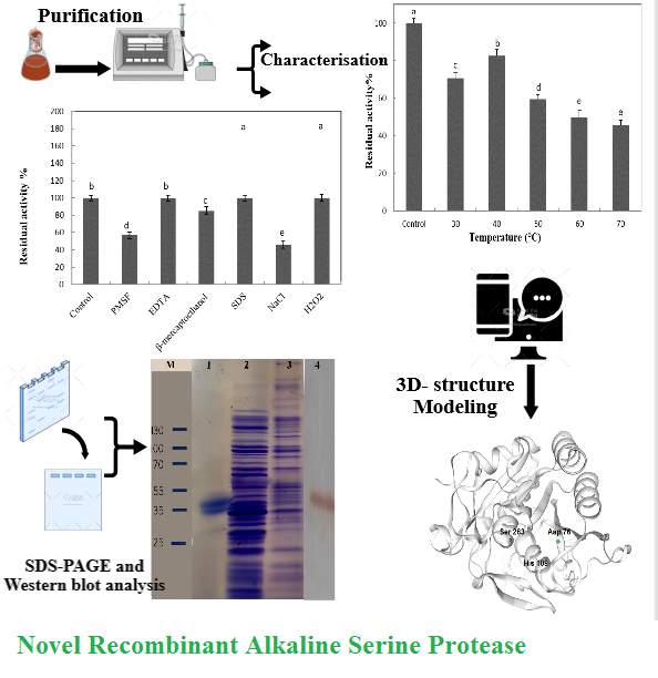
The recombinant enzyme was purified through cobalt affinity chromatography and the purification efficiency was assessed as 79.5%. The graph associated to purification is seen in Figure 1. The single long peak was associated to the release of the recombinant enzyme, which was eluted from the column at a concentration gradient of 90–100% elution buffer. The purified recombinant enzyme concentration included 130 µg ml-1. The specific activity of the purified recombinant enzyme was 0.12 U ml-1 mg-1, and the specific activity of the recombinant enzyme before purification was 0.066 U ml-1 mg-1. The enzyme samples extracted from the cell extract after induction, as well as the same sample after concentration with ammonium sulfate salt and the purified recombinant enzyme sample, were electrophoresed on 10% SDS-PAGE gels. Western blot analysis with anti-His tag antibodies revealed a distinct band at approximately 41 kDa, corresponding to the expected MW of the recombinant peptide. The six-His-tag, engineered at the C-terminal end of the recombinant protein for affinity purification, was still intact and was not cleaved from the peptide, indicating successful expression and detection of the full-length His-tagged recombinant protein (Figure 2). The three major elements in the successful production of recombinant protein included host condition, culture media condition and vector type. One of the appropriate microbial strains for exogenous gene expression was the E. coli strain [19].
The activity of the recombinant enzyme was assessed before and after purification. The quantity of absorbance after enzyme reaction at 280 nm included the absorbance of caseins broken by protease activity, which included a direct relationship with enzyme activity. Based on the definition, a unit is the quantity of enzyme that releases the fragments of peptides produced under standard conditions (0.001 A280 nm) within 30 min. In the present study, a novel recombinant serine alkaline protease derived from the native strain of Bacillus sp. RAM53 was expressed in a laboratory environment. The culture conditions were optimized for optimal production of this recombinant enzyme. The recombinant enzyme gene and protein with the scientific name of subtilase-like protease (MARF) were deposited in GenBank with accession numbers of OP922123.1 (https://www.ncbi.nlm.nih.gov/nuccore/OP922123.1) and WCO70948.1, respectively. The pure enzyme did not decrease its activity; instead, it increased its specific activity.
The specific activity of the purified recombinant enzyme was 0.12 U ml-1 mg-1, and the specific activity of the recombinant enzyme before purification was 0.066 U ml-1 mg-1. The concentration of the recombinant enzyme was assessed based on Bradford's standard method using a standard curve. The concentration of the recombinant enzyme was 0.3 mg ml-1 before purification and 0.13 mg ml-1 after purification.
Results of the thermal stability of the pure enzyme at various temperatures showed that the pure recombinant enzyme included relatively good stability for 3 h at 30 and 40 °C; hence, more than 70% of its activity were included. At 70 °C, 46% of the enzyme activity remained (Fig. 3). At all temperatures, a significant difference from the control was observed (p<0.05). The recombinant enzyme demonst-rated relatively good stability at high temperatures such as 60 and 70 °C without addition of stabilizing ions, compared to commercial enzymes. The subtilisin family of serine proteases represent an important class of industrial bio-catalysts widely used in the food industry [3]. Their extensive use is largely attributed to their favorable bio-chemical and catalytic characteristics. In addition, their high thermostability allows them to preserve activity under increased temperatures associated to industrial heat treat-ments [3,5,8,20]. The recombinant enzyme of the present study included good temperature resistance, compared to commercial enzymes.
Results of the stability to various pHs showed that the enzyme was stable at various pHs after 48 h of treatment, and its residual activity included 60–80% at neutral to alkaline pHs (Figure 4).
At all pHs, a significant difference with the control was observed (p < 0.05), and it showed similar stability at neutral to alkaline pHs, except for pH 9, and the highest stability was reported at pH 9. Subtilisin typically demonstrated significant stability under alkaline pH conditions, making it particularly appropriate for protein hydrolysis processes commonly used in food manufacturing [3,21].
Aryaei et al. identified and isolated an alkaline protease gene from a native strain of Bacillus sp. RAM and cloned the gene into pET28a+. The enzyme showed maximum activity at pH 9 and 40 °C [10]. Moradian et al. purified an alkaline serine protease from Bacillus sp. HR-08 and the protease activity was at pH 10 and 60 °C [18].
A thermostable serine-protease enzyme from Aeribacillus pallidus c10 strain was purified by Yildirim et al. and the optimal activity of this enzyme was at 60 °C and pH 9 [22]. Bervibacillus brostelensis isolated from a hot spring in Qeinerjeh produced an alkaline protease and showed maximum activity at 60 °C and pH 9 [23]. Alkaline proteases produced by Bacillus species are greatly important due to their thermal stability and stability at various pH levels, which are important in industries [1,24].
Results of pure enzyme treatment with 10-mM PMSF, 10-mM EDTA, 0.5% SDS, 0.5-M NaCl and 5% H2O2 after 1 h are reported in Figure 5. The EDTA did not affect the activity, and beta-mercaptoethanol decreased the activity of the enzyme a little with no significance (p < 0.05). The PMSF and NaCl decreased the enzyme activity by nearly 50% with significance (p < 0.05). In the presence of SDS and H2O2, almost all enzyme activity was preserved.
Several members of the S8 family (e.g. subtilisin) bind calcium for stability; inhibition can be seen with EDTA and EGTA, which are often reported as specific inhibitors of metallopeptidases. Calcium binding stabilizes these proteins in natural extracellular environments but poses a problem in industrial environments containing high concentrations of metal chelators. Subtilisin Carlsberg possesses three Ca2+ binding sites that stabilize its 3-D structure. The important industrial enzymes, subtilisin Carlsberg and subtilisin BPN, respectively included thermostability of 3.4 and 2.4 min at 50 °C and their thermal stability enhanced in presence of calcium [25,18]. These proteases are addressed as important commercial proteases and, therefore, the recombinant enzyme of the present study includes good temperature resistance, compared to commercial enzymes. In addition, the gene of this enzyme originates from B. mesophilus. A serine protease gene of B. megaterium, ispK, was cloned into E. coli. The thermostability increased 32.9-fold from 3.3 to 108.5 min at 60 °C in the presence of 2 mM of Ca2+ [26]. In the present study, the recombinant enzyme activity did not decrease in the presence of a chelating agent, resulting in calcium ions not neccessary for activity and stability in the structure. This characteristic is important for use in industry. Bacillus proteases have been shown stable in a wide range of pH and temperature [27]. A serine alkaline protease purified from Bacillus sp. HR08 was stable to hydrogen peroxide and its activity was complete [18].
The recombinant enzyme protein with the scientific name of subtilase-like protease was deposited in GenBank with the accession no. of WCO70948.1
(https://www.ncbi.nlm.nih.gov/protein/WCO70948.1). Based on the sequence alignment of S8 family peptidase (Bacillus) using Blast, NCBI, a sequence with reference number WP_000790938.1 was selected, which had nearly 86% identity to the recombinant enzyme sequence (Table 1). Then, the information on the amino acids of its active site was used to find amino acids in the active site of the recombinant alkaline serine protease. Based on the alignment and sequence homology, which included 86% identity with the subtilase family and available information on the active amino acids in the active site of the enzymes of this family, aspartic acid, histidine and serine (MEROPS - Peptidase Database) were assessed for the active site of the recombinant enzyme. The S8 termitase-like peptidases region is located at amino acids 40 to 270 in the recombinant enzyme.
In sequence analysis using ProScan (http://www.ebi.ac.uk/interpro/result/InterProScan/), the type of protease from the S8 subtilase family and the presence of a domain similar to thermitase were verified (Figure 6a). In addition, presence of Asp76, His109 and Ser263 in the active site of the enzyme was verified using ProScan and PROSITE (Figures 6b and 6c). Based on ProtParam prediction, the instability index (II) was computed as 18.38. This classified the recombinant protein as stable for more than 10 h in E.coli expression system. The aliphatic index of the recombinant enzyme was assessed as 71.52 as a thermostable protein. Several important subtilisins have been produced using various bacilli, including subtilisin Carlsberg from B. licheniformis; subtilisin BPN and Dj-4 from B. amyloliquefaciens; subtilisin E, NAT and J from B. subtilis; and subtilisin amylosacchariticus from B. amylosacchariticus [27]. The alkaline protease gene (Apr) from B. licheniformis 2709 was cloned into an expression vector of pET28b (+) and expressed in a high-expression strain of E. coli BL21 [29]. The recombinant enzyme included identities with S8 family peptidase from B. cereus, S8 family peptidase from Bacillus sp., S8 family peptidase from B. firmicutes and S8 family serine peptidase from B. thuringiensis as 86, 86, 86 and 85%, respectively. The subtilisin family is one of the largest serine peptidase families characterized to date. Structures have been assessed for several members of the subtilisin family that contain similar catalytic triad residues in an order of DHS (Asp, His, Ser)
(https://www.ebi.ac.uk/interpro/entry/InterPro/IPR034202/). Subtilisin possess broad substrate specificity, enabling efficient degradation of diverse protein substrates and demonstrates strong catalytic efficiency, which contributes to reduce processing times and improved production yields. Collectively, these characteristics highlight the significant potential of S8/thermitase-like proteases in various food processing uses [3,21]. The tertiary structure of the present alkaline serine protease was made based on a template selected by the Swiss model building. The template included thermitase with PDB code 1thm.1A, as well as sequence identity of 85% (Figure 7a). In Ramachandran plot, distribution of the backbone dihedral angles (ϕ and ψ) showed that a majority of residues were located in the most favored regions (dark green areas), primarily corresponding to α-helical and β-sheet conformations. A smaller fraction of residues was set within additionally allowed regions (light green areas), which was acceptable for certain amino acids in specific conformations. Overall, the plot suggests that the protein model includes good stereochemical quality, with most residues adopting energetically favorable conformations and a minimal number of outliers that are unlikely to affect the structural integrity significantly (Figure 7b). The local quality assessment in QMEAN, expressed as the predicted local similarity to the target, showed that the majority of residues included high similarity scores (> 0.8), indicating reliable modelling in most structural regions. Peaks close to 0.9–1.0 suggested well-defined secondary structures with high confidence in backbone conformation. However, a few distinct regions particularly near residues of 25–30 and 235–245 showed significantly lower quality scores (< 0.6), with the lowest approaching 0.3. These decreases often corresponded to loop regions, flexible termini or poorly conserved areas in the template alignment. Overall, the QMEAN profile supported a generally reliable global structure, with local weaknesses restricted to specific flexible or unresolved segments (Figure 7c). Using experimental data and ExPASY analysis as well as comparative modelling, this study predicted the protein 3-D structure and active site amino acids. Assessmentn of the predicted 3-D structure using Ramachandran plot revealed that a majority of residues were located in the most favored regions, with only a few residues in additionally allowed areas and a minimal number in disallowed regions. This distribution indicated that the backbone dihedral angles were largely in stereochemically favorable conformations, supporting the overall correctness of the model [29]. Collectively, these results suggest that the predicted protein model possesses good overall stereochemical quality and global reliability.
SWISS-MODEL is a fully automated protein structure homology-modelling server, accessible via the Expasy web server or DeepView program (Swiss Pdb-Viewer). Homo-logy modelling allows the protein engineer to design modifications of a protein before the determination of the 3-D structure using X-ray technique. Of all current theoretical approaches, comparative modelling is the best method that can reliably generate a 3-D model of a protein from its amino acid sequence [30,31]. The alkaline protease from thermophilic bacteria included an aliphatic index of 69.4 [32], while that of the recombinant enzyme was assessed as 71.52, showing that the recombinant enzyme included higher aliphatic index than the thermophilic type and thermal stability despite its mesophilic origin, compared to other thermophilic enzymes. This index should be addressed as a positive factor for increasing the thermal stability of globular proteins [33].
More than one million proteolytic enzyme sequences have been recorded in the Merops Database [34]. Therefore, identifying novel strains with the ability to produce enzymes appropriate for industrial uses and genes of these enzymes for the production of recombinant enzymes has always been useful. Especially in countries that buy industrial enzymes, this decreases the purchase costs. In addition to laboratory studies, bioinformatics studies for characterizing alkaline proteases help identify their industrial use and large-scale production. Therefore, protein-structure prediction tools are appropriate for the study of protein structures and functions [35].
- Conclusion
The current study focused on the biochemical characteristics and tertiary structure, physicochemical characteristics and catalytic potential of a novel recombinant alkaline serine protease. Despite lack of sufficient funding to sequence the N-terminal region of the purified protein that was a limitation of the present study, structural engineering methods can be used to improve enzyme production for the production of a novel high-quality recombinant enzyme on an industrial scale by predicting the tertiary structure of this enzyme using bioinformatics. Given its favorable biochemical characteristics, including pH stability over the range of 6–13, thermal stability at 60 °C, independent of Ca²⁺ and resistance to SDS and oxidizing agent of H₂O₂ with its classification as an alkaline protease, the enzyme demonstrates strong potential for use in various industrial sectors such as detergent, food and animal and poultry feed productions.
- Bioinformatics
- Expression
- Purification
- Recombinant alkaline protease
How to Cite
References
1. Abdul Razzaq A, Shamsi S, Ali A, Ali Q, Sajjad M, Malik A, Ashraf M. Microbial proteases applications. Front Bioeng Biotechnol. 2019; 7: 110. https://doi.org/10.3389/fbioe.2019.00110
2. Rao MB, Tanksale AM, Ghatge MS, Deshpande VV. Molecular and biotechnological aspects of microbial proteases. Microbiol Molecul Biol Rev. 1998; 62(3): 597-635. https://doi.org/10.1128/mmbr.62.3.597-635.1998
3. Santos Aguilar JG, Sato HH. Microbial proteases: Production and application in obtaining protein hydrolysates. Inst Food Sci Technol. 2018; 103: 253-262. https://doi.org/10.1016/j.foodres.2017.10.044
4. Saleem Sh, Akinola OM, Abdul Rahman MB, Leow ATCh, Muhd Noor ND, Salleh AB, Oslan SN. Appl Food Biotechnol. 2025; 12(1): 1-30(e19). https://doi.org/10.22037/afb.v12i1.48544
5. Ray A. Protease enzyme-potential industrial scope. Int J Tech. 2012; 2: 1-4.
6. Gupta S, Mehta M. Effect of cysteine protease in diet to reduce soybean meal without affecting performance of kadaknath birds in India. J Anim Res. 2016; 6(6): 951-955. https://doi.org/10.5958/2277-940X.2016.00136.4
7. Lee SA, Bedford MR, Walk CL. Meta-analysis: Explicit value of mono-component proteases in monogastric diets. Poult Sci. 2018; 97(6): 2078-2085. https://doi.org/10.3382/ps/pey042
8. Saleh AA, Ali H, Abdel-Latif MA, Emam MA, Ghanem R, El Hamid HAS. Exogenous dietary enzyme formulations improve growth performance of broiler chickens fed a low-energy diet targeting the intestinal nutrient transporter genes. PloS one. 2018; 13(5): e0198085. https://doi.org/10.1371/journal.pone.0198085
9. Alei A, Moradian F, Farhadi F, Rostami M. Identification, isolation, cloning and expression of a new alkaline serine protease gene from native Iranian Bacillus sp. RAM 53 for use in industry. Appl Biochem Biotechnol. 2023; 195(6). https://doi.org/10.1007/s12010-023-04585-9
10. Ariyaei A, Farhadi A, Moradian F, Rahimi Mianjiv G. Cloning, expression and characterization of a novel alkaline serine protease gene from native Iranian Bacillus sp.; A producer of protease for use in livestock. Gene 2019; 693: 10-15. https://doi.org/10.1016/j.gene.2019.01.020
11. Hadjidj R, Badis A, Mechri S, Eddouaouda K, Khelouia L, Annane R and et al. Purification, biochemical and molecular characterization of novel protease from Bacillus licheniformis strain K7A. Int J Biol Macromol. 2018; 114: 1033-1048. https://doi.org/10.1016/j.ijbiomac.2018.03.167
12. Singh S, Gupta P, Bajaj BK. Characterization of a robust serine protease from Bacillus subtilis K‐1. J Basic Microbiol. 2018; 58(1): 88-98. https://doi.org/10.1002/jobm.201700357
13. Joshi S and Satyanarayana T. Characteristics and applications of a recombinant alkaline serine protease from a novel bacterium, Bacillus lehensis. Bioresour Technol. 2013; 131: 76-85. https://doi.org/10.1016/j.biortech.2012.12.124
14. Kaur I, Kocher GS and Gupta VK. Molecular cloning and nucleotide sequence of the gene for an alkaline protease from Bacillus circulans MTCC 7906. Indian J Microbiol. 2012; 52(4): 630-637. https://doi.org/10.1007/s12088-012-0297-4
15. Sadeghi MM, Rabbani M, Naghitorabi M. Cloning of alkaline protease gene from Bacillus subtilis 168 H. Res Pharama Sci. 2009; 4: 43-46.
16. Gurunathan R, Huang B, Kumar Ponnusamy V, Hwang JSh and Dahms HU. Novel recombinant keratin degrading subtilisin like serine alkaline protease from Bacillus cereus isolated from marine hydrothermal vent crabs. Sci Rep. 2021; 11: 12007. https://doi.org/10.1038/s41598-021-90375-4
17. MohammadzadehS, Moradian F, Yeganeh S, Falahatkar B, Milla S. Design, production and purification of a novel recombinant gonadotropin-releasing hormone associated peptide as a spawning inducing agent for fish. Protein Expr Purif. 2020; 166: 105510. https://doi.org/10.1016/j.pep.2019.105510
18. Moradian F, Khajeh Kh, Naderi-Manesh H, Sadeghizadeh M. Isolation, purification and characterization of a surfactants, laundry detergents and organic solvents-resistant alkaline protease from Bacillus sp. HR-08. Appl Biochem Biotechnol. 2009; 159: 33-45. https://doi.org/10.1007/s12010-008-8402-1
19. Karray A, Alonazi M, Horchani Ben H, Bacha A. A novel thermostable and alkaline protease produced from Bacillus stearothermophilus isolated from olive oil mill sols suitable to industrial Biotechnology. Molecules 2021; 26: 1139. https://doi.org/10.3390/molecules26041139
20. Joseph BC, Pichaimuthu S, Srimeenakshi S, Murthy M, Selvakumar K, Ganesan M, Manjunath SR. An overview of the parameters for recombinant protein expression in Escherichia coli. J Cell Sci Ther. 2015; 6: 221. https://doi.org/10.4172/2157-7013.1000221
21. Shettar Sh S, Bagewadi ZK. Subtilisin: A bibliometric and comprehensive review of its structure, production and versatile applications. Discover Appl Sci. 2025; 7:1432. https://doi.org/10.1007/s42452-025-07895-1
22. Choi NSh, Chang KT, Maeng PJ, Kim SH. Cloning, expression and fibrin (ogen) olytic properties of a subtilisin DJ-4 gene from Bacillus sp. DJ-4. FEMS Microbiol Lett. 2004; 236: 325-331. https://doi.org/10.1016/j.femsle.2004.06.006
23. Lin S, Zhang M, Liu J, Jones GS. Construction and application of recombinant strain for the production of an alkaline protease from Bacillus licheniformis. J Biosci Bioeng. 2015; 119(3): 284-288. https://doi.org/10.1016/j.jbiosc.2014.08.002
24. Nasre Taheri M, Ebrahimipour G, Sadeghi H. Isolation and identification of alkaline protease-producing bacterium in the presence of washing-powder from Geinarje hot spring, Ardabil, Iran. Nova Biologica Reperta. 2019; 6(3): 292-301. https://doi.org/10.29252/nbr.6.3.292
25. Xue Y, Zeng L, Yan Q, Jiang Z. Efficient Production of a Novel Subtilisin-like Serine Protease in Aspergillus niger for the Preparation of DPP-IV Inhibitory Activity Peptides from Protein Byproducts J Agricul Food Chem. 2025;73(35):22014-22026. https://doi.org/10.1021/acs.jafc.5c06047
26. Yildirim V, Ozkan Baltaci M, Ozgencli I, Sisecioglu M, Adiguzel A, Adiguzel G. Purification and biochemical characterization of a novel thermostable serine alkaline protease from Aeribacillus pallidus C10: A potential additive for detergents. J Enzyme Inhib Med Chem. 2017; 32: 468-477. https://doi.org/10.1080/14756366.2016.1261131
27. Srimathi S, Jayaraman G, Narayanan PR. Improved thermodynamic stability of subtilisin Carlsberg by covalent modification. Enzyme Microb Technol. 2006; 39(2): 301-307. https://doi.org/10.1016/j.enzmictec.2005.10.027
28. Jeong YJ, Baek SC, Kim H. Cloning and characterization of a novel intracellular serine protease (IspK) from Bacillus megaterium with a potential additive for detergents. Int J Biol Macromol. 2018; 108: 808-816. https://doi.org/10.1016/j.ijbio mac.2017.10.173
29. Studer G, Rempfer C, Waterhouse AM, Gumienny R, Haas J, Schwede T. QMEANDisCo - distance constraints applied on model quality estimation. Bioinformatics. 2020; 36: 1765-1771. https://doi.org/10.1093/bioinformatics/btz828
30. Marti-Renom MA, Stuart AC, Fiser A, Sanchez R, Melo F, Sali A. Comparative protein structure modeling of genes and genomes. Annu Rev Biophys Biomol Struct. 2000; 29: 291-325. https://doi.org/10.1146/annurev.biophys.29.1.291
31. Tramontano A, Leplae R, Morea V. Analysis and assessment of comparative modeling predictions in CASP4. Proteins. 2001; 45(5): 22-38. https://doi.org/10.1002/prot.10015
32. Ikai AJ. Thermostability and aliphatic index of globular proteins. J Biochem. 1980; 88: 1895-1898.
33. Gasteiger E, Hoogland C, Gattiker A, Duvaud S, Wilkins MR, Appel RD, Bairoch A. Protein identification and analysis tools on the ExPASy server. In: Walker JM, editor. The Proteomics Protocols Handbook. Totowa (NJ): Humana Press; 2005. p. 571-607.
34. Verhamme IM, Leonard SE, Perkins V. Proteases: Pivot points in functional proteomics. Methods Mol Biol. 2020; 1871: 313-392. https://doi.org/10.1007/978-1-4939-8814-3_20
35. Mahmoud A, Kotb E, Alqosaibi AI, Al-Karmalawy AA, Al-Dhuayan IS, Alabkari H. In vitro and silico characterization of alkaline serine protease from Bacillus subtilis D9 recovered from Saudi Arabia. Heliyon 2021; 7: e08148. https://doi.org/10.1016/j.heliyon.2021.e08148
- Abstract Viewed: 183 times
- pdf Downloaded: 98 times