
Trilayer Cellulose Acetate-polyvinyl alcohol-cellulose acetate Sandwich Nanofibers for Enhanced Probiotic Viability under Thermal and Acidic Conditions
بیوتکنولوژی غذایی کاربردی,
دوره 12 شماره 1 (2025),
4 دی 2025
,
صفحه 1-12 (e11)
https://doi.org/10.22037/afb.v12i1.47378
چکیده
This study aimed to enhance probiotics thermal stability and viability in the digestive tract through encapsulation using hybrid fibers of cellulose acetate and polyvinyl alcohol with single-jet electrospinning. This study used Lactiplantibacillus plantarum NIMBB003 as an encapsulated probiotic strain in engineered sandwich nanofibers (cellulose acetate/polyvinyl alcohol and Lactiplantibacillus plantarum/cellulose acetate). Regarding nanostructure, polyvinyl alcohol and cellulose acetate nanofibers were spun independently; when these layers were set on top of each other, they could act as an integrated system. Results of scanning electron microscope images and Fourier transform infrared spectrometry have verified the micro/nanoencapsulation structure of probiotics. The layered structure demonstrated increased protection against environmental factors, particularly heat and acidity. Thermogravimetric analysis verified that cellulose acetate-polyvinyl alcohol and probiotic-cellulose acetate nanofibers maintained the structural stability up to 530 °C, while encapsulated probiotics showed 89.8% encapsulation efficiency or 9% improvement, compared to single-layer polyvinyl alcohol and probiotic fibers. Moreover, probiotic survival under simulated gastrointestinal conditions (75 °C and stomach acid exposure) was extended to 8 min, whereas unencapsulated probiotics were entirely destroyed within 5 min. Scanning electron microscopy and Fourier transform infrared spectroscopy validated the formation of nanofiber encapsulation and probiotic integration. This engineered nanofiber sandwich structure offers enhanced probiotic protection, making it a promising candidate for food and pharmaceutical uses.
- Introduction
Probiotics are living microorganisms that, when consumed in certain quantities, can include positive effects on the body. These microorganisms are critical in maintaining a balanced gut microbiome for overall wellbeing [1]. They can prevent the growth of harmful bacteria, maintain the health of the intestinal flora, decrease cholesterol, produce vitamins and antimicrobial substances and strengthen the immune system [2]. Decreasing constipation improves calcium absorption and treats digestive and non-digestive diseases (e.g. infection, constipation, inflammatory bowel diseases, colon cancer and cardiovascular and urinary diseases) [3]. Lactobacillus is a common probiotic genus that supports gut health, helps digestion and increases immunity. Temperature and oxygen levels significantly affect bacterial viability through manufacturing, storage and industry processes. The ideal growth temperature for most Lactobacillus species is 30–45 °C. However, high temperatures greater than 45 °C may decrease their viability during processing. While oxygen is not usually a major concern for Lactobacillus survival, low oxygen levels can help them grow well [4, 5]. The most common way for live probiotics to enter the body and reach the intestines is through consuming products containing probiotics in form of powders, capsules and granules [6]. Using various methods, probiotics can be protected through targeted delivery to specific parts of the digestive system. In the stomach, these tolerate highly acidic conditions (pH 1.5 - 3.5), which can destroy unprotected bacteria. Probiotics must tolerate bile salts and digestive enzymes to reach the small intestine with a further neutral pH (6-7). The colon, characterized by a slightly acidic environment (pH 5.5-6.5), is the primary site, where probiotics exert their beneficial effects by supporting microbiota balance, enhancing nutrient fermentation and promoting immune modulation [7].
One of these techniques is encapsulation, which makes these bacteria survive in the steps of entering, storing and passing through the human body. Characteristics of a shell polymer for probiotic compounds include high stability, immiscibility with active ingredients, non-toxicity, cost-effectiveness, biodegradability and compatibility with the host [8, 9]. Furthermore, effective factors in control and appropriate release of probiotic compounds include molecular weight, solubility, temperature and chemical structure, which can decrease the mobility of molecules and minimize their degradation over time and are widely used in dairy products [10]. Conventional methods of micro-encapsulation of probiotics such as emulsion, extrusion, spray drying and freeze-drying have been used for microencapsulation of probiotics. Emulsion and extrusion create problems in absorption operations by producing relatively large particles [11-14]. High temperature, crystallization and high pressure are disadvantages of spray and freeze drying [15]. Thus, methods that maximize bacterial viability should be used, when biopolymers are used to coat encapsulated strains for protection in the gastrointestinal tract or as carriers for direct encapsulation of microorganisms [16]. Recent studies on probiotic encapsulation highlight diverse polymer matrices and encapsulation techniques to enhance bacterial viability and stability. For example, cellulose-based polymers as well as trehalose, maltodextrin and vegetable wax combined with fluid bed encapsulation improved productivity during the encapsulation process and greater stability during storage for L. casei subsp. paracasei LMG P-21380 [17], while increasing survival rate of the microorganism during simulation for L. plantarum IS-10506 in cellulose and alginate-based polymers [18]. Spray drying encapsulation with whey protein-chitosan extended the shelf life of Kluyveromyces marxianus VM004 for 90 days at room temperature and enhanced its tolerance under simulated gastrointestinal conditions [19]. In contrast, maltodextrin-sucrose-sorbitol formulations improved the survival of Saccharomyces cerevisiae KTP, Issatchenkia occidentalis ApC, and Saccharomyces cerevisiae var. boulardii in simulated gastric environments while maintain-ing the stability of the encapsulation components [20].
Alginate beads coated with whey protein significantly protected L. plantarum spp. in alginate beads and extended viability of the encapsulated probiotics in the gastric environment, compared to free probiotics [21]. Micro-capsules from a mixture of alginate and modified starch based on emulsification encapsulation increased cell survival and resulted in greater storage capability for encapsulated probiotics, compared to free probiotics of L. acidophilus and Bifidobacterium lactis [22]. Electro-spinning polyvinyl alcohol (PVA) enabled the viability of L. gasseri encapsulated at -70 °C for long-term storage and inactivation of their metabolism [23]. These advances demonstrate the critical role of material selection and encapsulation methods in optimizing probiotic delivery.
Electrospinning is an easy, cheap green method that produces fibers by pumping a biopolymer solution containing probiotic bacteria under the effects of an electric field. When a drop is formed at the tip of the needle, a voltage of 5–30 kV is used to the device [24]. With the formation of positive charges on the droplet and electrostatic attraction, the droplet changes its shape into a Taylor cone and forms thin nanofibers on the collector during evaporation in the field [25]. This technology is appropriate for encapsulating probiotics as emulsions as well as susceptible, biocompatible and biodegradable compounds [26, 27]. Although various probiotic encapsulation techniques have been studied, most methods include limitations such as high-temperature degradation, low encapsulation efficiency (EE) and poor protection in acidic environments [28]. This electrospinning study focused on biocompatibility but was still under investigation in the dual-layered protective role of cellulose acetate (CA) for improving thermal stability. This study introduced a novel multiple-layered nanofiber composite of CA-PVA-CA designed to enhance probiotic viability under thermal and acidic stresses. The suggested structure has offered a promising solution for food and pharmaceutical uses by addressing key limitations of conventional encapsulation methods.
- Materials and Methods
The L. plantarum NIMBB003 isolated from camel milk and registered in GenBank of the National Centre for Biotechnology Information (NCBI) (code MT0121881) was received from Shams Bavaran Salamat Noor, Tehran, Iran. PVA polymer with a molecular weight of 72000 g mol-1 and 98% hydrolysis was purchased from Merck, Germany. The CA with a molecular weight of 32000 g mol-1 was purchased from Sigma-Aldrich, USA.
2.1 Microorganism and Media
The L. plantarum NIMBB003 was cultured in De Man-Rogosa-Sharpe (MRS) broth at 37 °C ±1 for 24 h and then centrifuged at 5000 rpm for 10 min. The harvested cells were washed twice with sterile phosphate-buffered saline (PBS, pH 7.4) and resuspended in similar buffer prior to mixing with the PVA solution for encapsulation.
2.2 Preparation of Polymer Solution
The PVA at 7% w/w concentration was dissolved in 80 °C deionized water by magnetic stirring for nearly 4 h [29]. The solutions were cooled to 20 °C. The critical point was the gentle heat to dissolve the powder, which was nearly 80 °C; further heat might cause burn and destroy of the material [30]. The CA was dissolved in acetone/dimethylformamide solvent in a 1:2 ratio at 20 °C for 30 min using magnetic stirrer [31].
2.3 Preparation of Probiotic Cells and Calculation of the Probiotic Encapsulation Efficiency
Standard plate count method was used to assess viability of the probiotic bacteria. Free cells were serially diluted in sterile peptone water, plated on MRS agar, incubated at 37 °C ±1 and enumerated after 48 h [32]. The percentage of probiotic EE (%) was assessed by counting the number of probiotic bacteria in the feed solution and the number of probiotic bacteria released by the nanofibers. Briefly, 10 mg of PVA nanofibers containing L. plantarum bacteria were suspended in 1 ml of phosphate-buffered saline diluent solution at pH 7.4. For engineered sandwich nanofibers (CA-PVA and L. plantarum-CA), samples were agitated for 4 h at 20 ºC. Following dilution and surface cultivation, the probiotic bacterial count was investigated, which indicated the number of released and unencapsulated probiotic bacteria. The EE (%) was calculated based on Eq. 1 [33]:
Eq. 1
2.4 Electrospinning Conditions of Polymer Solutions
For the electrospinning process, the following parameters were selected based on previous studies. Voltage was 20 kV, flow rate of the PVA solution was 1 ml.h-1 and flow rate of the CA solution was 1 ml.h-1. The distance from the tip to the collector was 15 cm [34]. Electrospinning was carried out in a controlled environment with a lab temperature of 25°C ±1 and relative humidity was set at 70–75%.
2.5 Encapsulation of Probiotics in Engineered Sandwich Nanofibers
All polymer solutions were electrospun from a 5 ml syringe tip and collected on a rotating cylindrical collector using electrospinning machine. All electrospinning was carried out using laboratory hood under standard laboratory temperature and humidity conditions. The samples included PVA nanofibers containing L. plantarum and engineered sandwich nanofibers (CA-PVA and L. plantarum-CA) with a control sample without probiotics. After spinning 2 ml of CA solution as the first layer and protector under optimal conditions, 2 ml of PVA solution with L. plantarum were spun as the middle layer under optimal conditions. Moreover, 2 ml of CA solution were spun again as the third layer. In this study, the quantity of material spun was consistent for each layer. This procedure was repeated three times for each sample.
2.6 Assessment of Thermal Resistance of Free and Encapsulated L. plantarum Probiotic Bacteria
To assess thermal resistance, yogurt soda (dough) was used as the test media with bacterial viability standardized to 10⁷ CFU (colony forming units) per gram or milliliter based on the food product requirements. The EE and fiber weight were incorporated into the experimental design to ensure that the bacterial concentration in 40-ml yogurt solution met this threshold. Three formulations were assessed, including (1) free probiotics (pro), (2) L. plantarum probiotic encapsulated in PVA nanofibers (PVA and pro) and (3) engineered sandwich nanofibers (CA/PVA and L. plantarum-CA). Samples were heat-treated for 0, 1, 3, 5 and 8 min with 1 ml of aliquots collected at each interval. Viable bacterial counts were carried out via serial dilution and agar plate cultivation. The study aimed to assess nanoencapsulation efficacy in protecting probiotics from thermal degradation, offering insights for optimizing heat-resistant probiotic food formulations.
2.7 Scanning Electron Microscopy
Scanning electron microscopy (SEM) (TESCAN MIRA3, Czech Republic) was used to investigate morphologies of the electrospinning nanofibers. The diameters of the nanofibers were calculated from the SEM images using image processing program [35].
2.8 Fourier Transform Infrared spectrometry
Fourier transform infrared spectrometry (FTIR) (THERMO IS50, Germany) was used for the chemical characterization of the nanofibers. The FTIR could detect impurities in the model. By identifying the impurity, its origin could be predicted and removed. Since each molecule included its identification peak, researchers could detect presence of impurities and pollutants by seeing additional peaks in the sample analysis [36].
2.9 Thermogravimetric analysis
Thermogravimetric analysis (TGA) (STA7300, Hitachi, Japan) was used to assess changes in a sample mass due to thermal decomposition, oxidation or reaction with other gases, represented as a percentage increase or decrease in weight on the plotted graph. This method assessed a specific quantity of materials based on temperature under controlled heating program and atmosphere. The sample was heated or cooled during the assessment using furnace, while its precise weight was assessed. The graph shows results for TGA of the samples.
- Results and Discussion
3.1 Morphology of Electrospinning Nanofibers
Based on Figure 1, the SEM results indicate that the diameter distribution of the nanofibers ranged 90-500 nm. This was totally verified definition of nanofibers and suggested that the synthesis was successful, yielding healthy fibers with a uniform diameter from a 7% by weight PVA solution under optimal electrospinning conditions. Regions at various nanofiber points have enhanced the structure crystallinity, significantly affecting the physical and mechanical characteristics of the scaffold produced during the electrospinning process.
Figure 2 shows 14% by weight, uniform, knot-free cellulose fibers with details during dissolution of cellulose acetate in acetone and dimethylformamide solvents (2:1) in nanometer dimensions [37]. Figure 3 shows the SEM image results for the optimal conditions of the engineered sandwich nanofibers (CA-PVA and L. plantarum-CA) production process. Figure 3a includes side view, illustrating three layers; Figure 3b includes top view and Figure 3c includes diameter distribution diagram of nanofibers. During the experiments, the engineered sandwich nanofiber images showed that L. plantarum NIMBB003 probiotic encapsulation was successfully carried out in PVA electrospun nanofibers by increasing the number of willows. It shows the entanglement of two composite nanofibers of PVA and CA. The morphology of electrospun fibers directly affected probiotic EE. Smaller nanofiber diameters (< 500 nm) enhanced protection and surface interactions, while porosity governed nutrient diffusion and release kinetics. Core-shell or aligned fibers further optimized targeted delivery and gastric survival. The nanofiber diameters in this study were similar with established ranges for similar PVA-based bioactive encapsulation systems. For PVA-CA nanofibers at concentration of 7% (w/v), voltage 22 kV, flow rate of the PVA and CA solution of 1.3 ml.h-1 and distance from the tip to the collector of 14 cm.
3.2 Fourier Transform Infrared Spectrometry Result
In FTIR analysis by assessing the electromagnetic wave interactions in the range of 0-4000 cm-1, it is possible to investigate the structure of molecules, functional groups and bonds in the samples. The samples were assessed in three states to analyze their chemical structure and crystallinity. As seen in Figure 2, the FTIR spectra of PVA nanofibers, PVA and probiotic nanofibers and CA-PVA and probiotics-CA nanofibers all included major peaks at 1100, 1700, 2750 and 13500 cm-1. The samples were assessed in three states to analyze their chemical structure and crystallinity. Four central regions with dominant peaks (A, B, C and D) can be seen in Figure 2. In the wavelength range of 3650-3200 cm-1 (Region A), a peak caused via absorption by free and bound OH groups were seen in the stretched vibration of PVA [38]. In the wavelength range of 2860-2730 cm-1 (Region B) in the spectra of PVA nanofibers with probiotics, the stretched vibration was due to the C-H aldehyde group. In the CA-PVA and probiotics-CA nanofibers range, the stretched pulse was due to C-H and O=C-H. In the stretched region from 1750 to 1700 cm-1 (Region C) of the PVA spectra, the extended pulse of the C-O-acetate groups of the polymer could be observed and in the region from 1140 to 1000 cm-1 (Region D) of the PVA spectra, the stretched vibration of the O-C-O group could be recorded [39, 40].
The FTIR spectra revealed four critical regions (A–D) that provided essential insights into the molecular structure and interactions in the analyzed nanofibers (Figure 4). Peak A (3200–3500 cm-¹), attributed to O-H stretching vibrations, indicated hydrogen bonding in PVA and potential moisture absorption with shifts reflecting polymer-probiotic interactions. Peak B (1700-1750 cm-¹), corresponding to C=O stretching, verified presence of CA in nanofibers and its intensity variations might reveal chemical modifications.
Peak C (1100–1200 cm-¹), associated with C-O-C or C-O stretching, showed polymer backbone structure and crystallinity changes, particularly in PVA-P70 NFs. Peak D (2800–3000 cm-¹), representing C-H stretching, helped monitor structural alterations in the polymer chains due to probiotics incorporation or layering. These peaks collectively served as molecular fingerprints as Peaks A and B were vital for assessing hydrogen bonding and esterification, while Peaks C and D explained chain packing and functional group integrity. Comparative analysis of these regions in the samples (e.g. PVA NFs against PVA-P70 NFs) enabled detection of probiotics EE, polymer-probiotic interactions and material stability, making them integrated for characterizing advanced encapsulation systems.
Therefore, FTIR spectroscopy analyzed molecular structure by measuring absorption of 400-4000 cm-¹. In this study, key peaks at 1100, 1700, 2750 and 13500 cm-¹ revealed critical functional groups, including O-H stretch (3200-3650 cm-¹, hydrogen bonds), C-H (2730-2860 cm-¹, aldehydes), C=O (1700-1750 cm-¹, acetate) and O-C-O (1000-1140 cm-¹, ether links). These functional groups were essential for identifying molecular interactions (e.g. PVA-probiotic hydrogen bonds) and crystallinity changes. The O-H peak shift indicated modified polymer-probiotic interactions, while C=O bands verified CA presence in layered fibers. Such analysis was vital for assessing encapsulation system stability and functionality.
3.3 Thermogravimetric Analysis Results
Based on the results from the TGA (Figure 5), the weighed sample was stable in the desired temperature range and the rapid decrease in the initial mass indicated drying or moisture removal of the nanofibers. The curves of all three nanofibers were used to investigate stoichiometry or to study reaction kinetics and show the decomposition of models in one-step with relatively stable intermediates. In addition, it could be seen that PVA and probiotics nanofibers were completely degraded in the temperature range of 500-530 °C. In CA/ PVA and probiotics-CA nanofibers, this process of degradation and decrease in weight percentage continued slowly over time; thus, addition of CA increased the thermal resistance of nanofibers. The thermal stability of the engineered sandwich nanofibers (CA-PVA and L. plantarum-CA) structure was better than that of traditional whey protein-alginate microcapsules, often showing decreased stability under high-temperature conditions.
3.4 Heat Stability of Free and Encapsulated L. plantarum Probiotic Bacteria
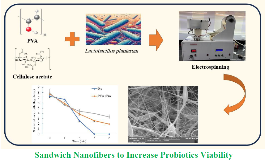
Thermal resistance was assessed in three conditions of probiotic bacteria (pro), probiotic enclosed in PVA nanofibers (PVA and pro) and PVA composite nanofibers containing probiotic bacteria between the two layers of cellulose acetate nanofibers from top and under (CA-PVA and pro-CA). As seen in Figure 6 in unencapsulated probiotics (pro), all bacteria were inactivated after 5 min of heat exposure. In contrast, PVA and Pro demonstrated enhanced thermal resistance with viable bacteria surviving longer than the free ones. The CA-PVA and Pro-CA composite showed the highest protective efficacy, preserving probiotic viability for up to 8 min at 75 °C. While conventional microencapsulation methods (e.g. spray drying) often decrease bacterial survival due to thermal stress [21], this study highlighted that the CA-PVA-CA dual-layer structure significantly improved heat tolerance. These findings challenged the general suggestion that encapsulated probiotics could not tolerate high-temperature food processing, offering a promising strategy for developing thermally stable probiotic products. This survival time is longer than that of previous studies using spray-dried probiotic microcapsules, which reported survival for 3-4 min under similar high-temperature conditions [23].
3.5 Encapsulation Efficiency Calculation and Results
Based on Table 1, the following results were achieved after counting the number of free and encapsulated bacteria using the stated efficiency formula. Data in Table 1 demonstrated that the CA-PVA and probiotics-CA nanofibers achieved significantly higher EE (89.8%), compared to that PVA and probiotics nanofibers did (81%) as shown by a lower quantity of unencapsulated bacteria (130,103 against 247,716 CFU.ml-1) despite similar initial bacterial concentrations (1,277,818 CFU.ml-1). This 8.8% absolute improvement (p < 0.05, assuming appropriate replicates) suggested that the CA outer layer enhanced probiotic retention, likely through better structural integrities or protective barrier characteristics. For robust statistical validation, triplicate experiments with standard deviation (SD) analysis verified if this difference was technically and statistically significant (p < 0.01). These results highlighted the potential of CA-PVA bilayer systems for industrial probiotic encapsulation, where high efficiency was critical.
The EE in this study was significantly higher than that in previous techniques such as spray drying and fluid bed encapsulation. For example, previous studies on whey protein-alginate encapsulation methods reported a survival rate of 60-75% under simulated gastrointestinal conditions [41]. The present study included higher values, demon-strating superior protections against heat and acidity.
- Conclusion
One of the major challenges of probiotics is their low thermal resistance, limiting their uses in heat-processed foods such as pasteurized dairy products. This study developed novel sandwich-structured nanofibers for the encapsulation of L. plantarum NIMBB003 using PVA and CA through an optimized electrospinning process. The resulting three-layer engineered sandwich nanofibers (CA-PVA and L. plantarum-CA) structure significantly improved probiotic viability, thermal stability and EE. This technology offers practical uses in food and pharmaceutical industries. It enables the production of heat-stable probiotics appropriate for pasteurized dairy products, functional beverages and baked goods while supporting the pharma-ceutical development of gastro-resistant probiotics capsules that ensure targeted delivery to the intestines. Key performance metrics highlight the effectiveness of this approach as EE of L. plantarum in engineered sandwich nanofibers (CA-PVA and L. plantarum-CA) reached 89.8%, representing a 9% improvement, compared to single-layer PVA and probiotics nanofibers. Furthermore, heat resistance assessment showed that the engineered sandwich nanofibers (CA-PVA and L. plantarum-CA) structure protected probiotics for up to 8 min at 75 °C, a significant increase in survival duration. The three-layer and morphology of excellent engineered sandwich nanofibers were verified through SEM, validating structural integrity of the nanofibers and successful incorporation of probiotics via FTIR. Results of this study demonstrate that the engineered sandwich nanofibers (CA-PVA and L. plantarum-CA) structure is a promising scalable method for enhancing probiotic survival and functionality within diverse industrial uses. While the engineered probiotic-loaded sandwich nanofibers demonstrate promising potentials for food preservation, key limitations must be addressed. The scalability of the three-layer CA-PVA electrospinning process needs rigorous assess to determine its feasibility for industrial-scale manufacturing. Future studies should prioritize pilot-scale production trials to optimize parameters such as nanofiber production speed and cost efficiency.
- Acknowledgements
This article is derived from Mr. Ahmadvand’s Master’s thesis at the University of Mazandaran. The authors would like to express their sincere gratitude to the thesis committee members for their valuable feedback and constructive suggestions.
- Conflict of Interest
The authors report no conflict of interest.
- Authors’ Contributions
- Ahmadvand: Conceptualization, Investigation, Perform the experiments, Writing – Original Draft. H. Ghafouri Taleghani: Supervised the research project, Data Curation, reviewed the manuscript and provided revisions. M. Shahavi: designed the experiments, Analysis, Methodology, Writing – Review & Editing. All authors read and approved the final manuscript.
- Using Artificial Intelligent Chatbots
No AI chatbots have been used in this study.
- Ethical Consideration
The authors declare no conflict of interest. This study did not involve human participants, animal experimentation, or personal data requiring ethical approval.
- Cellulose acetate
- Electrospinning
- Lactobacillus Sp.
- L. plantarum
- Nanofibers
- Polyvinyl alcohol
- Probiotics
ارجاع به مقاله
مراجع
Puspita AE, Pangastuti A, Listyawati S, Sari SLA. Investigating untapped potentials: Velvet beans as novel prebiotic sources and their effects on gut microbiota and short-chain fatty acid level. Appl Food Biotechnol. 2024; 11(1): e17. https://doi.org/10.22037/afb.v11i1.44643
Gallego CG, Salminen S. Novel probiotics and prebiotics: How can they help in human gut microbiota dysbiosis?. Appl Food Biotechnol. 2016; 3(2): 72-81. https://doi.org/10.22037/afb.v3i2.11276
Li C, Wang Z X, Xiao H, Wu FG. Intestinal delivery of probiotics: Materials, strategies, and applications. Adv Mater. 2024; 36(32): 2310174. https://doi.org/10.1002/adma.202310174
Kullar R, Goldstein EJC, Johnson S, McFarland LV. Lacto-bacillus Bacteremia and probiotics: A review. Microorgan-ism, 2023; 11(4): 896. https://doi.org/10.3390/microorganisms11040896
Jahanshahi M, Shahavi MH, Najafpour Darzi G. Advanced Downstream Processing in Biotechnology, In: Najafpour Darzi G, Editor. Biochemical Engineering and Biotechnology, 2nd Edition. Elsevier. 2015: 495-526.
Meng J , Liu S, Wu X. Engineered probiotics as live biotherap-eutics for diagnosis and treatment of human diseases. Crit Rev Microbiol. 2024; 50(3): 300-314. https://doi.org/10.1080/1040841X.2023.2190392
Chang X, Lambo MT, Liu D, Li X. The study of the potential application of nanofiber microcapsules loading lactobacillus in targeted delivery of digestive tract in vitro. LWT. 2021; 148: 111692. https://doi.org/10.1016/j.lwt.2021.111692
Gholian MM, Babaei A, Zendeboodi F, Mortazavian AM, Koushki V. Effect of different inactivation condition on Lactobacillus gasseri and Lactobacillus plantarum: Culturability, cell integrity and morphology. LWT. 2024; 197: 115915. https://doi.org/10.1016/j.lwt.2024.115915
Shahavi MH, Najafpour Darzi G, Jahanshahi M. Hydrodyn-amic behaviour and biochemical characterization of a simple custom expanded bed column for protein purification. Afr J Biotechnol. 2008; 7(23): 4336-4344. https://www.ajol.info/index.php/ajb/article/download/59585/47875
Li Z, Li C, Lin F, Yan L, Wu H, Zhou H, Guo Q, Lin B, Xie B, Xu Y, Lin Z, Liu W, Huang Y. Duck compound probiotics fermented diet alters the growth performance by shaping the gut morphology, microbiota and metabolism. Poult Sci. 2024; 103(6): 103647. https://doi.org/10.1016/j.psj.2024.103647
Azizkhani M, Jafari Kiasari F, Tooryan F, Shahavi MH, Partovi R. Preparation and evaluation of food-grade nanoemulsion of tarragon (Artemisia dracunculus L.) essential oil: Antioxidant and antibacterial properties. J Food Sci Technol. 2021; 58(4): 1341-1348. https://doi.org/10.1007/s13197-020-04645-6
Shahavi MH, Hosseini M, Jahanshahi M, Meyer RL, Darzi GN. Evaluation of critical parameters for preparation of stable clove oil nanoemulsion. Arab J Chem. 2019; 12(8): 3225-3230. https://doi.org/10.1016/j.arabjc.2015.08.024
Shahavi MH, Hosseini M, Jahanshahi M, Meyer RL, Darzi GN. Clove oil nanoemulsion as an effective antibacterial agent: Taguchi optimization method. Desalin Water Treat. 2016; 57(39): 18379-18390. https://doi.org/10.1080/19443994.2015.1092893
Shahavi MH, Hosseini M, Jahanshahi M, Najafpour Darzi G. Optimization of encapsulated clove oil particle size with bio-degradable shell using design expert methodology. Pak J Biotechnol. 2015; 12(2): 149-160. https://pjbt.org/index.php/pjbt/article/view/158
Broeckx G , Vandenheuvel D, Claes IJJ, Lebeer S, Kiekens F. Drying techniques of probiotic bacteria as an important step towards the development of novel pharmabiotics. Int J Pharm. 2016; 505(1): 303-318. https://doi.org/10.1016/j.ijpharm.2016.04.002
Gheorghita R, Anchidin-Norocel L, Filip R, Dimian M, Cova-sa M. Applications of biopolymers for drugs and probiotics delivery. Polymers, 2021. 13(16): 2729. https://doi.org/10.3390/polym13162729
Semyonov D, Ramon O, Kovacs A, Friedlander L, Shimoni E. Air-Suspension fluidized-bed microencapsulation of prob-iotics. Dry Technol. 2012; 30(16): 1918-1930. https://doi.org/10.1080/07373937.2012.708692
Surono I, Verhoeven J, Verbruggen S, Venema K. Microencapsulation increases survival of the probiotic Lactobacillus plantarum IS‐10506, but not Enterococcus faecium IS‐27526 in a dynamic, computer‐controlled in vitro model of the upper gastrointestinal tract. J Appl Microbiol. 2018; 124(6): 1604-1609. https://doi.org/10.1111/jam.13740
Suryabhan P, Lohith K, Anu-Appaiah KA. Sucrose and sorbi-tol supplementation on maltodextrin encapsulation enhance the potential probiotic yeast survival by spray drying. LWT. 2019; 107: 243-248. https://doi.org/10.1016/j.lwt.2019.03.002
Vanden Braber NL, Díaz Vergara LI, Rossi YE, Aminahuel CA, Mauri AN, Cavaglieri LR, Montenegro MA. Effect of microencapsulation in whey protein and water-soluble chitosan derivative on the viability of the probiotic Kluyveromyces marxianus VM004 during storage and in simulated gastrointestinal conditions. LWT, 2020. 118: 108844. https://doi.org/10.1016/j.lwt.2019.108844
Gbassi GK, Vandamme T, Ennahar S, Marchioni E. Micro-encapsulation of Lactobacillus plantarum spp in an alginate matrix coated with whey proteins. Int J Food Microbiol. 2009; 129(1): 103-105. https://doi.org/10.1016/j.ijfoodmicro.2008.11.012
Kailasapathy K. Survival of free and encapsulated probiotic bacteria and their effect on the sensory properties of yoghurt. LWT, 2006; 39(10): 1221-1227. https://doi.org/10.1016/j.lwt.2005.07.013
Fung W-Y, Yuen K-H, Liong M-T. Agrowaste-Based nanofibers as a probiotic encapsulant: Fabrication and charac-terization. J Agr Food Chem. 2011; 59(15): 8140-8147. https://doi.org/10.1021/jf2009342
Valizadeh R, Zandi M, Ganjloo A, Dardmeh N. Electrospun zein fibers loaded with pomegranate flower extract: Characterization and release behavior. Food Biosci. 2024; 62: 105217. https://doi.org/10.1016/j.fbio.2024.105217
Valizadeh R, Zandi M, Ganjloo A, Dardmeh N. Eco-friendly nanocomposite biofilm based on sage seed gum/gelatin/TiO2: Fabrication and characterization. Int J Biol Macromol. 2024; 283: 137558. https://doi.org/10.1016/j.ijbiomac.2024.137558
Damani MH, Partovi R, Shahavi MH, Azizkhani M. Nanoemulsions of Trachyspermum copticum, Mentha pulegium and Satureja hortensis essential oils: Formulation, physicochemical properties, antimicrobial and antioxidant efficiency. J Food Meas Charact. 2022; 16(3): 1807-1819. https://doi.org/10.1007/s11694-022-01294-5
Soleymani Lashkenrai A, Najafi M, Peyravi M, Jahanshahi M, Mosavian MTH, Amiri A, Shahavi MH. Direct filtration procedure to attain antibacterial TFC membrane: A facile developing route of membrane surface properties and fouling resistance. Chem Eng Res Des. 2019; 149: 158-168. https://doi.org/10.1016/j.cherd.2019.07.003
Valizadeh R, Zandi M, Ganjloo A, Dardmeh N. Bioactive multilayer film based on the sage seed gum-gelatin, TiO2 and electrospun zein fibers encapsulating pomegranate peel extract. Int J Biol Macromol. 2024; 283: 137826. https://doi.org/10.1016/j.ijbiomac.2024.137826
Elkasaby M, Hegab HA, Mohany A, Rizvi GM. Modeling and optimization of electrospinning of polyvinyl alcohol (PVA). Adv Polym Technol. 2018; 37(6): 2114-2122.
Wu J-Y, Wang C-Y, Chen K-H, Lai Y-R, Chiu C-Y, Lee H-C, Chang Y-K. Electrospinning of quaternized chitosan-poly-(vinyl alcohol) composite nanofiber membrane: Processing optimization and antibacterial efficacy. Membranes. 2022; 12(3): 332. https://doi.org/10.3390/membranes12030332
Tarus B, . Fadel N, Al-Oufy A, El-Messiry M. Effect of polymer concentration on the morphology and mechanical characteristics of electrospun cellulose acetate and poly (vinyl chloride) nanofiber mats. Alex Eng J. 2016; 55(3): 2975-2984. https://doi.org/10.1016/j.aej.2016.04.025
Çanga EM, Dudak FC. Improved digestive stability of probiotics encapsulated within poly(vinyl alcohol)/cellulose acetate hybrid fibers. Carbohydr Polym. 2021; 264: 117990. https://doi.org/10.1016/j.carbpol.2021.117990
Duman D, Karadag A. Inulin added electrospun composite nanofibres by electrospinning for the encapsulation of pro-biotics: Characterisation and assessment of viability during storage and simulated gastrointestinal digestion. Int J Food Sci Technol. 2021; 56(2): 927-935. https://doi.org/10.1111/ijfs.14744
Teixeira MA, Amorim MTP, Felgueiras HP. PVA/CA based electrospun nanofibers: Influence of processing parameters in the fiber diameter. IOP Conference Series: Material Sci Eng., 2019; 634(1): 012040.
https://doi.org/10.1088/1757-899X/634/1/012040
Ashori A, Sepahvand S Jonoobi M. Development of biodegradable nanofiber filters based on surface-modified cellulose nanofibers with graphene oxide for high removal of airborne particulate matter. Int J Biol Macromol. 2024; 261: 129687. https://doi.org/10.1016/j.ijbiomac.2024.129687
El Fawal GF, Abu-Serie MM. Preparation of poly(vinyl alcohol) nanofibers containing disulfiram-copper complex by electro-spinning: A potential delivery system against melanoma. Daru J Pharm Sci. 2024; 32: 573-583. https://doi.org/10.1007/s40199-024-00527-w
Niksefat Abatari M, Sarmasti Emami MR, Jahanshahi M, Shahavi MH. Superporous pellicular κ-Carrageenan–Nickel composite beads; Morphological, physical and hydrodynamics evaluation for expanded bed adsorption application. Chem Eng Res Des. 2017; 125: 291-305. https://doi.org/10.1016/j.cherd.2017.07.012
Sánchez-Machado DI, López-Cervantes J, Vega-Cázarez CA, Hernández-Ruiz KL, Campas-Baypoli ON, Soto-Cota A, Madera-Santana TJ. Functional and antibacterial characterization of electrospun nanofiber membranes made of chitosan and polyvinyl alcohol. Results Chem. 2024; 7: 101314. https://doi.org/10.1016/j.rechem.2024.101314
Shahavi MH, Selakjani PP, Abatari MN, Antov P, Savov V. Novel biodegradable poly (lactic acid)/wood leachate composites: investigation of antibacterial, mechanical, morphological, and thermal properties. Polymers. 2022; 14(6): 1227. https://doi.org/10.3390/polym14061227
Jahanshahi M, Najafpour G, Ebrahimpour M, Hajizadeh S, Shahavi MH. Evaluation of hydrodynamic parameters of fluidized bed adsorption on purification of nano-bioproducts. Physica Status Solidi C. 2009; 6(10): 2199-2206. https://doi.org/10.1002/pssc.200881737
Kailasapathy K. Survival of free and encapsulated probiotic bacteria and their effect on the sensory properties of yoghurt. LWT, 2006; 39(10): 1221-1227
- چکیده مشاهده شده: 477 بار
- pdf (English) دانلود شده: 198 بار