
Temperature-dependent Modulation of yenI and yenR Quorum Sensing Gene Expression in Yersinia enterocolitica by probiotic Bifidobacterium Species
Applied Food Biotechnology ,
Vol. 12 Núm. 1 (2025),
4 enero 2025
,
Página 1-8 (e13)
https://doi.org/10.22037/afb.v12i1.47464
Resumen
Background and Objective: The process of coordination and communication between cells through diffusible signaling molecules is known as quorum sensing. Quorum sensing affects several factors in pathogens, including pathogenicity, adhesion, motility, biofilm production and cell aggregation. By controlling these signaling molecules, pathogenic factors can be controlled.
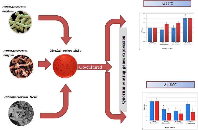
Material and Methods: In this study, effects of Bifidobacterium lactis, Bifidobacterium bifidum and Bifidobacterium longum on the expression of quorum sensing genes in Yersinia enterocolitica were studied at two various temperatures of 37 and 32 °C. First, Bifidobacterium spp. were activated in brain infusion broth and then cultured simultaneously with Yersinia enterocolitica, RNA extraction was carried out and yenI and yenR gene expression was assessed using real-time polymerase chain reaction.
Results and Conclusion: Results revealed significant differences in gene expression at 37 and 32 °C. When Bifidobacterium sp. was co-cultured with Yersinia enterocolitica at 32 °C, the bacterial quorum sensing gene expression significantly decreased in all treated cells, except yenI in Bifidobacterium bifidum co-culture, compared to the control. Bifidobacterium longum showed the highest decreasing effect in quorum sensing gene expression in Yersinia sp. by 64%. In contrast, co-culturing Bifidobacterium sp. with Yersinia enterocolitica at 37 °C revealed that Quorum sensing gene expression levels in Yersinia enterocolitica did was not change significantly in all cultures, except that with Bifidobacterium bifidum; almost similar to the control samples. These findings indicated that the interactions between probiotic bacteria and pathogens varied under various temperature conditions, demonstrating a temperature-dependent pattern.
Keywords: Bifidobacterium spp., Gene expression, Gene regulation, Quorum sensing, Yersinia enterocolitica
- Introduction
Quorum sensing (QS) refers to the process of coordination and communication between cells, involving production of diffusible extracellular molecules and hence regulation of specific gene expression. The QS signals are called autoinducers because they are produced by the cells and typically induce their own synthesis, creating a positive feedback loop. Various types of QS systems reported in microorganisms include 1) LuxI/R type, which is commonly used by Gram-negative bacteria and uses acyl-homoserine lactone (AHL (molecules. This system is used for intraspecies communication due to its high specificity; 2) peptide signaling system, used by Gram-positive bacteria; 3) AI-2/LuxS signaling system, which facilitates interspecies communication and is detected in all bacteria and 4) AI-3 signaling system, which operates through epinephrine-norepinephrine and is used for interspecies and interterritorial communications [1,2,3]. In Gram-negative bacteria, QS can be inhibited through three methods of 1) inhibiting synthesis of AHLs, 2) degrading AHL molecules and 3) interfering with the receptors, which can be achieved through compounds produced by plants and microorganisms [4].
Yersinia enterocolitica, as a member of the Enterobacteriaceae family, is a Gram-negative, short rod-shaped, facultative anaerobic non-spore-forming bacterium. It multiplies at 0–45 °C, which distinguishes it from other enteric pathogenic bacteria [5]. Studies have shown that the three pathogenic species of Yersinia produce AHLs QS signals. Protein of LuxI, which synthesizes AHL as well as LuxR as a regulator, is detected in all three pathogenic Yersinia spp. However, Y. enterocolitica includes a unique pair of LuxRI (YenRI), whereas the other two species include two pairs. The YenI is responsible for synthesizing two types of AHLs, one of which is N-hexanoyl homoserine lactone (C6-HSL) and the other is N-3-oxo-hexanoyl homoserine lactone (3-OXO-C6-HSL). These two compounds are produced at a 1:1 ratio in Y. enterocolitica [6,7,8].
Gut microflora of humans and animals consists of several probiotic species such as Bifidobacterium. Typically, population of Bifidobacterium in the intestine of healthy adults ranges 10¹⁰-10¹¹ CFU g-1; however, this decreases with age. Most species of Bifidobacterium are rod-shaped, anaerobic, non-motile and non-spore-forming species. Dairy fermentation products such as yogurt, cheese and fermented butter are addressed as major sources of probiotics [9]. For QS inhibition, extensive studies have been carried out on various pathogenic bacteria. In one study carried out on bacteria isolated from fish, 200 bacterial species were isolated from fish and ability of three selected species to inhibit QS system in Aeromonas hydrophila was verified. The study revealed that the bacteria belonged to the Bacillus genus and possessed an enzyme capable of degrading AHLs. The effect of this bacterium on A. hydrophila in fish showed that the control group of fish developed skin lesions over time and died after 12 d. However, fish that consumed the isolated strain survived and skin damage significantly decreased [10]. In another study investigating the effect of stress on signal production in Listeria spp., it was reported that stress induced by nisin and lactic acid did not play a role in production of AI-2 as an adaptive response to the environment and no dependency between these two factors was observed. However, this study concluded that QS could generally help L. monocytogenes cells adapted to stresses such as nisin and lactic acid [11].
Further studies on the inhibitory effect of various Bifidobacterium strains on EHEC (Enterohemorrhagic Escherichia coli) showed that B. longum, B. adolescentis and B. breve were the most inhibitory strains. For biofilm formation, most Bifidobacterium strains did not show a significant inhibitory effect, with only B. longum decreasing biofilm formation by 36%. Pathogenicity decreased in the presence of B. longum and results indicated that it regulated seven types of proteins in Escherichia coli [12]. Study of the effect of lactic acid produced by Pediococcus acetilactis on QS in Pseudomonas aeruginosa showed that lactic acid included an inhibitory effect on motility and short-chain HSL, elastase, protease, pyocyanin and biofilm productions. However, concentration of lactic acid needed to be precisely assessed for effective QS regulation [13]. Generally, the present study aimed to investigate the effect of co-culturing B. lactis, B. bifidum and B. longum on the expression of QS genes, yenI and yenR, in Y. enterocolitica under various temperature conditions.
- Materials and Methods
2.1. Bacterial Strain Preparation and Activation
The bacterial strains of B. lactis ATCC 19435, B. bifidum ATCC 29521, B. longum ATCC 15707 and Y. enterocolitica ATCC 23715 were provided by the Iranian Research Organization for Science and Technology (IROST). The Bifidobacterium strains were cultured in brain heart infusion (BHI) media (Merck, Germany) at 37 °C for 48 h under absolute anaerobic conditions using type A gas pack, anaerobic jar and indicators. Moreover, Y. enterocolitica was cultured in BHI media at 29 °C for 48 h [12]. For co-culturing Bifidobacterium spp. with Y. enterocolitica, fresh 24-h cultures of each Bifidobacterium strain were first adjusted to a cell density of 0.5 McFarland standard. Then, 100 µl of each adjusted Bifidobacterium culture were added to 10 ml of fresh Y. enterocolitica culture media and incubated at the specified temperature for each treatment [12].
2.2. Simultaneous Cultivation of Bifidobacterium sp. with Yersinia enterocolitica
The Y. enterocolitica was cultured in BHI media with three various probiotic bacteria separately at 32 and 37 °C for 48 h under anaerobic conditions. To standardize cell populations and ensure addition of a consistent microbial cell count in the assays, 0.5 McFarland standard was used with fresh 24-h culture that experienced two previous passages [12].
2.3. Primer Design and Polymerase Chain Reaction for the Verification of yenI and yenR Gene Presence
Primers were designed for the analysis of yenI and YenR genes as well as the housekeeping gene (Table 1). Briefly, DNA extraction from Y. enterocolitica was carried out using boiling method. Then, polymerase chain reaction (PCR) was carried out (ASTEC G02, Japan) and the PCR products were analyzed via gel electrophoresis.
2.4. RNA Extraction and Reverse-transcriptase Polymerase Chain Reaction Analysis
Yersinia enterocolitica and each Bifidobacterium sp. were simultaneously incubated at 32 and 37 °C for 48 h under anaerobic conditions. After incubation, cultures were centrifuged and the supernatant was discarded. The cell pellet was collected using 1.5-ml microtubes for RNA extraction. The RNA extraction was carried out using extraction kit (Kiagen Fanavar Arya, Iran) based on the manufacturer’s instructions and the RNA concentration was assessed using NanoDrop spectrophotometer (Thermo Scientific NanoDrop 2000, USA). Furthermore, cDNA synthesis was carried out using cDNA synthesis kit (Yekta Tajhiz Azma, Iran) and gene expression analysis was carried out using reverse-transcriptase polymerase chain reaction (RT-PCR) machine (Corbett Rotor Gene-RG 3000, Australia). Gene expression was analyzed using 2^-ΔΔCt method [14]. This method involved assessing gene expression level and normalizing it against a reference gene. Normalization corrected for variations in amplification efficiency, extraction conditions and initial sample volumes. Gene expression levels in treated and untreated samples were investigated and the difference in Ct values was calculated. Similar procedure was used to the reference sample. The relative gene expression changes were calculated by dividing the gene expression changes in the target gene by those of the reference gene. The calculation method and formulas were as follows [15]:
∆Ct = Ct target gene - Ct housekeeping gene, ∆∆Ct = ∆Ct experiment - ∆Ct Control, Fold gene = 2-∆∆Ct
Where, Ct was the threshold cycle.
2.5. Statistical analysis
The mean Ct values of the treated samples were compared to those of the controls using one-way analysis of variance (ANOVA) to investigate if there was a statically significant difference between them (p < 0.05). All experiments were carried out in triplicate.
- Results and Discussion
3.1. Verification of the presence of yenI and yenR genes in Yersinia enterocolitica
First, presence of the yenI and yenR genes in Y. enterocolitica strain must be verified. Results of the PCR amplification of yenI and yenR genes in Y. enterocolitica and their verification on agarose gels are shown in Figures 1 and 2. Figure 1 verified presence of the yenI gene and Figure 2 verified presence of the yenR gene.
3.2. Analysis of yenI and yenR gene expression in Yersinia enterocolitica co-cultured with Bifidobacterium strains at 37 and 32 °C
The T-PCR analysis results Fold change for the yenI and yenR genes in comparison to the housekeeping gene are shown in Tables 2 and 3 and Figures 3 and 4. Fold change is a metric that expresses the extent of change between two measurements. In gene expression analysis, such as microarray or RNA-Seq analysis, it refers to the ratio of expression between samples or between groups. Moreover, melting curve and amplification of the target sequence in the treatments during RT-PCR are shown in Figure 5. In results achieved at 32 °C, adding Bifidobacterium to Y. enterocolitica culture decreased QS gene expression. At 37 °C, results completely varied. Results showed differences in expression of the studied genes at 37 and 32 °C. At 32 °C, presence of Bifidobacterium sp. decreased the expression of signal-producing genes, which was significant in most samples. At 37 °C unlike 32 °C, most samples did not differ significantly from the control samples. Studies have shown that 37 °C significantly affects regulation of genes associated to pathogenesis such as secretion systems and toxins and helps bacteria multiply better in host conditions.
In Y. enterocolitica, gene expression was affected by temperature, particularly at 37 °C. These temperature-dependent changes in gene expression assisted the bacteria effectively evaded the host immune system and enhanced its virulence. Specifically, at this temperature, expression of key genes associated with the bacterial virulence factors increased. These genes included those involved in the secretion of specific proteins, toxin production and responses to environmental stresses. At 37 °C, Y. enterocolitica efficiently used type III secretion system (T3SS), which was essential for the transfer of toxic proteins into host cells and expression of genes linked to this system increased significantly. Genes involved in production of specific toxins that helped in the bacterial toxic activities and proteins that prevented the host cell’s immune responses and regulatory systems for survival and stress conditions were significantly upregulated [16-18].
Technically, Y. enterocolitica, genes responsible for the production of AHLs can be regulated in response to environmental changes and host conditions [8]. Studies have shown that at 37 °C as the natural body temperature of humans, changes in the expression of these genes occur. Compounds encoded by these genes are involved in regulating collective behaviors such as toxin production, biofilm formation, pathogen-linked behaviors, host interactions and responses to environmental stress. At 37 °C, AHL production may increase, as this temperature specifically mimics the natural conditions of the human body and is critical for regulating pathogenic activities of the bacteria. Therefore, AHL-producing genes may become more active at this temperature, enabling the bacteria to regulate their collective behaviors, including toxin production and expression of other virulence factors [19, 20].
Naturally, Y. enterocolitica is an enteric pathogen that grows rapidly at 37 °C. At 37 °C, Y. enterocolitica typically reaches its maximum growth within 6–12 h, while bifidobacteria needs 12–24 h. Due to their high sensitivity to environmental conditions, bifidobacteria grow more slowly than that Y. enterocolitica does at 37 °C. Since Y. enterocolitica grows faster and reaches higher numbers within a shorter time at 37 °C compared to bifidobacteria, and due to the increases in expression of signal-producing genes involved in QS, toxin production, biofilm formation, pathogenic behavior and host interaction, it seems that Y. enterocolitica proliferates sufficiently and produces necessary beneficial compounds before bifidobacteria can reach sufficient numbers and begin production of inhibitory compounds. Additionally, these compounds (e.g. acidic substances) may further inhibit rapid growth of bifidobacteria. Once Y. enterocolitica dominates the environment, bifidobacterial growth becomes restricted. Due to the high sensitivity of bifidobacteria to environmental compounds, there is a possibility of no or weak growth of these bacteria. Secreted compounds needed for the bifidobacterial growth at 37 °C may be used by Y. enterocolitica due to the high number of Y. enterocolitica in the environment; thus, accelerating the bacterial growth [21,22,23].
In contrast, growth dynamics of the two bacterial species differ at 32 °C under strict anaerobic conditions, with bifidobacteria growing slightly faster than Y. enterocolitica. This faster growth of bifidobacteria leads to the production of growth-inhibitory compounds, limiting Y. enterocolitica proliferation and hence affecting expression of QS genes. At this temperature, expression of QS genes is less than that at 37 °C, as pathogenic activity is optimal at the latter temperature [24,25]. Based on these findings, presence of significant differences in the expression of QS genes at 32 °C and absence of significant differences of these genes at 37 °C can be justified. At the two temperatures, B. longum showed the minimum gene expression averagely. Previous studies on B. longum have shown that this bacterium is a clinically versatile probiotic strain and can control growth of pathogenic bacteria [26,27].
- Conclusion
This study assessed temperature-dependent effects (37 against 32 °C) of various Bifidobacterium probiotic species on QS gene expression in Y. enterocolitica during co-culturing. Results showed that temperature changes included distinct effects on gene expression and pathogenic and probiotic behaviors of the highlighted bacteria. Additionally, results demonstrated that temperature variations significantly altered gene expression patterns, which directly modulated pathogenic behaviors of Y. enterocolitica and probiotic effects of Bifidobacterium strains. At 32 °C, Bifidobacterium sp. produced inhibitory compounds that suppressed proliferation of Y. enterocolitica and downregulated QS-associated genes. Pathogenic activities of Y. enterocolitica were significantly terminated at this temperature, with decreased expression of QS genes. This suggested that at 32 °C, Y. enterocolitica could not fully activate its virulence pathways, weakening its ability to escape immune defenses and infect the host. In contrast, no significant differences were observed between the controls and the treated samples at 37 °C. This study highlights the critical roles of temperature in modulating bacterial interactions and potentially their pathogenic characteristics. At 32 °C, Bifidobacterium sp. effectively suppressed QS gene expression in Y. enterocolitica. At 37 °C, QS gene levels were largely unchanged in all cultures, similar to those in control samples. These findings have provided valuable highlights for developing probiotic-based strategies to prevent and/or help treat infections caused by Y. enterocolitica and other pathogens. However, further studies seem necessary.
- Bifidobacterium spp.
- Gene expression
- Gene regulation
- Quorum sensing
- Yersinia enterocolitica
Cómo citar
Citas
Garmyn D, Gal L, Lemaitre JP, Hartmann A, Piveteau P. Communication and autoinduction in the species Listeria monocytogenes: A central role for the agr system. Commun Integr Biol. 2009; 2(4): 371-374. https://doi.org/10.4161/cib.2.4.8610
Nicola C Reading, Vanessa Sperandio. Quorum sensing: The many languages of bacteria. FEMS Microbiology Letters. 2006; 254(1): Pages 1-11. https://doi.org/10.1111/j.1574-6968.2005.00001.x
Rutherford S T, Bassler B L. Bacterial quorum sensing: Its role in virulence and possibilities for its control. CSH Perspectives in Medicine. 2012; 2(11): a012427. https://doi.org/10.1101/cshperspect.a012427
Machado I, Silva L R, Giaouris E D, Melo L F, & Simoes M. Quorum sensing in food spoilage and natural-based strategies for its inhibition. Food Res Int. 2020; 127: 108754. https://doi.org/10.1016/j.foodres.2019.108754
Fabrega A, Vila J. Yersinia enterocolitica: Pathogenesis, virulence and antimicrobial resistance. Enferm Infecc Microbiol Clin. 2012; 30(1): 24-32. https://doi.org/10.1016/j.eimc.2011.07.017
Medina-Martinez M S, Uyttendaele M, Meireman S, Debevere J. Relevance of N-acyl-L-homoserine lactone production by Yersinia enterocolitica in fresh foods. J Appl Microbiol. 2007; 102(4): 1150-1158. https://doi.org/10.1111/j.1365-2672.2006.03143.x
Throup J P, Camara M, Briggs G S, Winson M K, Chhabra S R, Bycroft B W, Williams P, Stewart G S. Characterisation of the yenI/yenR locus from Yersinia enterocolitica mediating the synthesis of two N-acylhomoserine lactone signal molecules. Mol Microbiol. 1995; 17(2): 345-356. https://doi.org/10.1111/j.1365-2958.1995.mmi_17020345.x
Ng Y K, Grasso M, Wright V, Garcia V, Williams P, Atkinson S. The Quorum Sensing System of Yersinia enterocolitica 8081 Regulates Swimming Motility, Host Cell Attachment and Virulence Plasmid Maintenance. Genes. 2018; 9(6): 307. https://doi.org/10.3390/genes9060307
Anandharaj M, Sivasankari B, Parveen Rani R. Corrigendum to effects of probiotics, prebiotics and synbiotics on hypercholesterolemia: A review. Chin J Biol. 2020; 2014: 572754. https://doi.org/10.1155/2014/572754
Chu W, Lu F, Zhu W, & Kang C. Isolation and characterization of new potential probiotic bacteria based on quorum-sensing system. J Appl Microbiol. 2011; 110(1): 202-208. https://doi.org/10.1111/j.1365-2672.2010. 04872.x
Turovskiy Y, Rosenberg L, Chikindas M. Autoinducer-2-mediated quorum sensing in not involved in listeria monositogenes adaptive responses to the food preservatives lactic acid and nisin. J Food Saf. 2007; 27: 386-399. https://doi.org/10.1111/j.1745-4565.2007.00087.x
Kim Y, Lee J W, Kang S G, Oh S, Griffiths M W. Bifidobacterium spp. influences the production of autoinducer-2 and biofilm formation by Escherichia coli O157:H7. Anaerobe. 2012; 18(5): 539-545. https://doi.org/10.1016/j.anaerobe.2012.08.006
Kiymaci M, Altanlar N, Gumustas M, Ozkan S, Akin A. Quorum sensing signals and related virulence inhibition of Pseudomonas aeruginosa by a potential probiotic strain's organic acid. Microb Pathog. 2018; 121: 190-197. https://doi.org/10.1016/j.micpath.2018.05.042
Adams G. A Beginner’s Guide to RT- PCR, qPCR and RT- qPCR. University of Leicester, UK. Published by Portland Press. 2020.
Pfaffl M W. A new mathematical model for relative quantification in real-time RT-PCR. Nucleic acids research. 2001; 29(9): e45. https://doi.org/10.1093/nar/29.9.e45
Platt-Samoraj A. Toxigenic properties of Yersinia enterocolitica biotype 1A. Toxins (Basel). 2022; 14(2): 118. https:// doi.org/10.3390/toxins14020118
Truchado P, Giménez-Bastida JA, Larrosa M, et al. Inhibition of quorum sensing (QS) in Yersinia enterocolitica by an orange extract rich in glycosylated flavanones. J Agric Food Chem. 2012; 60(36):8885-8894. https://doi.org/10.1021/jf301365a
Kapatral V, Olson JW, Pepe JC, Miller VL, Minnich SA. Temperature-dependent regulation of Yersinia enterocolitica Class III flagellar genes. Mol Microbiol. 1996; 19(5):1061-1071. https://doi.org/10.1046/j.1365-2958.1996.452978.x
Atkinson S, Chang CY, Sockett RE, Cámara M, Williams P. Quorum sensing in Yersinia enterocolitica controls swimming and swarming motility. J Bacteriol. 2006; 188(4): 1451-1461. https://doi.org/10.1128/JB.188.4.1451-1461.2006
Atkinson S, Sockett RE, Cámara M, Williams P. Quorum sensing and the lifestyle of Yersinia. Curr Issues Mol Biol. 2006; 8(1): 1-10.
Lindberg CW, Borch E. Predicting the aerobic growth of Y. enterocolitica O:3 at different pH-values, temperatures and L-lactate concentrations using conductance measurements. Int J Food Microbiol. 1994; 22(2-3): 141-153. https://doi.org/10.1016/0168-1605(94)90138-4
Yehualaeshet T, Graham M, Montgomery M, Habtemariam T, Samuel T, Abebe W. Effects of temperature on the viability, growth and gene profile of Yersinia pseudotuberculosis and Yersinia enterocolitica inoculated in milk. Food Control. 2013. https://doi.org/10.1016/j.foodcont.2013.05.025
Fredriksson A M. Yersinia enterocolitica. In; Foodborne Diseases. Elsevier. 2017; pp 223–233.
Garro M, Valdez G, Savoy G. Temperature effect on the biological activity of Bifidobacterium longum CRL 849 and Lactobacillus fermentum CRL 251 in pure and mixed cultures grown in soymilk. Food Microbiol. 2004; 21: 511-518. https://doi.org/10.1016/j.fm.2004.01.001
Ruiz L, Ruas-Madiedo P, Gueimonde M, de Los Reyes-Gavilán CG, Margolles A, Sánchez B. How do bifidobacteria counteract environmental challenges? Mechanisms involved and physiological consequences. Genes Nutr. 2011; 6(3): 307-318. https://doi.org/10.1007/s12263-010-0207-5
Schopping M,Zeidan AA, Franzen CJ. Stress Response in Bifidobacteria. Microbiol Mol Biol Rev. 2022; 86: e00170-21. https://doi.org/10.1128/mmbr.00170-21
Li Yiran, Satoshi Arai, Kumiko Kato, Sadahiro Iwabuchi, Noriyuki Iwabuchi, Natsumi Muto, Hideki Motobayashi, Shukuko Ebihara, Miyuki Tanaka, Shinichi Hashimoto. The potential immunomodulatory effect of Bifidobacterium longum subsp. longum BB536 on healthy adults through plasmacytoid dendritic cell activation in the peripheral blood. Nutrients. 2024; 16(1): 42. https://doi.org/10.3390/nu16010042
- Resumen ##plugins.themes.ojsPlusA.frontend.article.viewed##: 364 ##plugins.themes.ojsPlusA.frontend.article.times##
- pdf (English) ##plugins.themes.ojsPlusA.frontend.article.downloaded##: 95 ##plugins.themes.ojsPlusA.frontend.article.times##