
Characterization of the Chitosan Microencapsulation of Rosa damascena Mill. Extract and (Lacticaseibacillus casei and Bifidobacterium longum) Probiotics
التكنولوجيا الحيوية الغذائية التطبيقية,
مجلد 12 عدد 1 (2025),
4 كانون الثاني 2025
,
الصفحة 1-13 (e32)
https://doi.org/10.22037/afb.v12i1.46674
الملخص
Background and Objective: Rosa damascena Mill. possesses bioactive compounds, including flavonoids and anthocyanins, which are addressed for their antioxidant and anti-inflammatory characteristics. This study aimed to develop and optimize a microencapsulation system for rose extract and probiotics. It focused on particle size and morphological characteristics analyzed via particle size analysis, scanning electron microscopy-energy dispersive X-ray spectroscopy and , gas chromatography-mass spectrometry and further assessed the bioavaliability and bioaccessibility of the encapsulated probiotics.
Material and Methods: Lacticaseibacillus casei and Bifidobacterium longum were cultivated in De Man, Rogosa, and Sharpe broth. The petals of Rosa damascena Mill. were extracted with ethanol 70% 1:1 aqueous solution. The microencapsulation involved dissolving the extract and probiotics, followed by the addition of chitosan and sodium tripolyphosphate to form stable colloids. The particle size was analyzed using dynamic light scattering and the morphology of microcapsules was investigated using scanning electron microscopy coupled with energy-dispersive X-ray spectroscopy. Detection of ethanol was carried out using gas chromatography-mass spectrometry. Probiotic viability was assessed after storage at 4 °C for 0, 14 and 28 d and bioaccessibility was assessed using in vitro gastrointestinal simulation method.
Results and Conclusion: The microencapsulation process resulted in spherical microcapsules with a mean particle size of 3107 nm ±273.2. Scanning electron microscopy analysis verified uniform morphology, indicating effective encapsulation. The probiotic count for microencapsulated samples was 5.16 ±0.37 log cfu ml-1. Gas chromatography-mass spectrometry data showed that the ethanol content was 2.53% ±0.21 (v/v). Microencapsulation of R. damascena Mill. and the probiotics increased the recovery of anthocyanin by 9%. These findings have suggested that combining microencapsulation with probiotic strains provides viable strategy to improve the functional delivery of anthocyanin-rich botanicals in nutraceutical uses.
Keywords: Microencapsulation, Rosa damascena Mill. extract, Lacticaseibacillus casei, Bifidobacterium longum, Chitosan
- Introduction
Rosa damascena Mill., a member of the genus Rosa (Rosaceae), comprises more than 200 species worldwide. This species is extensively cultivated not only as an ornamental plant but also as a valuable source of raw materials for cosmetic and pharmaceutical industries due to its characteristic aroma and diverse bioactive compounds, including tannins, flavonoids, terpenes, polyphenols, carotenoids, phenylethyl alcohol and vitamins B, C, E and K. Of these constituents, anthocyanins (a major flavonoid pigments in red rose petals) demonstrates strong antioxidant activity, lessening oxidative stress, enhancing immune defense and providing additional biological benefits such as anti-inflammatory, antidiabetic, anticancer, cardiopro-tective and neuroprotective effects [1-2].
Anthocyanins can alter gut microbiota composition by promoting the growth of beneficial bacteria such as Bifidobacteria and Lacticaseibacillus, which are essential for maintaining gut barrier health. [1, 3]. Anthocyanins modulate the expression of tight junction proteins such as occludin, claudin and ZO-1, which are essential for forming and maintaining intestinal epithelial barrier integrity. Tight junctions act as selective barriers that restrict paracellular permeability to harmful molecules andregulation of these proteins by anthocyanins enhances preservation of intestinal barrier function [1, 3].
Lacticaseibacillus casei is addressed for its resilience in acidic environments such as the stomach and intestines and its ability to produce lactic acid, which inhibits the growth of pathogenic bacteria in the gut. The L. casei has demonstrated positive effects in enhancing intestinal function, reliving gastrointestinal (GI) symptoms and decreasing inflammation within the digestive system. Additionally, research shows that L. casei can improve muscle strength and physical function in animal models and help lessening oxidative stress; thereby, promoting overall health [4]. Bifidobacterium longum provides anti-inflammatory and antioxidant benefits that decreasing liver lipid accumulation and improve muscle and cognitive functions in aging animal models. The B. longum has been shown to enhance intestinal barrier function and improve the gut microbiota composition [5]. The L. casei and B. longum demonstrate immunomodulatory effects by enhancing the body immune response to infections. In studies on Plasmodium infection, B. longum was detected effective in decreasing parasitemia and inflammation, indicating its capability to strengthen immune defense by modulating gut immune responses [5-6]. The L. casei and B. longum can lessendysbiosis or gut microbiota imbalances caused by infections. The bioactive components of R. damascena and probiotic functions of L. casei and B. longum present promising ways for improving overall health, specifically through enhanced antioxidant activity, immune response and gut microbiota balance, making them valuable for therapeutic uses.
Microencapsulation is a process that involves coating a compound or particle to form microcapsules. This microencapsulation concept enables the separation of compounds and probiotic cells from their environment through a protective layer. The characteristics of this protective system are designed to safeguard the cell core and release it in a controlled manner under specific conditions, while allowing the transport of small molecules through the membrane [7]. Chitosan is effective as a coating agent due to its advantages, including non-toxicity, appropriateness for drug delivery, biodegradability and biocompatibility. Chitosan is a natural linear biopolyamino sugar derived through the deacetylation of chitin, with a non-linear chain formula (C₆H₁₁NO₄)n that is odorless and white[8]. Chitosan microencapsulation of R. damascena has successfully been carried out, demonstrating its efficacy in preserving flower quality and extending shelf life, offering a sustainable and effective approach for postharvest storage in commercial uses [9]. Studies have shown that probiotic bacteria can survive after being encapsulated with chitosan. This is because encapsulation efficiency significantly increases with higher concentrations of the biopolymer [10]. The survival rate of chitosan-encapsulated probiotics is high after incubation at low pH, although the population decreases slightly. The decrease in bacterial count during gastric acid simulation is attributed to the highly acidic stomach pH, which affects the strength of the sodium alginate-chitosan polymer as a matrix for encapsulating lactic acid bacteria (LAB) [11]. Moreover, microencap-sulation techniques, particularly those using chitosan, provide promising ways for enhancing the stability and bioavailability of these probiotics in GI environments; thereby, improving their therapeutic potentials. Microen-capsulation of R. damascena Mill. and probiotics (L. casei and B. longum) (MERP) underscores the value of R. damascena bioactives and probiotic encapsulation in preventive and therapeutic uses for managing oxidative stress, inflammation and gut health.
The research identified several key gaps in the existing literature. Previous studies have largely focused on the antioxidant and anti-inflammatory characteristics of R. damascena Mill. and the probiotic benefits of L. casei and B. longum individually. However, a limited attention has been paid to synergizing these bioactives and probiotics within a stable microencapsulation system to enhance their bioavailability. The present findings, however, provided additional insight and might serve as a valuable reference for further development of a chitosan-sodium tripolyphos-phate (STPP) microencapsulation system for the synergistic co-encapsulation of R. damascena Mill. bioactives (anthocyanins and flavonoids) and probiotics (L. casei and B. longum). Combining antioxidant-rich rose extracts with probiotics in a robust encapsulation matrix, this study uniquely linked further nutraceutical uses with gut health and inflammation management, highlighting its potentials for advancing targeted delivery in the functional food system.
- Materials and Methods
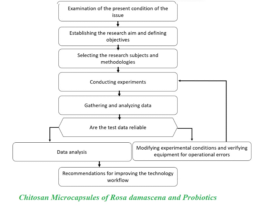
Chitosan was purchased from Merck (Germany). Solvents, including distilled water used as a solvent obtain from Brataco (Indonesia) and ethanol used as a co-solvent, obtained from Merck (Germany). All other chemicals and solvents used in this study were of analytical grade. The procedures for Rosa damascena extraction, probiotic culturing, and microencapsulation using the chitosan–STPP system are illustrated in Figure 1.
2.1. Probiotic Isolation Stage
The L. casei and B. longum were purchased from the Food and Nutritional Culture Collection (FNCC), Gadjah Mada University, Indonesia. Colonies were sampled using sterile inoculation needle and each bacterium was inoculated into De Man, Rogosa, and Sharpe (MRS broth). The cultures were then incubated at 37 °C for 24 h. After incubation, their optical density and colony counts were recorded using spectrophotometer (Thermoscientific, USA). Gram staining was carried out to ensure purity of the bacteria [5].
2.2. Red Rose Extraction Stage
Rose petals were extracted using 70% ethanol pro analis (Merck, Germany) and aliquoted 1:1 [12]. The extract was then filtered through filter paper or a vacuum filtration system to separate the solid from the liquid. The extract was evaporated using rotary evaporator (Materia Medica Batu, Indonesia). The resulting liquid extract was collected using sterile glass bottle. The extract was freeze-dried (Martin Christ, Germany).
2.3. Microencapsulation Preparation
The Microencapsulation was carried out using chitosan-STPP matrix. The stages of MERP formulation included preparation of chitosan (Merck, Germany) in 2% acetic acid (Merck, Germany), preparation of 0.1% NaTPP (Brataco, Indonesia) in distilled water (DW), preparation of 40% rose extract in absolute ethanol:aliquot (3:1) and synthesis of MERP with the composition of 10 ml of rose extract, suspension of L. casei and B. longum each as much as 5 × 10⁹ CFU ml-1, 8 ml of NaTPP and 32 ml of chitosan. The samples were stirred for 60 min at 400 rpm and stored at 4 °C. Then, the sample was freeze-dried [13]. This formulation was used in in vivo studies. The microencapsulation formulation contained 80 mg of rose extract, 5 × 10⁹ L. casei and 5 × 10⁹ B. longum ml-1. The selected dose was based on prior evidence showing the anti-inflammatory effects of R. damascena Mill extract at doses ranging from 500 to 1000 mg kg-1. Additionally, L. casei and B. longum have been reported to include anti-inflammatory effects within a dose range of 5 × 10⁶ to 5 × 10⁹ CFU for L. casei and 10⁸ to 10¹⁰ CFU for B. longum [21-22].
2.4. Viability Assessment of Microencapsulation
The viability of the microencapsulation was assessed by storing the encapsulated probiotics in PBS and 2% citric acid at 4 °C for 0, 14 and 28 d, with each treatment assessed four times. After these storage intervals, samples were cultured in MRS broth to assess bacterial survival under cold storage conditions. After the culture, the samples were incubated at 37 °C for 24 h, allowing for bacterial growth. Gram staining and spectrofotometry were carried out on the incubated samples to verify the presence and viability of the encapsulated bacteria, ensuring the microencapsulation preserved bacterial integrity within the storage rime [17]. A comparison was made between free cells Lacticaseibacillus casei (FCL), free cells Bifidobacterium longum (FCB) and free cells Lacticaseibacillus casei and Bifidobacterium longum (FCLB), revealing significant differences in stability [18].
2.5. Particle Size Analysis
The particle size of the microencapsulated samples was assessed using dynamic light scattering (DLS) and particle size analyzer (PSA) (Malvern Panalytical, UK), assessed two times at the Department of Chemical Engineering, Institut Teknologi Surabaya, Surabaya, Indonesia. This technique provided accurate size distribution data essential for assessing encapsulation quality. Samples were prepared by diluting them with deionized water in a 2:1 ratio to ensure optimal measurement conditions. The analysis was carried out at a controlled temperature of 25 °C, facilitating reliable results on the particle size distribution within the microencapsulation matrix [19].
2.6. Scanning Electron Microscopy and Energy Dispersive X-ray
Scanning electron microscopy and Energy Dispersive X-ray (SEM-EDX) (JSM-6510LA) analyses were carried out at UGM Integrated Research and Testing Laboratory (Laboratorium Penelitian dan Pengujian Terpadu, Indonesia) to investigate catalyst characteristics. The SEM analysis provided detailed insights into the shape and size of the catalyst particles, which were critical for catalytic efficiency. Moreover, SEM-EDX was carried out to record the morphological characteristics of the microcapsule and to show semi-quantitative information on the elemental composition of the microcapsules [19].
2.7 Gas Chromatography-Mass Spectrometry
The microencapsulated sample was assessed three times using gas chromatography-mass spectrometry (GC-MS). The measurements was carried out using Shimadzu GCMS-QP2020NX, Japan, equipped with a quadrupole mass spectrometry detector. A SH-I-624Sil MS column (30-m length, 0.25-mm i.d. and 1.4-µm film thickness (Shimadzu, Japan) was used for the separation of target ethanol and other volatile compounds. Injections were carried out in a split mode (ratio of 1:50). Nitrogen was used as a carrier gas. The injector temperature was 250 °C. The oven temperature was set at 40 °C for 5 min, then increased to 240 °C at a rate of 30 °C min-1 and set isothermally for 4 min. All samples were equilibrated to 70 °C for 5 min with agitation of 500 rpm using autosampler before injecting 100 μl of headspace onto the column. The mass spectrometer was operated in single ion monitoring mode with the following ions monitored as ethanol, 1.6–1.8 min m/z 31, 45 and 46; 2-methyl-1-propanol, 2.4–2.5 min m/z 33, 43 and 74; 1-butanol, 2.85–3.3 min m/z 41, 56 and 73 (internal standard); and 2-methyl-1-butanol, 3.7–3.8 min m/z 41, 57 and 87. Dwell time of 200 ms was used for each ion. The transfer line to the mass spectrometer was heated to 240 °C, the source temperature was set at 230 °C and the quadrupole was set at 150 °C [20].
2.8. In Vitro Bioaccessibility Assessment
In vitro bioaccessibility assessment was carried out to assess the availability of active compounds in rose extract and MERP through a modified two-stage (GI) digestion simulation based on previous methods [21]. In the stomach stage, simulated gastric fluid (SGF) was prepared by dissolving 0.1 g of NaCl and 0.35 ml of 37% HCl in 50 ml of DW; then, adding 0.16 g of pepsin and adjusting pH to 1.2. A total of 0.5 ml of the extract was dissolved in 50 ml of SGF, pH was adjusted to 2.5 and the mixture was incubated at 37 °C for 2 h at 100 rpm using shaker incubator. In the intestinal stage, 30 ml of the sample from the stomach phase were incubated at 37 °C for 10 min using water bath; then,pH increased to 7.0. Then 1 ml of CaCl₂ solution (750 mM) and 2.5 ml of lipase (1.6 mg/ml) were added to the mixture and incubated using shaker incubator (100 rpm, 37 °C, 2 h). After the digestion process, the sample was centrifuged (4000 rpm, 25 °C, 40 min) and the micelle phase (middle layer) was filtered using 0.45-µm microfilter. The filtrate was mixed with 70% ethanol in a 1:1 ratio and centrifuged (1750 rpm, 25 °C, 10 min). The supernatant was analyzed using UV-vis spectrophotometer to assess concentration of the active compound (anthocyanin) and then proportion of bioaccessibility was calculated based on the ratio of the compound content in the micelle phase to the total compound in the initial sample.
(1)
Where, C1 was the active compound concentration after GI simulation (in the micelle phase) and Co was the active compound content before GI simulation (in the entire sample).
2.9. Data Analysis
Data analysis involved descriptive interpretation of results from PSA, SEM, EDX and GC-MS. The PSA results provided quantitative data on microencapsulation particle sizes, essential for assessing uniformity and encapsulation quality. The SEM imaging offered detailed visual information on particle morphology, while EDX analysis assessed elemental composition, contributing to insights into structural integrity. The GC-MS data calculated quantities of ethanol in MERP. Results of the viability assessment were shown as averages with SD for thrice measurements. The averages were recorded using two-way analysis of variance (ANOVA) with Tukey's multiple comparison test including a significance level of 0.05 [95% confidence interval (CI)]. Differences in bioaccessibility proportions between the sample groups were analyzed using independent T-test (p-value < 0.05).
- Results and Discussion
3.1. Scanning Electron Microscopy and Energy Dispersive X-ray
This study demonstrated the efficacy of chitosan-STPP encapsulation in delivering bioactive compounds and probiotics, particularly R. damascena extracts and probiotic strains of L. casei and B. longum. The SEM characterization of the microencapsulated rose extract showed distinct spherical morphology within the particles. Image a (1000× magnification) illustrates that the microcapsules were well-dispersed without signs of aggregation, verifying effective encapsulation. Image B, captured at higher magnification (10,000×), reveals a smooth surface structure with a uniform spherical shape, suggesting structural integrity and encapsulation stability. The particle diameters varied within a narrow range, approximately between 2.8 and 3.4 µm, indicating minimal size heterogeneity. This morphology is essential for optimal encapsulation performance, providing a consistent surface area and stability for targeted uses. The final quantity of probiotics in foods and their viability in the gastrointestinal tract (GIT) are affected by encapsulation efficiency [22]. The particles were observed as nearly spherical, similar to those from UV–vis spectral analysis. It is important to state that the size of these microencapsules plays a critical role in their characteristics and biological activity [23].
The SEM-EDX analysis was carried out to investigate elemental composition of the microcapsules created from rose extract and probiotics, encapsulated within a chitosan matrix that was crosslinked by sodium tripolyphosphate (TPP). Three various spectra (Figure 2) were analyzed, each representing separate regions of interest on the microcapsule surface. These spectra allowed for a comparison of the X-ray energy dispersion and provided a comprehensive map of the elemental distribution within the microcapsule. The EDX mapping detected key peaks corresponding to carbon (C), oxygen (O), sodium (Na), chlorine (Cl and potassium (K) in the K-series, verifying the elemental makeup of the microcapsule structure. The high intensity of the carbon peak (C Ka) indicated that carbon was the most frequent element in the microcapsules. The carbon (and oxygen) peaks became the major characteristics. This increase in the carbon peak reflected the presence of low‑molecular‑weight (LMW) chitosan, a C‑rich polysaccharide [24]. This suggested the presence of organic compounds derived from the rose extract and probiotic ingredients, the two of which contributed to the core composition of the microcapsules. This frequency of carbon verified that organic constituents from the rose extract and probiotic components formed the primary constituents of the microcapsule core, aligning with their intended encapsulation design.
Additionally, the oxygen peak (O Ka) was prominent, likely associated with organic constituents such as polysaccharides in the rose extract. Polysaccharides and other oxygen-rich compounds are integral to forming the encapsulation matrix, enhancing the structural stability and bioactivity of the microcapsule. Sodium (Na Ka), chlorine (Cl Ka and potassium (K Ka) peaks were documented as well, with sodium likely originating from sodium TPP (the crosslinking agent), while chlorine and potassium might be originated from mineral components in the rose extract or probiotics. Detection of these elements not only verified the successful encapsulation of rose extract and probiotic materials within the chitosan network but also indicated effective crosslinking with sodium TPP, which strengthened the microcapsule structure and ensured the stability of its bioactive compounds.
Presence of these distinct elemental peaks, particularly for carbon and oxygen, highlighted the organic-rich nature of the microcapsule, tailored to support the encapsulated compound controlled release and bioavailability in targeted uses. This structural and compositional integrity, verified by SEM-EDX, underscored the microcapsule potentials for delivering therapeutic agents efficiently, regarding its protective matrix that shields susceptible bioactive compounds while allowing the gradual release in specific environments such as the GIT. These characteristics of microcapsule as an innovative platform for drug delivery system target oxidative stress, inflammation and gut health enhancement through controlled bioactive release [25].
The SEM-EDX analysis verifie high presence of carbon and oxygen, indicating a robust organic matrix structure appropriate for gradual and targeted release in the GIT. Chitosan-STPP encapsulation introduced sodium and chlorine, which reinforced cross-linking and structural stability, aligning with previously reported benefits of cross-linked encapsulation matrices [26]. These findings were similar to those of Shavisi, who reported that the inclusion of STPP enhanced the physical stability characteristics of chitosan-based encapsulations [27].
The structural integrity and compositional characteristics verified using SEM-EDX suggested that these microcapsules were promising vehicles for delivering therapeutic agents efficiently. Previous studies have demonstrated that alginate/chitosan nanoparticles (ALG/CS-NPs) demonstrated superior stability in simulated environmental conditions and modulated fucoxanthin (FX) release kinetics within simulated GI environments, highlighting their potentials as promising candidates for FX delivery systems in diverse uses spanning nutraceutical, functional food and pharmaceutical formulations [28].
- Particle Size Analyzer
The microencapsulation PSA indicated a Z-average of 3107 nm ±273.2 demonstrating significant particle sizes. This distribution included heterogeneity within the encapsulated particles, but the size homogeneity was close to the threshold, enhancing encapsulation stability (Figure 4). The narrow distribution indicated that most particles included a similar size range, highlighting effectiveness of the encapsulation process in maintaining homogeneity [29]. The intensity data underscored the quality and uniformity of particle distribution essential for effective encapsulation performance.
The PSA result indicated that the particles were smaller than those reported in similar studies. Microparticles of L. casei achieved through spray-drying and chitosan–Ca-alginate complexation typically reached an average size of nearly 11 µm, while multilayer microcapsules were reported in the range of 6.2–12.2 µm. For B. longum, larger capsules were observed, with sizes between 2.8 and 3.1 mm in alginate-dairy matrices [30-31]. The microcapsule size in the present study was significantly smaller, which might offer advantages for enhanced bioavailability and stability during GI transition. Consistent particle size distribution helps in dosing precision; similar to studies suggesting that uniform particle sizes improve probiotic survival rates in GI conditions [26, 32]. The homogeneous particle size distribution verifies findings on high encapsulation efficiency in systems, where particle uniformity supports survival rates in the GIT [33].
- Gas Chromatography-Mass Spectrometry Analysis
The GC separation was optimized using conditions detailed in the Methods section and enabled separation of all target components (Figure 5). Good separation was achieved for all target compounds with retention times of 1.706, 2.440, 3.175 and 3.753 min, respectively. It could be seen that the ethanol signal showed lower peak height and area, indicating that ethanol from the extract decreased during microencapsulation preparation. However, the ethanol content was calculated of 2.53% ±0.21 (v/v). The quantity of ethanol was considered relatively high, which was possibly due to the high concentration of chitosan-STPP, which decreased pore size of the interface of microparticles and made ethanol entrapped in the microencapsulation system. The concentration of chitosan-STPP or other variables should be optimized in further studies to decrease the quantity of ethanol in the microencapsulated sample.
- Viability Assessment of Microencapsulation
The viability assessment results indicated that the encapsulated probiotics were viable and capable of proliferation at 0, 14 and 28 d of cold storage at 4 °C. Post-storage, samples cultured in MRS broth demonstrated significant bacterial growth following a 24-h incubation at 37 °C. Gram staining further verified structural integrity and viability of the bacteria, with cells demonstrate the characteristic purple color indicative of Gram-positive bacteria.
The viability of MERP was assessed over 28 d. On Day 0, the highest mean value was observed in FCLB (11.15 ±0.45), while the lowest mean value was in MERP (5.16 ±0.37). On Day 14, the values were stable for all groups, with FCL showing a slight increase to 10.57 ±0.27, while MERP included a similar value at 5.18 ±0.28. Moreover, FCLB was relatively high at 10.88 ±0.64. On Day 28, the values slightly decreased but were still within a similar range, with FCLB including the highest value at 10.89 ±0.30 and MERPincluding the lowest at 5.16 ±0.38. The differences between groups were statistically significant (p < 0.05), as indicated by the various superscript letters. Details of bacterial viability assessments within several days can be seen in Table 1.
The results verified that the bacteria in MERP were viable and capable of proliferation, despite a decrease in their numbers after reculturing. However, this decrease in bacterial count could be attributed to various factors such as the GC-MS analysis revealed an ethanol concentration of 2.53%. Based on the previous studies, B. longum shows limited ethanol tolerance, being viable only at 2–5% (v/v) and completely inhibited at 8% or greater. In contrast, L. casei demonstrated greater ethanol resistance, sustaining growth up to 8–10%. Ethanol suppressed essential glycolytic and citric cycle enzymes, diminishing ATP synthesis and inducing cellular energy depletion [34-35]. Similar studies have shown that after microencapsulation, bacterial growth during reculturing may not reach optimal levels. This is often due to the protective coating, which, while safeguarding the bacteria, can limit their interaction with nutrients, impacting their growth potential [18].
The viability results of encapsulated probiotics in this study might include real-world uses, particularly in the fields of food matrices and pharmaceuticals. The preservation of probiotic stability within a 28-d storage time, as demonstrated using viability assessments, suggested that chitosan-STPP microencapsulation could effectively protect probiotics during food processing and storage, ensuring their delivery in a viable state to the GIT. In food uses, this microencapsulation technology could be used in functional foods such as dairy products (e.g., yogurt and kefir), fermented foods (e.g., sauerkraut and kimchi) and snack foods enriched with probiotics. Encapsulation helps protect probiotics from environmental factors such as heat, acidity and moisture, which often decrease their viability in conventional food products [27]. Microencapsulation of probiotics may integrated into pharmaceutical formulations, ensuring that live bacteria are protected until they reach the target site, such as the intestines. This system could be used in the development of oral probiotics and prebiotic supplements aimed at gut microbiota modulation, which is associated with a variety of health benefits, including immune modulation and decrease of GI disorders [26].
- Gastrointestinal Simulation Assessment and Bioaccessi-bility Proportion
The GI simulation showed that raw rose extract before digestion contained 42.2 ±7.66 mg l⁻¹ anthocyanins, which decreased to 7.1 ±1.23 mg l⁻¹ after the simulated GI condition, corresponding to a bioaccessibility of 16.82% ±0.86 (Table 1). The MERP formulation was detected to start with 7.23 ±0.80 mg l-¹ anthocyanins, decreasing to 1.87 ±0.50 mg l-¹ post simulation and yielding a significantly higher bioaccessibility of 25.80% ±4.13. The results of an independent samples t-test verified that the difference in the bioaccessibility of the two groups was statistically significant at p = 0.024
Lower initial anthocyanin values of MERP represented effective microencapsulation; however, higher bioaccessibility values of MERP demonstrated that the microencapsulation media and probiotic coculturing helped anthocyanins resist degradation in the harsh gastric phase and release further easily in the intestine. Using probiotics as a fermentation aid can help in anthocyanin stabilization as well as increasing the recovery of anthocyanin by 9% (absolute values of bioaccessibility increases) (Table 2). The p-value was significant, verifying that this increase was not a result of chance differences and supported the hypothesis that MERP effectively countered conventional anthocyanin decrease detected in raw preparations. The results of this study were similar to those of various studies showing that increased bioaccessibility of active compounds such as anthocyanins in rose extract could be explained by the protective and controlled release mechanisms provided by microencapsulation and probiotic co culture systems. The presence of biopolymer matrices such as whey protein, inulin, casein, alginate and chitosan can act as a physical barrier that shields bioactive materials from the acidic environment of the stomach; thus, limiting the extent of degradation in the stomach and optimizing the release in the intestine phase [36-39]. Moreover, co-microencapsulation with L. casei and L. rhamnosus probiotics has been effective in preserving anthocyanin compound stability through microenvironmental regulation, which inhibits oxidation and enzymatic inactivation [37, 40].
Simulation studies involving GI phases have revealed that the co-microencapsulation strategy is effective in increasing anthocyanin release in the two gastric phases of simulation; thereby, increasing absolute bioaccessibility values significantly by 9-10% [36-38, 40]. This not only improves bioavailability, viability, enzymatic activity and functional activity of probiotics, as well as beneficial metabolic values of short chain fatty acids (SCFA), contributing to make nutraceutical preparations involving the rose extract a further potent approach to enhance the overall use of rose extracts in functional nutraceuticals by strengthening bioavailability of bioactive compound functionalities as well as boosting functional use activity of bioactive compounds in nutraceutical preparations [36], [40]. Further in vivo studies should focus on assessing the stability, release kinetics and therapeutic benefits of chitosan-STPP encapsulated probiotics and R. damascena extracts in real GI environments. These findings suggest that combining microencapsulation with probiotic strains offers a viable strategy to improve the functional delivery of anthocyanin-rich botanicals in nutraceutical uses.
- Conclusion
This study successfully engineered and optimized a chitosan-STPP microencapsulation system, characterized using PSA and SEM-EDX. The system demonstrated favorable physicochemical characteristics, including uniform spherical morphology with smooth surfaces and verified the effective co-encapsulation of rose extract and probiotics. Ethanol levels were still within tolerance limits in L. casei and B. longum strains. The MERP protected probiotics during formulation and storage and represented effective increase in bioaccessibility of anthocyanins. These findings suggest that combining microencapsulation with probiotic strains offers a viable strategy to improve the functional delivery of anthocyanin-rich botanicals in nutraceutical uses.
- Acknowledgements
This research study was supported by the State University of Malang (grant no. 3.4.93/UN32/KP/2024).
- Declaration of competing interest
The authors report no conflict of interest.
- Authors’ Contributions
Conceptualization, R.K and A.R.; methodology, R.K and R.A.; software, D.R.; validation, T.H., R.A. and R.K.; formal analysis, D.R.; investigation, R.A.; resources, R.A. and A.R; data curation, D.R.; writing—original draft preparation, R.A. and D.R; writing—review and editing, D.R.; visualization, R.A.; supervision, R.K.; project administration, R.A.; funding acquisition.
- Using Artificial Intelligent Chatbots
Artificial intelligence tools were used to support language refinement and readability, while the manuscript was prepared by the authors.
- Ethical Consideration
This study involved no human participants or animals, and all research procedures were conducted in accordance with institutional laboratory safety regulations and established principles of good scientific practice.
- Microencapsulation
- Rosa damascena Mill. extract
- Lacticaseibacillus casei
- Bifidobacterium longum
- Chitosan
كيفية الاقتباس
المراجع
1. Akram M., Riaz M., Munir N., Akhter N., Zafar S., Jabeen F., Ali Shariati M., Akhtar N., Riaz Z., Altaf S.H., Daniyal M. Chemical constituents, experimental and clinical pharmacology of Rosa damascena: A literature review. J. Pharm. Pharmacol. 2020; 72(2): 161-174. https://doi.org/10.1111/jphp.13185
2. Salehi B., Sharifi-Rad J., Cappellini F., Reiner Ž., Zorzan D., Imran M., Sener B., Kilic M., El-Shazly M., Fahmy N.M., Al-Sayed E. The therapeutic potential of anthocyanins: Current approaches based on their molecular mechanism of action. Front. Pharmacol. 2020; 11: 1300. https://doi.org/10.3389/fphar.2020.01300
3. Matar A, Damianos JA, Jencks KJ, Camilleri M. Intestinal barrier impairment, preservation and repair: An update. Nutrients. 2024; 16(20): 3494. https://doi.org/10.3390/nu16203494
4. Ni Y., Yang X., Zheng L., Wang Z., Wu L., Jiang J., Yang T., Ma L., Fu Z. Lactobacillus and Bifidobacterium improve physiological function and cognitive ability in aged mice by the regulation of gut microbiota. Mol Nutr Food Res. 2019; 63(22): 1900603. https://doi.org/10.1002/mnfr.201900603
5. Fitri L.E., Sardjono T.W., Winaris N., Pawestri A.R., Endharti A.T., Norahmawati E., Handayani D., Kurniawan S.N., Azizah S., Alifia L.I., Asiyah R. Bifidobacterium longum administration diminishes parasitemia and inflammation during Plasmodium berghei infection in mice. J Inflamm. Res. 2023; 16: 1393. https://doi.org/10.2147/JIR.S400782
6. Winaris N., Pawestri A.R., Azizah S., Alifia L.I., Asiyah R., Ayuningtyas T.R., Fitri L.E., Sardjono T.W. Plasmodium infection and dysbiosis: A new paradigm in the host-parasite interaction. Parasite Immunol. 2023; 45: e12980. https://doi.org/10.1111/pim.12980
7. Corona-Hernandez R.I., Álvarez-Parrilla E., Lizardi-Mendoza J., Islas-Rubio A.R., de la Rosa L.A., Wall-Medrano A. Structural stability and viability of microencapsulated probiotic bacteria: A review. Compr Rev Food Sci Food Saf. 2013; 12(6): 614-628. https://doi.org/10.1111/1541-4337.12030
8. Jayakumar R., Prabaharan M., Nair S.V., Tokura S., Tamura H., Selvamurugan N. Novel carboxymethyl derivatives of chitin and chitosan materials and their biomedical applications. Prog Mater Sci. 2010; 55(7): 675-709. https://doi.org/10.1016/j.pmatsci.2010.03.001
9. Ali E.F., Issa A.A., Al-Yasi H.M., Hessini K., Hassan F.A.S. The efficacies of 1-methylcyclopropene and chitosan nanoparticles in preserving the postharvest quality of Damask rose and their underlying biochemical and physiological mechanisms. Biology. 2022; 11(2): 242. https://doi.org/10.3390/biology11020242
10. Sandoval-Castilla O., Lobato-Calleros C., García-Galindo H.S., Alvarez-Ramírez J., Vernon-Carter E.J. Textural properties of alginate–pectin beads and survivability of entrapped Lb. casei in simulated gastrointestinal conditions and in yoghurt. Food Res Int. 2010; 43(1): 111-117. https://doi.org/10.1016/j.foodres.2009.09.010
11. Widaningrum., Miskiyah ., Indrasti D., Hidaya H. Improvement of viability of Lactobacillus casei and Bifidobacterium longum with several encapsulating materials using extrusion method. J Ilmu Ternak Dan Vet. 2019; 23(4): 189. https://doi.org/10.14334/jitv.v23i4.1547
12. Chen K., Zhang Q., Yang S., Zhang S., Chen G. Comparative study on the impact of different extraction technologies on structural characteristics, physicochemical properties and biological activities of polysaccharides from seedless chestnut rose (Rosa sterilis) fruit. Foods. 2024; 13(5): 772. https://doi.org/10.3390/foods13050772
13. Acosta-Piantini E., Villarán M.C., Martínez Á., Lombraña J.I. Examining the effect of freezing temperatures on the survival rate of micro-encapsulated probiotic Lactobacillus acidophilus LA5 using the flash freeze-drying (FFD) strategy. Microorganisms. 2024; 12(3): 506. https://doi.org/10.3390/microorganisms12030506
14. Lazarenko L.M., Babenko L.P., Gichka S.G., Sakhno L.O., Demchenko O.M., Bubnov R.V., Sichel L.M., Spivak M.Ya. Assessment of the safety of Lactobacillus casei IMV B-7280 probiotic strain on a mouse model. Probiotics Antimicrob Proteins. 2021; 13(6): 1644-1657. https://doi.org/10.1007/s12602-021-09789-1
15. Zhou X., Mao B., Tang X., Zhang Q., Zhao J., Zhang H., Cui S. Exploring the dose–effect relationship of Bifidobacterium longum in relieving loperamide hydrochloride-induced constipation in rats through colon-released capsules. Int J Mol Sci. 2023; 24(7): 6585. https://doi.org/10.3390/ijms24076585
16. Lin Y., Ren Y., Zhang Y., Zhou J., Zhou F., Zhao Q., Xu G., Hua Z. Protective role of nano-selenium-enriched Bifidobacterium longum in delaying the onset of streptozotocin-induced diabetes. R Soc Open Sci. 2018; 5(12): 181156. https://doi.org/10.1098/rsos.181156
17. Mokarram R.R., Mortazavi S.A., Najafi M.B.H., Shahidi F. The influence of multi-stage alginate coating on survivability of potential probiotic bacteria in simulated gastric and intestinal juice. Food Res Int. 2009; 42(8): 1040-1045. https://doi.org/10.1016/j.foodres.2009.04.023
18. Rengadu D., Gerrano A.S., Mellem J.J. Microencapsulation of Lactobacillus casei and Bifidobacterium animalis enriched with resistant starch from Vigna unguiculata. Starch - Stärke. 2021; 73(7-8): 2000247. https://doi.org/10.1002/star.202000247
19. Akorede A.O., Adeyemi M.M., Ado K., Abdullahi I. Model optimization and characterization of epoxy and amine microcapsules as dual self-healing mechanism. Chem Afr. 2024; 7(6): 3233-3246. https://doi.org/10.1007/s42250-024-00993-4
20. Korban A., Charapitsa S., Čabala R., Sobolenko L., Egorov V., Sytova S. Advanced GC–MS method for quality and safety control of alcoholic products. Food Chem. 2021; 338: 128107. https://doi.org/10.1016/j.foodchem.2020.128107
21. Shah B.R., Zhang C., Li Y., Li B. Bioaccessibility and antioxidant activity of curcumin after encapsulated by nano and Pickering emulsion based on chitosan-tripolyphosphate nanoparticles. Food Res Int. 2016; 89: 399-407. https://doi.org/10.1016/j.foodres.2016.08.022
22. Fazilah N.F., Hamidon N.H., Ariff A.B., Khayat M.E., Wasoh H., Halim M. Microencapsulation of Lactococcus lactis Gh1 with gum Arabic and Synsepalum dulcificum via spray drying for potential inclusion in functional yogurt. Molecules. 2019; 24(7): 1422. https://doi.org/10.3390/molecules24071422
23. Peron S., Hadi F., Azarbani F., Ananda Murthy H.C. Antimicrobial, antioxidant, anti-glycation and toxicity studies on silver nanoparticles synthesized using Rosa damascena flower extract. Green Chem Lett Rev. 2021; 14(3): 518-532. https://doi.org/10.1080/17518253.2021.1963492
24. Erdélyi L., Fenyvesi F., Gál B., Haimhoffer Á., Vasvári G., Budai I., Remenyik J., Bereczki I., Fehér P., Ujhelyi Z., Bácskay I. Investigation of the role and effectiveness of chitosan coating on probiotic microcapsules. Polymers. 2022; 14(9): 1664. https://doi.org/10.3390/polym14091664
25. Rostami E. Chitosan-based nanoparticles for drug delivery. In: Nanoengineering of Biomaterials for Drug Delivery and Biomedical Applications. Vol. 2. 2021:1 32. https://doi.org/10.1002/9783527832095.ch1
26. Shavisi N., Shahbazi Y. Chitosan-gum Arabic nanofiber mats encapsulated with pH-sensitive Rosa damascena anthocyanins for freshness monitoring of chicken fillets. Food Packag Shelf Life. 2022; 32: 100827. https://doi.org/10.1016/j.fpsl.2022.100827
27. de Campos T.A.F., de Marins A.R., da Silva N.M., Matiucci M.A., Dos Santos I.C., Alcalde C.R., de Souza M.L.R., Gomes R.G., Feihrmann A.C. Effect of the addition of the probiotic Bifidobacterium animalis subsp. Lactis (BB-12) in free and microencapsulated form and the prebiotic inulin to synbiotic dry coppa. Food Res Int. 2022; 158: 111544. https://doi.org/10.1016/j.foodres.2022.111544
28. Sorasitthiyanukarn F.N., Muangnoi C., Rojsitthisak P., Rojsitthisak P. Stability and biological activity enhancement of fucoxanthin through encapsulation in alginate/chitosan nanoparticles. Int J Biol Macromol. 2024; 263: 130264. https://doi.org/10.1016/j.ijbiomac.2024.130264
29. Choudhury N., Meghwal M., Das K. Microencapsulation: An overview on concepts, methods, properties and applications in foods. Food Front. 2021; 2(4): 426-442. https://doi.org/10.1002/fft2.94
30. Ivanovska T.P., Petruševska-Tozi L., Kostoska M.D., Geškovski N., Grozdanov A., Stain C., Stafilov T., Mladenovska K. Microencapsulation of Lactobacillus casei in chitosan–Ca–alginate microparticles using spray-drying method. Maced J Chem Chem Eng. 2012; 31(1): 115-123. https://doi.org/10.20450/mjcce.2012.64
31. Prasanna P.H.P., Charalampopoulos D. Encapsulation of Bifidobacterium longum in alginate-dairy matrices and survival in simulated gastrointestinal conditions, refrigeration, cow milk and goat milk. Food Biosci. 2018; 21: 72-79. https://doi.org/10.1016/j.fbio.2017.12.002
32. Luca L., Oroian M. Influence of different prebiotics on viability of Lactobacillus casei, Lactobacillus plantarum and Lactobacillus rhamnosus encapsulated in alginate microcapsules. Foods Basel Switz. 2021; 10(4): 710. https://doi.org/10.3390/foods10040710
33. Yuan C., Hu R., He L., Hu J., Liu H. Extraction and prebiotic potential of β-glucan from highland barley and its application in probiotic microcapsules. Food Hydrocoll. 2023; 139: 108520. https://doi.org/10.1016/j.foodhyd.2023.108520
34. Kang S., Long J., Park M.S., Ji G.E., Ju Y., Ku S. Investigating human-derived lactic acid bacteria for alcohol resistance. Microb. Cell Factories. 2024; 23(1): 118. https://doi.org/10.1186/s12934-024-02375-4
35. Vinay-Lara E., Wang S., Bai L., Phrommao E., Broadbent J.R., Steele J.L. Lactobacillus casei as a biocatalyst for biofuel production. J Ind Microbiol Biotechnol. 2016; 43(9): 1205-1213. https://doi.org/10.1007/s10295-016-1797-8
36. Li X., et al. Microencapsulation with fructooligosaccharides and whey protein enhances the antioxidant activity of anthocyanins and their ability to modulate gut microbiota in vitro. Food Res Int. 2024; 181: 114082. https://doi.org/10.1016/j.foodres.2024.114082
37. Enache I.M., Vasile A.M., Enachi E., Barbu V., Stănciuc N., Vizireanu C. Co-microencapsulation of anthocyanins from black currant extract and lactic acid bacteria in biopolymeric matrices. Molecules. 2020; 25(7): 1700. https://doi.org/10.3390/molecules25071700
38. Bolea C.-A., Cotârleț M., Enachi E., Barbu V., Stănciuc N. Co-microencapsulated black rice anthocyanins and lactic acid bacteria: Evidence on powders profile and in vitro digestion. Molecules. 2021; 26(9): 2579. https://doi.org/10.3390/molecules26092579
39. Rosales-Chimal S., Navarro-Cortez R.O., Bello-Perez L.A., Vargas-Torres A., Palma-Rodríguez H.M. Optimal conditions for anthocyanin extract microencapsulation in taro starch: Physicochemical characterization and bioaccessibility in gastrointestinal conditions. Int J Biol Macromol. 2023; 227: 83-92. https://doi.org/10.1016/j.ijbiomac.2022.12.136
40. Enache I.M., Vasile M.A., Crăciunescu O., Prelipcean A.M., Oancea A., Enachi E., Barbu V.V., Stănciuc N., Vizireanu C. Co-microencapsulation of anthocyanins from cornelian cherry (Cornus mas L.) fruits and lactic acid bacteria into antioxidant and anti-proliferative derivative powders. Nutrients. 2022; 14(17): 3458. https://doi.org/10.3390/nu14173458
- الملخص المشاهدات: 74 الأوقات
- pdf (English) التنزيلات: 44 الأوقات