Editorial


Commentary


Reviews


Molecular Genetics of Bartter Syndrome

Hassan Fazilaty, Babak Behnam

Journal of Pediatric Nephrology, Vol. 2 No. 1 (2014), 31 January 2014, Page 6-14
https://doi.org/10.22037/jpn.v2i1.5610

Bartter syndrome (BS) is a heterogeneous disorder, caused by mutations in several genes which mostly encode proteins involved in ions transportation across renal cells in the thick ascending limb of the nephron. It is characterized by deficient renal reabsorption of sodium and chloride, which results in a group of certain symptoms. Different types of BS can be distinguished from different clinical manifestations, and most importantly, via analyzing possible affected gene(s) for its confirmation. A close associated syndrome which was primarily considered as a mild variant of BS, Gitelman syndrome (GS), is characterized by hypokalemic metabolic alkalosis with hypocalciuria, and hypomagnesemia. In this review, we discuss different features of BS and also GS, including clinical and genetic alterations which correspond to each type.

 Keywords: Bartter Syndrome; Molecular Genetics; Child.

Special Reports


Original Research Papers


Comparison of Two Diagnostic Methods of Vesicoureteral Reflux in Children with Urinary Tract Infection

Ahmadali Nikibakhsh, Shams Vazirian, Abolhassan Seyedzadeh, Mahmood Jalili, Negare Hoseinzade

Journal of Pediatric Nephrology, Vol. 2 No. 1 (2014), 31 January 2014, Page 21-25
https://doi.org/10.22037/jpn.v2i1.4347


Introduction: Urinary tract infection is common in children and vesicoureteral reflux is one of its predisposing factors. Contrast cystography and radionuclide cystography are two common methods for the diagnosis of vesicoureteral reflux. This study compared two methods, indirect radionuclide cystography (IRC) with voiding cycling method and voiding cystoureterography with contrast (VCUG).

Materials & Methods: This analytical study was conducted on 55 children with urinary tract infection who were referred to our nephrology clinic in six months. In order to diagnose urinary reflux, 109 ureters (one child had a single kidney) were evaluated using IRC and VCUG methods with a one-month interval. Kappa coefficient was used to determine the agreement rate, and the McNemar’s test was employed to compare the ability of two methods in the diagnosis of VUR.

Results: The mean age of the children was 5.4 years (range: 6 months to 13 years). A total of 38 children (69%) were female and 17 (30.9%) were male. Seventy percent of the children older than three years old had urinary control. From 109 ureters, 29 (26.4%) with urinary reflux were detected by the IRC method, whereas only 15 (13.6%) were diagnosed using VCUG.

 Statistically, the two methods did not have agreement in the diagnosis of VUR (Kappa: 0.556, p< 0.001) and the IRC method had more power to diagnose VUR in comparison with VCUG.

Conclusions: Although we observed a significant difference in the diagnostic value of two methods, the choice of diagnostic method depends on specific technical conditions. However, in ideal conditions, the IRC method is suggested to be performed since it is more powerful in the diagnosis of urinary reflux.

Keywords: Vesico-Ureteral reflux; Urinary Tract Infections; Pediatrics; Diagnostic Imaging.

Is Urinary Interleukin-8 a Marker of Vesicoureteral Reflux in Children?

Abolhassan Seyedzadeh, Alireza Eskandarifar, Seyed Hamid Madani, Parisa Amoori, Akram Soleimani

Journal of Pediatric Nephrology, Vol. 2 No. 1 (2014), 31 January 2014, Page 26-29
https://doi.org/10.22037/jpn.v2i1.5117

Introduction: Developing non-invasive but accurate methods to diagnose vesicoureteral reflux (VUR) is in progress. Cytokines, such as interleukin-8 (IL-8), are important mediators in inflammatory responses and are demonstrated to change during UTI and pyelonephritis, as well. Therefore, we attempted to evaluate the differences of IL-8 in children with UTI compared to children with and without VUR to assess if it can be hypothesized to be an appropriate diagnostic marker in children with VUR.

Materials and Methods: We evaluated urine levels of IL-8 in 41 children aged 1 to 60 months who recovered from UTI for a minimum duration of 2-3 weeks. They were divided into 2groups: A and B (with and without VUR, respectively). Additionally, a group of normal children was considered as the control group (group C). Urine IL-8 levels were measured for the three groups and corrected for urine creatinine (Cr) (IL-8/Cr). Afterwards, they were compared using One-Way ANOVA test.

Results: The mean IL-8/Cr level was 81.7 ± 90.1 in group A, 289.8±640.2 in group B, and 9.6 ± 12.2 in group C with no significant difference (p=0.056).

Conclusions: Our finding suggests there is no significant difference in urine IL-8/Cr levels between patients with and without VUR and therefore, we cannot propose IL-8 as a diagnostic marker for VUR. 

Keywords: Interleukin-8; Pediatrics; Pyelonephritis; Urinary Tract Infections; Vesico-Ureteral Reflux.

The Incidence of Renal Scarring and its Related Factors in Children with First Pyelonephritis

Behnaz Falakaflaki, Saeideh Mazloomzadeh, Ziba Molaei

Journal of Pediatric Nephrology, Vol. 2 No. 1 (2014), 31 January 2014, Page 30-34
https://doi.org/10.22037/jpn.v2i1.5334

Introduction:Urinary tract infection (UTI) is common in children. UTIs are important in view of the morbidity and risk of scarring. Several factors have been reported to be responsible for progression to scarring. The aim of this study was to determine the incidence of scar and its related factors.

Materials and Methods:In this study, 26 males and 77 females (3 months - 12 years) with first pyelonephritis were evaluated. All patients underwent ultrasound, cystourethrography, and Dimercaptosuccinic acid scan. A follow-up scan was performed 6 months later. Age, gender, organism, presence, and grade of vesicoureteral reflux (VUR), delay in treatment, total white blood cell counts (WBC), erythrocyte sedimentation rate (ESR), and C-reactive protein (CRP) levels on admission were recorded. Logistic regression analysis was used to evaluate the association between the variables and scar.

Results:Of 103 patients, 47.6% had VUR. Scar was detected in 38.8%. There were significant associations between delay in treatment (p=0.0001), grade of VUR (p=0.03) and elevated ESR (p= 0.006), CRP (p=0.002) and WBC (p=o.oo5) with scar. No association was established with age, sex, VUR, and organism. On multivariate analysis, delay in treatment was independently associated with scar.

Conclusions:We found that the grade of VUR, delay in treatment, and increased ESR, CRP and WBC were important factors related to scar.

Keywords: Child; Pyelonephritis; Renal Scar.

Frequency of Massive Proteinuria in Childhood Pyelonephritis and the Response to Antibiotic Therapy

Masoumeh Mohkam, Ghazaal Shabestanipour, Farshid Kompani, Fatemeh Abdollah Gorji, Nasrin Esfandiar, Mostafa Sharifian, Reza Dalirani

Journal of Pediatric Nephrology, Vol. 2 No. 1 (2014), 31 January 2014, Page 35-38
https://doi.org/10.22037/jpn.v2i1.5667

Introduction: Urinary tract infection (UTI) is one of the most common bacterial infections in childhood which can contribute to high blood pressure and renal failure later in life. There are diffident methods for evaluation of a child with UTI for differentiation of cystitis from acute pyelonephritis. One of which is measuring protein in urine. The aim of this study is to investigate the role of UTI in provoking proteinuria.

Material and Methods: This is Quasi- experimental study, before and after, in patients with acute pyelonephritis in Mofid Children's Hospital during 2004-2006. All pyelonephritic patients were treated by intravenous ceftriaxone for at least for 10 days. Random urine samples were taken from all patients at the onset of admission before starting the antibiotic and at the ninth day of treatment for the evaluation of urine protein and creatinine.

Results: 152 children between 1 to 2 years of age entered the study. The prevalence of proteinuria in the acute phase of pyelonephritis was 94.8%. According to our study the prevalence of proteinuria during pyelonephritis is higher in children less than 2 years old (97.3%) and 20% of patients showed nephrotic range of proteinuria. In all cases random urine samples were normal after completion of treatment (p<0.005).

Conclusions: the results of this study illustrate that proteinuria has a high frequency during UTI and acute pyelonephritis. Proteinuria during pyelonephritis may be massive and in the nephrotic range but should not be the cause of concern because in the majority of cases it disappears following treatment.

Keywords: Pyelonephritis; Proteinuria; Antibiotics; Child.

Case Reports


Typical Hemolytic Uremic Syndrome with Diffused Brain Ischemia as a Complication: A Case Report of a Child in Iran

Faezeh Javadi Larijani, Azadeh Sayarifard, Farnia Javadi Larijani, Nematolla Ataei, Atieh Pajouhi

Journal of Pediatric Nephrology, Vol. 2 No. 1 (2014), 31 January 2014, Page 39-42
https://doi.org/10.22037/jpn.v2i1.4979

Hemolytic uremic syndrome includes hemolytic anemia, acute kidney injury, and thrombocytopenia. In this article, a child with hemolytic uremic syndrome (HUS) and acute nervous system involvement is reported. The patient was a 22-month-old boy who presented with hemorrhagic diarrhea and anuria. He was admitted by the impression of HUS and received supportive care such as acute peritoneal dialysis and packed cell infusion. During recovery period and after the beginning of the fifth session of dialysis, the child suddenly experienced loss of consciousness and focal convulsion in some extremities which led to hemiparesis of the left side of the body, and speech and swallowing impairment. The patient underwent a diagnostic and therapeutic process and was followed for one year. Any organ damage such as nervous system involvement and an appropriate period of follow up should be considered in a typical HUS syndrome, especially in pediatric patients.


Keywords:Hemolytic-Uremic Syndrome; Brain Ischemia; Infarction; Child; Iran.

A Rare case of HUS and ADEM

Mastaneh Moghtaderi, Nahid Rahimzadeh, Hamid Eshaghi, Amin Shahrokhi, Mehryar Mehrkash

Journal of Pediatric Nephrology, Vol. 2 No. 1 (2014), 31 January 2014, Page 43-45
https://doi.org/10.22037/jpn.v2i1.4965

A 3-year-old girl was admitted with typical hemolytic uremic syndrome (HUS) and conservative treatments were initiated. During hospitalization, she had seizures, right hemiparesis, and loss of consciousness. Initial MR of the brain showed changes of acute disseminated encephalomyelitis (ADEM). She was treated with intravenous methylprednisolone and immunoglobulin. Upon improvement of her clinical condition, she was discharged with oral prednisolone which was tapered after two months. After one year fallow-up, the child now has a normal renal function and normal neurodevelopment.

A case of Non-compaction of Left Ventricle Coexistent with Juvenile Nephronophthisis. Is this Another Presentation of Ciliopathy?

Hossein Emad Momtaz, Asadolah Tanasan, Mojdeh Afkhami

Journal of Pediatric Nephrology, Vol. 2 No. 1 (2014), 31 January 2014, Page 46-49
https://doi.org/10.22037/jpn.v2i1.5276

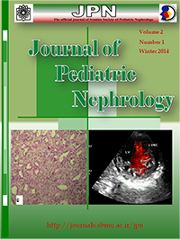
Nephronophthisis is a chronic tubulo-interstitial nephritis which can progress to end-stage renal disease. Juvenile nephronophthisis is the most common type of nephronophthisis, which accounts for 5-10% of the cases of pediatric end stage renal diseases. Left ventricular non-compaction (LVNC), a rare form of cardiomyopathy, is the result of intrauterine arrest of compaction of the endomyocardial morphogenesis. Clinical manifestations of LVNC range from asymptomatic child to a progressive deterioration in the cardiac function, congestive heart failure, arrhythmias, systemic thromboembolism and sudden cardiac death. This report presents a case of juvenile nephronophthisis with LVNC. A 15-year-old boy was referred to our nephrology outpatient clinic with a 6-month history of non-specific complaints such as lethargy, anorexia, polydipsia, polyuria, and pallor. Abdominal sonography showed a generalized increase in the parenchymal echo of kidneys. Renal biopsy was performed for him which showed nephronophthisis. Echocardiography was done and revealed LVNC. He was discharged with training for careful follow-up. Our reported case had nephronophthisis and LVNC. To the best of our knowledge, there is no report of this combination in the literature. This suggests that LVNC may be another presentation of cilia involvement. The clinical coexistence of LVNC and nephronophthisis could guide us to better localize and discover the underlying genetic mutations and the role of ciliopathies in various human diseases; Therefore, further research with a special focus on potential common derangement of cilia and protein products in these diseases is recommended.

 Keywords: Juvenile nephronophthisis; Isolated Noncompaction of the Ventricular Myocardium; Ciliary Motility Disorders

Photo quiz