Regeneration


Cell-Surface Interaction and Biological Behavior of Cells: An Overview

Parviz Ranjbarvan, Ali Golchin, Symzar Hosseinzadeh, Adel Marzban

Journal of "Regeneration, Reconstruction & Restoration" (Triple R), Vol. 4 No. 4 (2019), 14 March 2020, Page 121-129
https://doi.org/10.22037/rrr.v4i4.29799

Generally, cell interactions with the surface of biomaterials are mediated by chemical groups and biological intermediates. Cell behavior includes adhesion, proliferation, migration, differentiation, and function can be under the impression of cell-surface interaction. Therefore, Knowledge of cell-surface interaction is useful in the engineering and production of ideal cell-tissue based products. According to this important, this study was designed to review the interaction between cells and the surface of polymeric structures in cell culture conditions. This study gave an overview of the effects of key parameters on cell attachment, proliferation, migration, differentiation, and cell fate in culture. The cell-material surface interactions focus on the communication between surface properties of the substrate, protein/water adsorption, cell adhesion, and its function. Surface modification as compared to non-modified surface improves the intrinsic properties of a surface which yields a convenience of the biocompatible construct. However, the quality of notable parameters between cell and surface express the main role in tissue engineering, and subsequent can be led to high- quality cell-tissue based products.
Comparison of Enamel Matrix Derivatives and Platelet-Rich Fibrin on Human Gingival Fibroblasts Proliferation and Viability

Ardeshir Lafzi, Shahram Vaziri, Arash Ghafouri Rankouhi, Mahdi Kadkhodazadeh, Tohid Sabetmoghaddam

Journal of "Regeneration, Reconstruction & Restoration" (Triple R), Vol. 4 No. 4 (2019), 14 March 2020, Page 130-134
https://doi.org/10.22037/rrr.v4i4.29625

Introduction: Biologic mediators such as platelet-rich fibrins (PRF) or enamel matrix derivatives (Emdogain®) are used for the regeneration of the soft and hard tissues. The present study aimed to compare the efficacy of PRF and Emdogain® on the proliferation and differentiation of gingival fibroblasts in vitro.

Materials and Methods: In this randomized controlled in vitro trial, blood samples were obtained from a healthy person and Choukroun’s PRF was prepared according to the standard protocol. Fibroblast cells were cultured with PRF and different concentration of Emdogain®. MTT test were used to measure fibroblast cells proliferation after 24 and 72 hours.

Results: As compared to negative control (FBS 1%), Emdogain® 50μg/ml significantly increased cells proliferation up to 123.2% and 111.2% after 24 and 72 hours. The values were 214.9% and 121.9% for Emdogain® 100μg/ml. The increased proliferation values were significant for both groups after 24 hours (P<0.001) while the values were significant for 100 μg/ml Emdogain after 72 hours (P<0.001). Cells exposure to PRF showed significant effect on their proliferation after 24 hours compared to the negative controls (P<0.001; 21±1.73%). After 72 hours; PRF showed reverse effect on the proliferation and decreased them up to 38% and 60% regarding negative controls.

Conclusion: the most proliferation and viability of fibroblast cells were obtained after exposure to Emdogain® and the differences between Emdogain® and PRF were significant.

Influence of Whole-Body Vibration on Dynamic Response of Lumbar Spine after Transformal Lumbar Interbody Fusion

Samaneh Nemati, Siamak Khorramymehr, Mohammad-Mehdi Khani, Hekmat Farajpour

Journal of "Regeneration, Reconstruction & Restoration" (Triple R), Vol. 4 No. 4 (2019), 14 March 2020, Page 135-141
https://doi.org/10.22037/rrr.v4i4.26825

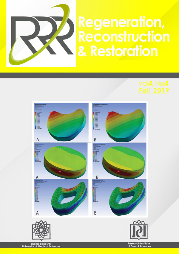
Introduction: Occupational whole body vibration (WBV) plays a major role in determining dynamic responses of the lumbar spine. WBV has been shown to cause low-back problems and degenerative disc diseases. Fusion surgery such as trans-formal lumbar inter-body fusion (TLIF) have been widely utilized to treat such disorders.

Materials and Methods: In this study, finite element method (FEM) was used to investigate dynamic responses of the lumbar spine due to WBV with the frequency in the range of regular physiologic activities after TLIF. A FE model of the L1-L5 lumbar spine was modeled and cyclic loading with the frequency of 1 Hz and 5 Hz were exerted to the model. Then, the disc bulge and stress distribution on the annual ground substance and vertebral bodies were measured.

Results: It was observed that the maximum disc bulge (MDB) and maximum von-Mises stress (MMS) occurred in proportion to the loading frequency; overall, in the 5 Hz model, MDB and MMS were detected to happen 5 times more frequently as compared to the 1 Hz model. However, the magnitude of MDB and MMS were not generally affected by the loading frequency.

Conclusions: It can be concluded that different frequency of WBV, although in the physiologic range, can alter dynamic responses of the lumbar spine and, thus, their fatigue behavior. In the results can be of assistance to broaden the understanding regarding the dynamic responses of the lumbar spine during WBV after TLIF.

Reconstruction


Orbital and Preseptal Cellulitis: Epidemiology, Etiology, and Management

Dariush Hasheminia, Navid Naghdi, Sobhan Pourarz

Journal of "Regeneration, Reconstruction & Restoration" (Triple R), Vol. 4 No. 4 (2019), 14 March 2020, Page 142-147
https://doi.org/10.22037/rrr.v4i1.26606

Introduction: Considering the little evidence regarding peri-orbital infections, this study was aimed to obtain information about the epidemiology, etiology, and management of orbital infections.

Material and Methods: In this retrospective investigation, all patients with peri-orbital infection who were hospitalized in a tertiary ophthalmologic center in AL-Zahra hospital, Isfahan, Iran from 2008 up to 2018 were identified. Documented data and radiographic images were extracted. The data regarding epidemiology, etiology and disease course was analyzed.

Results: Sixty nine patients (35 males, 34 females) with the age range between 3 months to 85 years were included. Preseptal cellulitis was recorded in 53 cases (76.8%) and orbital cellulitis was seen in 16 cases (23.2%), and the proportion of preseptal to orbital was 3.3 to 1. The mean duration of hospitalization in patients with preseptal cellulitis was 6.38 ±, 4.59 days and in patients with orbital cellulitis was 12.44 ±, 9.63 days. Most patients with preseptal cellulitis were treated by medication therapy (71.7%), while the orbital cellulitis were often treated by surgical procedures (56.2%). Sinusitis was the main cause of both preseptal and orbital cellulitis in all age groups, except infants under 1 year, which dacryocystitis was identified as primary factor.

Conclusions: The prevalence of peri-orbital infection was higher in children. The prevalence ratio of preseptal to orbital cellulitis was 3.3:1. The main etiologic factor was sinusitis. There is no agreement for the treatment modalities of peri-orbital infections and the timing of surgical intervention. Continuous evaluation of treatment course, both clinically and radiographically is important. Reassessment is recommended when improvement is not seen after 6-7 days of treatment.


The Modified Continuous Double Locking Suture: A Novel Suturing Approach in Dental Implant Surgery

Mohammad Reza Talebi Ardakani, Zeinab Rezaei Esfahrood, Shireen Shidfar

Journal of "Regeneration, Reconstruction & Restoration" (Triple R), Vol. 4 No. 4 (2019), 14 March 2020, Page 148-150
https://doi.org/10.22037/rrr.v4i4.29761

Introduction: An alternative to interrupted sutures for implant surgery at sites of edentulous ridges is the continuous locking suture, however it tends to loosen and time-consuming as the result.

Methods and Materials: The modified continuous double locking suture is a modification of continuous locking suture, which is designed to approximate flap margins of an edentulous area.

Results: Application of modified continuous double locking suture, reduces the chance of suture loosening by adding an extra knot at each area.

Conclusion: By utilizing modified continuous double locking suture, it is easier to manipulate flap margins; nonetheless, more studies are needed to evaluate its efficacy in dental practice.

Evaluation of Greater Palatine Canal and Foramen Anatomical Variation on Cone-beam CT Radiography

Hossein Tavassolizadeh, Azadeh Torkzadeh, Lida Kheiri, Parisa Negahdar

Journal of "Regeneration, Reconstruction & Restoration" (Triple R), Vol. 4 No. 4 (2019), 14 March 2020, Page 151-155
https://doi.org/10.22037/rrr.v4i4.29763

Introduction: A known method for maxillary nerve block is through the greater palatine canal (GPC). Poor knowledge about the anatomy might result in undesired conditions such as perforation of the arteries, nerve damage and blindness. This study aimed at determining variations of the GPC and greater palatine foramen (GPF) on cone beam computed tomography (CBCT) images.

Materials and Methods: This cross-sectional descriptive study was carried out at Islamic Azad University dental school and private clinics of Isfahan in 2018. A total of 92 CBCT images of adults over 18 years old were evaluated and GPC length, GPF diameter and its distance to the anterior nasal spine (ANS), posterior nasal spine (PNS) and mid-maxillary suture (MMS) were measured. The data were analyzed using SPSS 18.

Results: The mean distance of GPF to ANS (47.96 mm in men and 45.36 mm in women), to PNS (18.9 mm in men and 17.21 mm in women), and to MMS (15.93 mm in men and 14.73 mm in women), as well as the diameter of the GPF (6.07 mm in men and 5.01 mm in women) were significantly higher in men on both right and left sides (P<0.05). The mean GPC length in men and women was 34.36±2.5 and 31.39±1.9 mm, respectively, which was significantly larger in men. The more common canal pathway in sagittal plane was inferior/inferior-anterior (70%) in both genders. The inferior/inferior-anterior pathways were more common in both genders in coronal plane; however, this difference was not significant (P>0.05).

Conclusion: Since the maxillary nerve block technique might cause irreparable complications through GPC, it is very important to know the position of the foramen and the canal direction. Therefore, it is advisable to use CBCT in order to better understand the GPF and GPC anatomy.

Excision of Large Mandibular Complex Odontoma through Unilateral Sagittal Split Osteotomy

Mahboube Hasheminasab, Reza Sharifi, Nazanin Mahdavi, Arezoo Javani

Journal of "Regeneration, Reconstruction & Restoration" (Triple R), Vol. 4 No. 4 (2019), 14 March 2020, Page 156-161
https://doi.org/10.22037/rrr.v4i4.29339

Introduction: There are various alternative surgical approaches for removal of large benign lesions in the posterior part of the mandible. This study describes three cases of large mandibular mixed odontogenic lesions which were excised via sagittal split approach.

Report of Cases: All three cases were treated by surgical excision via unilateral sagittal split osteotomy under general anesthesia following a 2 weeks period of intermaxillary fixation.

Results: The lesions were enucleated completely and all three osteotomy sites healed with no consequences with an intact neurosensory function.

Conclusion: This article suggests the sagittal split osteotomy approach for conservative treatment of large mandibular lesions based on excellent access, limited bone removal and reducing further complications.

Restoration


Comparison of Different Orthognathic Surgeries Impact on Pharyngeal Airway Space in CL III Patients

Mahtab Nouri, Sara Alijani, Golnaz Nahvi

Journal of "Regeneration, Reconstruction & Restoration" (Triple R), Vol. 4 No. 4 (2019), 14 March 2020, Page 162-169
https://doi.org/10.22037/rrr.v4i4.29305

Introduction: This study sought to compare the changes in the pharyngeal airway space, hyoid bone position, tongue, soft palate, and retro-lingual soft tissues following three different orthognathic surgeries on class III patients.

Materials and Methods: This quasi-experimental study was conducted on 75 skeletal class III patients. The predictor variable was the type of orthognathic surgery namely mandibular setback, maxillary impaction & mandibular setback and maxillary advancement & mandibular setback (25 patients in each group). The primary outcome variables were the dimensions of the pharyngeal airway space and retrolingual region, measured preoperatively and at least one year postoperatively. Cephalometric landmarks were also measured. Pre- and post-operative cephalograms were superimposed based on the S-N line with registration at the S-point.

Results: When considering the changes in the whole sample (75 cases), significant changes were found in oropharyngeal length (UMPW, VLPW), tongue height (H-VT), horizontal position of vallecula (V_C3) and horizontal position of the hyoid bone. The only significant difference noted, was the tongue height and vertical position of hyoid between the three groups.

Conclusion: Orthognathic surgery significantly affects the oropharyngeal space and position of the tongue and hyoid bone. The three types of Orthognathic surgeries on class III patients caused different effects on oropharyngeal space, tongue and hyoid position with a significant impact on tongue height in maxillary advancement plus mandibular setback.

Full-mouth Rehabilitation of an Amelogenesis Imperfecta Case with Skeletal Open Bite

Vahideh Nazari, Marzieh Alikhasi

Journal of "Regeneration, Reconstruction & Restoration" (Triple R), Vol. 4 No. 4 (2019), 14 March 2020, Page 170-176
https://doi.org/10.22037/rrr.v4i4.29675

Introduction: Amelogenesis imperfect (AI) refers to a group of autosomal dominant disorders primarily affecting the enamel and can present a variety of clinical appearances. This report describes a multidisciplinary approach for oral rehabilitation of a young man patient with hypoplastic AI with severe skeletal anterior open bite (AOB).

Case Report: A 22-year-old male patient was presented with compromised masticatory efficiency and dissatisfaction with his teeth appearance. Based on the clinical and radiographic examination, the patient was confirmed as AI (hypoplastic type) with severe AOB. The treatment included orthognathic surgery (Le Fort I osteotomy and genioplasty) for correcting AOB, long face, and limited space in the posterior region, followed by crown lengthening and prosthetic rehabilitation. Before surgery, a mock-up of all teeth was performed to facilitate alignment and stabilization of the dental arches during the Le Fort I osteotomy.

Results: Solid tooth interdigitation that provided with mock-up, helped to adjust the stable skeletal position of the maxilla during the surgery.

Conclusion: preoperative mock-up appears to be an efficient fixation option to achieve an acceptable occlusion after prosthetic rehabilitation in AI patients with severe AOB.