Regeneration


Fetal Cells Homing in Maternal Bony Defects: A Preliminary in vivo Investigation

Sepanta Hosseinpour, Saeed Reza Motamedian, Seyyed Mehrshad Jafari, Sara Soudi, Masoud Soleimani, Arash Khojasteh

Journal of "Regeneration, Reconstruction & Restoration" (Triple R), Vol. 4 No. 2 (2019), 14 March 2020, Page 41-45
https://doi.org/10.22037/rrr.v4i2.29281

Introduction: Fetal cells are present in maternal tissue during pregnancy as well as post-partum. Although their clinical significance is not clear, these cells can be found in injured, diseased and normal tissue. In this study, the authors sought to assess the possibility of fetal cells’ homing in iatrogenic maternal jawbone defects.

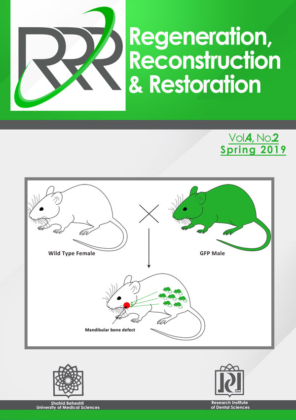
Materials and Methods: Eight wild female mice were bred with eight male mice carrying the green fluorescent protein (GFP). Two mice with the same specification were bred with non GFP male mice. A two-millimeter defect was created in pregnant mice mandibles on day 12.5 of pregnancy. The mice were euthanized 4 days later.

Results: GFP+cells were investigated in mandibular defects using immunoflurescent, immunohistochemical staining, and quantitative polymerase chain reaction. GFP+cells were present at defect margins of four cases by all evaluations. GFP+cells were absent in normal tissues and in control mice.

Conclusion: Fetal cells were distinguishable at the margin of iatrogenic jaw bone defect in the mice but their function remain to be elucidate.

Effect of Tetracycline Dressing in Reducing Post-Operative Complications after Impacted Third Molar Surgery

Amirali Badri, Mahboube Hasheminasab, Masoud Jamshidian, Younes Ghoreyshi

Journal of "Regeneration, Reconstruction & Restoration" (Triple R), Vol. 4 No. 2 (2019), 14 March 2020, Page 46-49
https://doi.org/10.22037/rrr.v4i2.29237

Introduction Surgical removal of mandibular impacted third molars is a routine dental procedure which is associated with the complications such as trismus, swelling, alveolar osteitis, and pain. Furthermore, tetracycline dressings are possibly useful to prevent some of these complications, although this finding requires more investigations. This study determined the effects of post-surgical placement of tetracycline impregnated gauze in dental socket to prevent the incidence of the complications related to the surgical removal of impacted third molars in patients referred to School of Dentistry, Tehran University of Medical Sciences, Tehran, Iran.

Materials and Methods: In this split-mouth randomized clinical trial, 27 patients were enrolled. All the patients had bilateral impacted mandibular third molars. A single surgeon extracted all teeth. At the end of surgery, a gauze pack was placed on socket in one side and a tetracycline impregnated mesh in the socket of the other side. The patients were called at 3rd day after the surgery and the maximum mouth opening and pain were measured and the incidence of alveolar osteitis was recorded. The patients' pain scores were calculated at 2nd, 3rd and 7th days post-surgery by 10-scaled VAS system. The patients' pain scores and the amount of maximum mouth opening were subjected to Wilcoxon Signed Ranks test.

Results: There was no incidence of alveolar osteitis in any of the patients. The mean pain scores of the patients after using tetracycline impregnated mesh and gauze pack were 6.07 and 6.59 on 2nd day, 3.78 and 4.07 on 3rd day, and 0.19 and 0.33 on 7th day post-operatively. No statistically significant differences were observed regarding pain scores of the patients in both groups on 2, 3 and 7 days post-operatively. The mean maximum mouth opening of the patients in tetracycline dressing and control groups were found to be 24.11mm and 21.74mm after 3rd day post-operatively which was significantly higher in the tetracycline group than control group (P=0.03).

Conclusion: It seems that there is no need to use of tetracycline impregnated mesh in dental sockets following surgical removal of mandibular third molars.

Surgical Treatment of Pyogenic Granuloma by 980 Diode Laser

Javad Sarabadani, Ali kouhrokhi, Hanieh Jadidi, Nasim Chiniforush

Journal of "Regeneration, Reconstruction & Restoration" (Triple R), Vol. 4 No. 2 (2019), 14 March 2020, Page 50-53
https://doi.org/10.22037/rrr.v4i2.25024

Introduction: Pyogenic granuloma is a pseudo-tumor which has a great tendency to attached gingiva. Conservative surgery is a treatment choice for this lesion. Among different laser wavelengths used for oral surgery including CO2, Nd:YAG, Erbium family and diode laser, diode can be the best option due to providing blood less area that make better view for surgeon to remove it completely.

Case Report: A 14-year-old girl with a chief complaint of exophytic lesion in lower jaw was referred. The surgery was done by 980 diode laser with output power of 1 W in continuous mode. The lesion was sent to pathology laboratory for further investigation. The patient was advised to keep oral hygiene instructions.

Results: After 1 weeks, complete healing was observed.

Conclusion: Diode laser can be a treatment choice for this kind of lesions due to diminished infection and bacteremia following surgery, reduction of relapse and bone degeneration, less invasive technique and reduced chair time.

Reconstruction


Outcome of Pectoralis Major Mayocutaneous Flap in Reconstruction of Maxillofacial Defects

Reza Tabrizi, Birkan Taha Ozkan, Mohammad Hassani, Behrouz Barati

Journal of "Regeneration, Reconstruction & Restoration" (Triple R), Vol. 4 No. 2 (2019), 14 March 2020, Page 54-58
https://doi.org/10.22037/rrr.v4i2.30030

Introduction: The aim of the present study was to evaluate outcomes and complications related to pectoralis major mayocutaneous (PMM) flaps in the reconstruction of maxillofacial defects.

Materials and Methods: Subjects who underwent pectoralis major mayocutaneous flaps to reconstruct maxillofacial defects due to trauma or tumor resection were studied. Complications were considered in two stages: early and late. A modified version of the University of Washington Quality of Life questionnaires, version 4, was used to evaluate the functional outcome of patients who underwent free flap or PMM flap reconstruction. The survey is composed of 6 domains: pain, appearance, activity, swallowing, chewing, and speech. We scored domains on a scale ranging from 0 (worst) to 100 (best). We considered scores higher than 70 in the normal range for appearance, activity, swallowing, chewing, and speech.

Results: Thirty-one subjects were studied. The most common defect sites were oral floor (48.3%) and buccal soft tissue (32.2%). The least common was neck (19.3%). Immediate complications consisted of dehiscence in 3 cases (9.6%) and flap necrosis in 2 cases (6.4%). Late complications included dehiscence in 3 cases, partial necrosis of skin without muscle necrosis in 1 case, and donor site infection in one case. The most common complications seen in both stages were dehiscence (19.3%) and necrosis (9.6%).In the immediate phase, patients mostly complained of pain. In the late stage, pain significantly decreased. Activity, swallowing, and speech improved in the late stage.

Conclusion: Our study indicated a significant improvement in patients' functional outcomes after discharge from the hospital. The pain was a major complaint of patients immediately after reconstruction with PMM flap, which should be considered as a significant concern for maxillofacial surgeons.

A Case of Gingivobuccal Coplex Cancer of Anterior Maxilla in a 14 Year Old Child

Shishir Mohan Devki, Venkatesh Balaji Hange

Journal of "Regeneration, Reconstruction & Restoration" (Triple R), Vol. 4 No. 2 (2019), 14 March 2020, Page 59-62
https://doi.org/10.22037/rrr.v4i2.26523

Introduction: Squamous cell carcinoma (SCC) of the oral cavity ranks as the 12th most common cancer in the world and the 8th most frequent in males. SCC is extremely unusual in the pediatric population and researchers believe that the pathology of SCC in pediatric patients is a separate entity different from SCC in the adult population.

Materials and Methods: A 14 year old girl patient with ill-defined swelling with 1.5 x 4cm ,ulcerated surface texture along with tiny bleeding points was came to department of Oral and Maxillofacial Surgery K.D.Dental College & Hospital Clinically the lesion appears to be a benign soft tissue tumor with provisional diagnosis of peripheral giant cell granuloma. Pre-surgical investigations which were carried out were found to be within normal range. Radiographic examination did not reveal any significant bone loss in relation to the maxillary right anterior teeth or bone.

Results: SCC of gingiva by histopathological examination was confirmed and patient referred to nearby cancer institute for definitive treatment.

Conclusion: Despite the fact that SCC has been frequently reported in old male patient, here, we reported a case of young female patient. Since the early detection of SCC is vital, presence of SCC in younger patients also needs to be considered by paediatrics.

Restoration


The Impact of Crown-to-Implant Ratio on Marginal Bone Loss: A Meta-Analysis on Human Prospective Clinical Trials

Vahid Khoshkam, Guo-hao Lin, Marzieh Alikhasi, Homayoun H. Zadeh

Journal of "Regeneration, Reconstruction & Restoration" (Triple R), Vol. 4 No. 2 (2019), 14 March 2020, Page 63-71
https://doi.org/10.22037/rrr.v4i2.29353

Introduction: Multiple systematic reviews have documented the efficacy of short implants in alveolar sites with moderate vertical height atrophy. Short implants in sites with alveolar ridge atrophy are often restored with elongated restoration, leading to relatively high crown-to-implant (C/I) ratio. It is important to determine whether the increased C/I ratio has any detrimental biological effects on peri-implant bone. To that end, this systematic review sought to examine the long-term effects of C/I ratio on peri-implant marginal bone loss, as reported by prospective clinical trials.

Materials and Methods: Electronic search of 3 databases and hand searching were performed to identify prospective human trials that had reported both crown/implant ratio and associated peri-implant marginal bone loss with the follow-up of at least 36 months. Random-effect meta-analysis was performed to analyze weighted mean difference (WMD) and confidence interval (CI) for recorded variables according to PRISMA guidelines. Weighted linear regression model was used to evaluate R2 value.

Results: Publication search yielded 1134 records and after evaluating titles, abstracts and full-texts, 8 prospective trials were included for quantitative data synthesis. Meta-analysis for the comparison of MBL among selected studies showed a WMD of 0.01 mm, with a 95% CI= 0.08 mm to -0.07 mm (p= 0.90). Results failed to detail any significant correlation between C/I ratio and marginal bone loss. Weighted linear regression model and low (0.214) R2 ratio suggested a lack of correlation between MBL and C/I ratio.

Conclusion: Basad on the available evidence, there is no relationship between C/I ratio within the range tested and marginal bone loss.

The Effect of Application Time on Effectiveness of Two Caries Disclosing Agents

Kasra Tabari, Fatemeh Mashhadi Abbas, Morteza Jahanbani, Fazel Anjamrouz, Sina Rahmanpanah, Soudabeh Koulivand

Journal of "Regeneration, Reconstruction & Restoration" (Triple R), Vol. 4 No. 2 (2019), 14 March 2020, Page 72-75
https://doi.org/10.22037/rrr.v4i2.29581

Introduction: The main goal of treating dental caries is complete removal of carious tissues with maximum preservation of sound dentin before restoring the cavity. The aim of this study was to evaluate the effect of application time on efficacy of an experimental caries detector dye (with polypropylene glycole as the solvent) and SNOOP (with monopropylene glycole as the solvent).

Materials and Methods: In this experimental in vitro study, 100 extracted human molars and premolars with occlusal caries, were divided into five groups. In each group, the caries were removed by conventional method (optical and tactile sence) or following caries detector dye application for 10 and 20 seconds. Histological assessment was used to evaluate bacterial existance after caries excavation. Data were analyzed using Fisher’s exact test. Alpha was set at 0.05.

Results: Statistical analysis revealed that there was significant difference between clinical removal of caries and using caries disclosing dyes. However, no significant difference between removal of carious dentin after 10 or 20 seconds application of the both caries disclosing dyes was observed.

Conclusion: Using caries disclosing dyes for 10 or 20 seconds, improves efficacy of caries removal in comparison with conventional procedure.

Effect of Curing Time on Micro shear Bond Strength of Enamel and Dentin Bonding Agents to Enamel

Amir Ghasemi, Mostafa Alam, Kamyar Abbasi, Farhood Massoumi, Shervin Shafiei

Journal of "Regeneration, Reconstruction & Restoration" (Triple R), Vol. 4 No. 2 (2019), 14 March 2020, Page 76-80
https://doi.org/10.22037/rrr.v4i2.25438

Introduction: The bond strength to enamel may be simply improved by increasing the polymerization rate via prolonging the curing time. This study sought to assess the effect of increasing the curing time on microshear bond strength of enamel (Heliobond) and dentin (Excite) bonding agents to enamel.

Materials and Methods: 90 extracted, sound human molar teeth randomly divided into six groups. In group 1, the enamel surfaces etched 30 seconds and were rinsed for 30 seconds. After drying, Heliobond was applied and light cured for 10 seconds, in groups 2 and 3, the curing time was 20 and 40 seconds, respectively. In group 4, specimens were prepared as in group 1 except that Excite dentin bonding agent was used.  Groups 5 and 6 were prepared as in group 4 but the curing time was 20 and 40 seconds, respectively. The microshear bond strength of specimens was measured using micro tensile tester and analyzed. The mode of failure of specimens was evaluated under a microscope.

Results: Two-way ANOVA showed significant differences in bond strength of Heliobond and Excite specimens cured for different times. However, no significant difference was noted in microshear bond strength of specimens cured for 10 and 20 seconds (neither in Excite nor in Heliobond groups). Pairwise comparison of groups revealed significant differences between specimens cured for 10 and 20 seconds with 40 seconds curing times. The overall microshear bond strength of specimens in Heliobond groups was significantly higher than that of Excite groups. The mode of failure of groups bonded with Heliobond and Excite was not significantly different.

Conclusion: Within the limitations of this study, prolonging the curing time up to the certain time significantly increased the micro shear bond strength of both enamel (Heliobond) and dentin (Excite) bonding agents to enamel.