Mechanical Vulnerability of L3-L4 Spinal Segment after Fusion Surgery Utilizing Dynamic Stress Analysis
Regeneration, Reconstruction & Restoration (Triple R),
Vol. 4 No. 1 (2019),
2 February 2020
,
Page 15-19
https://doi.org/10.22037/rrr.v4i1.26277
Abstract
Introduction: Pedicle screw-based spine fusion has been employed as a felicitous approach for treatment of degenerative lumbar spinal diseases. Although the pedicle screw designs and fixation techniques have progressed, many clinical studies have reported the adjacent segment degeneration (ASD) and several other adverse effects after surgery. In the case of fixation techniques, the use of semi-rigid rods such as Polyarylether ether ketone (PEEK) rods can be an appropriate substitute for rigid fusion instrumentation. However, the biomechanical effects of using viscoelastic PEEK rods for use in clinical studies are still unclear.
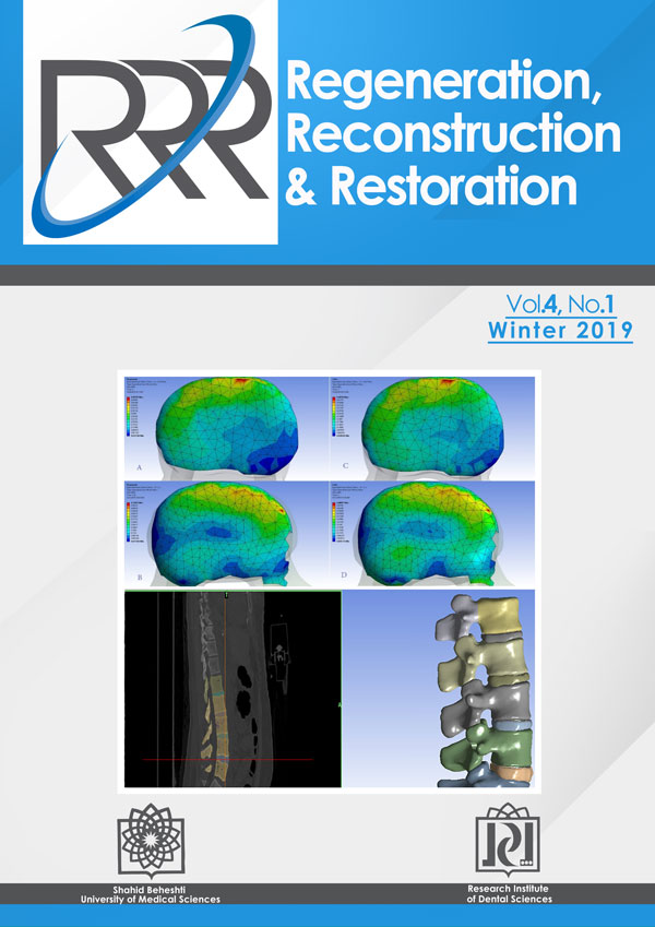
Materials and Methods: In this study, the effects of using two pedicle screw-based stabilization systems, i.e. viscoelastic PEEK rods and elastic Titanium rods, on the stress distribution in L3-L4 intervertebral disc and the surrounding osseous tissue were investigated. An L1-L5 lumbar region was modeled. Subsequently, a mild degenerative disc disease was simulated in the L3-L4 lumbar level. Next, an axial cyclic torque was applied to the model.
Results: The results showed that, at the end of the loading cycle, the maximum von-Mises stress in the L3-L4 intervertebral disc as well as the overall value of stress in the surrounding osseous tissue were slightly greater in the viscoelastic model as compared to the elastic one. Nonetheless, the stress distribution contours and the location of maximum stress were different in the two models.
Conclusion: It can be postulated that the vulnerable areas of lumbar bone are different in two models.
- Dynamic Stress
- Fusion Surgery
- Finite Element Analysis
- Mechanical Vulnerability
How to Cite
- Abstract Viewed: 312 times
- PDF Downloaded: 117 times