Original Article


A Prospective Clinical Study on Blood Mercury Levels Following Endodontic Root-end Surgery with Amalgam

Masoud Saatchi, Elham Shadmehr, Seyed Morteza Talebi, Mohsen Nazeri

Iranian Endodontic Journal, Vol. 8 No. 3 (2013), 28 July 2013, Page 85-88
https://doi.org/10.22037/iej.v8i3.3597

Introduction: The purpose of this clinical study was to compare the blood mercury levels before and after endodontic surgery using amalgam as a root-end filling material. Materials and Methods: Fourteen patients requiring periradicular surgery participated in this prospective clinical study. A zinc-free amalgam was employed as root-end filling material. Blood samples were collected at three intervals: immediately before, immediately after and one week postoperatively. Mercury content of the blood was determined using gold amalgamation cold-vapor atomic absorption spectrometry. Obtained data were analyzed using analysis of variance for repeated measures and paired t-test. Results: The mean (SD) of blood mercury levels was 2.20 (0.24) ng/mL immediately before surgery, 2.24 (0.28) ng/mL immediately after surgery and 2.44 (0.17) ng/mL one week after the periradicular surgery. The blood mercury level one week post-operative was significantly higher than both blood mercury levels immediately before (P<0.001) and immediately after (P=0.005) the surgery. Conclusion: Placement of an amalgam retroseal during endodontic surgery can increase blood mercury levels after one week. The mercury levels however, are still lower than the toxic mercury levels. We suggest using more suitable and biocompatible root-end filling materials.
Evaluation of the Quality of Four Root Canal Obturation Techniques Using Micro-Computed Tomography

Mandana Naseri, Ali Kangarlou, Atefeh Khavid, Mostafa Goodini

Iranian Endodontic Journal, Vol. 8 No. 3 (2013), 28 July 2013, Page 89-93
https://doi.org/10.22037/iej.v8i3.3642

Introduction: One of the key factors in successful endodontic therapy is to adequately fill the root canals. The aim of this in vitro study was to compare the quality of four different root canal obturation techniques: cold lateral condensation (CLC), warm vertical condensation (WVC), Obtura II (OII) and Gutta Flow (GF) by using micro-computed tomography (micro CT). Materials and Methods: A total of 20 extracted maxillary first molars prepared with ProTaper files, were randomly divided into four groups. Micro CT was used to measure the internal volume of root canals. Following application of AH26 sealer to canal obturation, new micro-CT images were taken and the volume percentage (VP) of voids, gutta-percha and sealer at different levels were calculated with CT software. Data was statistically analyzed using Kruskal-Wallis and Mann-Whitney U tests. Results: The highest percentage of filling material was observed in GF group followed by OII with no statistically significant difference (P>0.05). These two groups had a significantly more acceptable filling than WVC and CLC groups (P<0.05). Voids were detected in all samples. There was a significant difference between the highest and the lowest percentage of voids in CLC (19.6%) and GF groups (6.7%), respectively. In the apical third, CLC and OII showed the highest and the lowest percentage of voids (5.5% and 2.6%) and the lowest and highest percentage of gutta-percha (76.52% and 94.26%), respectively. These differences were statistically significant (P<0.05). Conclusion: None of the root canal filled teeth were gap-free. GF and CLC techniques showed the highest and lowest VP of obturation materials, respectively.

Residual Dentin Thickness of Bifurcated Maxillary Premolars Following Two Post Space Preparation Methods

Jamileh Ghoddusi, Maryam Forghani, Ali Bagherpour, Fatemeh Mahmudabadi, Majid Sarmad

Iranian Endodontic Journal, Vol. 8 No. 3 (2013), 28 July 2013, Page 94-98
https://doi.org/10.22037/iej.v8i3.3707

Introduction: The aim of this in vitro study was to compare the effect of Gates-Glidden and Peeso reamer drills on residual dentin thickness during post space preparation in order to discover which method has minimum root structure damage. Materials and Methods: Thirty extracted human maxillary premolars with bifurcations at root middle were horizontally cut 15 mm coronal to the apical end after root canal treatment. The samples were scannedby Cone Beam Computed Tomography (CBCT) before and after preparing the post space. Residual dentin thicknesses were measured at 4-, 6-, and 8-mm levels from the apex. Data were analyzed using repeated measured ANOVA. Results: Endodontic therapy and post space preparations removed more dentin within the bifurcation of both roots compared to outer dentin. The difference in residual dentin thickness was highly significant regarding stage (before and after post space preparation) in all levels and stage × device in coronal and middle levels (P<.05). This in vitro study emphasizes the minimal dentin width in the buccal root of maxillary premolars, especially near the bifurcation. Conclusion: Lack of adequate residual dentin thickness after post space preparation implies that the use of posts in maxillary first premolars should be limited. When mandatory, it is recommended that post space be prepared with Gates-Glidden drill in the palatal root of maxillary first premolars and use of Peeso reamer be avoided.

The Prevalence of Bilateral Three-Rooted Mandibular First Molar in Indian Population

Rupali Karale, Champa Chikkamallaiah, Jayshree Hegde, Srirekha Aswathanarayana, Lekha Santhosh, Kusum Bashetty, Shwetha Rajanna Susheela, Srinivas Panchajanya

Iranian Endodontic Journal, Vol. 8 No. 3 (2013), 28 July 2013, Page 99-102
https://doi.org/10.22037/iej.v8i3.2956

Introduction: The purpose of the present study was to evaluate the prevalence of bilateral three-rooted mandibular first and second molars in Indian population. Materials and Methods: A total of 215 patients were screened bilaterally for mandibular first and second molar and 430 samples of periapical radiographs were obtained. The gender, symmetry, and prevalence of three-rooted mandibular first and second molars were recorded. The correlation between left and right side occurrences and distribution were recorded and analysed using Z-test. Results: The results showed that 33 teeth had three-rooted mandibular first molars, 16 male and 17 female (P=0.442). Overall, 21 teeth of right jaw and 12 teeth of left jaw (P=0.103) showed presence of an extra-root. The prevalence of three-rooted mandibular first molars was 7.67% and second molar was 0.23%. The bilateral frequency distribution was 3.72% for the first molar. There was no statistically significant difference between right side and left side mandibular molars. Also, gender did not show a significant relationship with this variant. Conclusion: The endodontic treatments of first mandibular molars require a careful clinical approach in Indian population as a high racial prevalence of 7.67% three-rooted molars was found. However, in the same population, 0.23% mandibular second molars had three roots.

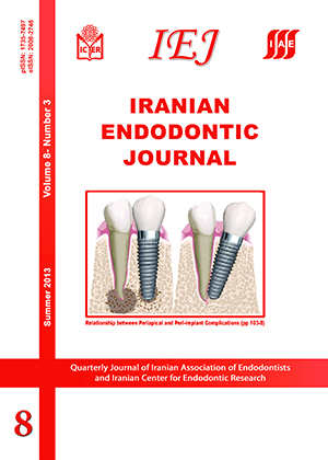
Introduction: There are numerous studies supporting the high success rate of dental implants used for reconstruction of missing teeth. However, complications like mucositis and peri-implantitis are increasingly reported. Placement of dental implants in partially edentulous patients is associated with the risk of peri-implant diseases, especially when an old or a new inflammatory lesion is present adjacent to the implant site. Although no consensus has been reached on the difference in prevalence of peri-implant mucositis and peri-implantitis between fully and partially edentulous patients, available data clearly show that the combination of periodontal lesion and peri-implantitis is a possible risk factor for further complications. Several classification systems have been suggested for determination of the severity of disease around dental implants. However, no classification has been proposed for combined biological complications around teeth and implants. This study reviews the possible pathologic communication routes between natural dentition and the implants installed adjacent to them. Furthermore, we introduce a new classification system for the peri-implant disease in association with natural teeth called “PIST”. This system was designed based on the origin of the defects in order to clarify the different pathological situations which can be detected around dental implant. Using this classification system can help improve diagnosis, comparison and subsequent selection of the best treatment option.

Sealing Ability of Orthograde MTA and CEM Cement in Apically Resected Roots Using Bacterial Leakage Method

Saeed Moradi, Reza Disfani, Kiarash Ghazvini, Mahdi Lomee

Iranian Endodontic Journal, Vol. 8 No. 3 (2013), 28 July 2013, Page 109-113
https://doi.org/10.22037/iej.v8i3.3589

Introduction: The aim of this in vitro study was to determine the sealing ability of orthograde ProRoot mineral trioxide aggregate (MTA) and calcium enriched mixture (CEM) cement as root-end filling materials. Materials and Methods: Fifty four extracted single-rooted human teeth were used. The samples were randomly divided into 3 experimental groups. In group A and B, 4 mm of WMTA and CEM cement were placed in an orthograde manner and 3 mm of apices were resected after 24 hours. In group C the apical 3 mm of each root was resected and the root-end prepared with ultrasonic tips to a depth of 3 mm and subsequently, then filled with MTA. The apical sealing ability was performed with bacterial leakage method. Statistical analysis was carried out with Chi-square test. Results: There were no significant differences in the extent of bacterial leakage between the three experimental groups (P>0.05). Conclusion: Based on the limitations of this in vitro study, we concluded that MTA and CEM cement can be placed in an orthograde manner when there is a potential need for root-end surgery.

An In Vitro Comparison of Propex II Apex Locator to Standard Radiographic Method

Kalyan Vinayak Chakravarthy Pishipati

Iranian Endodontic Journal, Vol. 8 No. 3 (2013), 28 July 2013, Page 114-117
https://doi.org/10.22037/iej.v8i3.3523

Introduction: The aim of this in vitro study was to compare the accuracy of radiography in assessing working length to Propex II apex locator. Materials and Methods: Thirty single canal extracted human teeth with patent apical foramen were selected. Access cavities were prepared. Anatomic length (AL) was determined by inserting a K-file into the root canal until the file tip was just visible at the most coronal aspect of the apical foramen; subsequently 0.5 mm was deducted from this measured length. Working length by radiographic method (RL) was determined using Ingle’s method. Propex II apex locator was used to determine the electronic working length (EL). From these calculated lengths, AL was deducted to obtain D-value. D-value in the range of +/-0.5 mm was considered to be acceptable. Results: The percentage accuracy of RL and Propex II apex locator was 76.6% and 86.6%, respectively. Paired t-test revealed significant difference between the RL and Propex II apex locator (P<0.05). Conclusion: Under these in vitro conditions, Propex II apex locator has determined working length more accurately than radiographic method.

The Effect of Deproteinized Bovine Bone Mineral on Saos-2 Cell Proliferation

Arash Khojasteh, Mohammad Hossein Ghahremani, Seyed Nasser Ostad, Mohammad Eslami, Pourya Motahhary, Golnaz Morad, Shireen Shidfar

Iranian Endodontic Journal, Vol. 8 No. 3 (2013), 28 July 2013, Page 118-122
https://doi.org/10.22037/iej.v8i3.3198

Introduction: Deproteinized bovine bone mineral (Bio-Oss) is a xenogenic bone substitute, widely used in maxillofacial bone regeneration. The aim of this in vitro study was to investigate its influence on the growth behavior of human osteosarcoma cell line, Saos-2 culture, and compare it with the physiologic dose of Dexamethasone, an inductive factor for osteoblasts. Materials and Methods: Human osteosarcoma cells, Saos-2, were cultured on Bio-Oss and their growth rate was compared to Saos-2 cultures treated with Dexamethasone 10-7 M in contrast to cells cultivated in PBS, in the control group. Assessment of proliferation was performed after 24, 36, and 48 hours by counting cells using trypan blue exclusion method. Alkaline phosphatase was measured spectrophotometrically at 405 nm with paranitrophenol buffer. Results: After 48 hours, the number of Saos-2 cells increased significantly when subcultured with Bio-Oss. Bio-Oss was more effective on the enhancement of proliferation of Saos-2 cells when compared to the physiologic dose of Dexamethasone (P<0.05). Alkaline phosphatase activity increased in cells grown on Bio-Oss and dexamethasone 10-7M in contrast to cells cultivated in PBS control group. The greatest level of activity was observed in the group containing Bio-Oss after 48 hour. Conclusion: The significant increase of cell proliferation and alkaline phosphatase activity in cells cultured on Bio-Oss, compared to Dexamethasone-treated cells, suggests the important role of this bone substitute in promoting bone regeneration.

Prevalence of Toothache and Associated Factors: A Population-Based Study in Southeast Iran

Shahla Kakoei, Masoud Parirokh, Nouzar Nakhaee, Forogh Jamshidshirazi, Maryam Rad, Sina Kakooei

Iranian Endodontic Journal, Vol. 8 No. 3 (2013), 28 July 2013, Page 123-128
https://doi.org/10.22037/iej.v8i3.3520

Introduction: This study was carried out to estimate toothache prevalence among adult residents in Kerman. Materials and Methods: This cross-sectional, population-based study was conducted among individuals aged over 18 years (n=1800). The relevant data on the prevalence of toothache and associated factors were collected by interviewing the individuals in their homes and filling out a questionnaire designed by the examiners. Prevalence of toothache and associated factors that patients recalled previous to their interview were analyzed by chi-square test and multivariate logistic regression analysis. Results: Nine hundred ninety-one individuals (55.1%) reported toothache during the 6 months before the interview. The participants who flossed daily, had regular dental visits, and had higher education showed a significantly lower prevalence of toothache (P<0.05), whereas regular tooth brushing and economic level of residency had no significant effect on the prevalence of toothache. Individuals between the ages of 26 and 45 [odds ratio (OR)=2.0], with a family size of more than 4 (OR=1.5), not using dental floss (OR=1.5), or having a mental or psychological illness (OR=1.5) were more likely to have a history of toothache. Conclusion: High prevalence of toothache (more than half) among residents of Kerman shows a serious and major public health problem. Toothache prevalence in middle aged adults, lower education, bigger family size, lower dental hygiene habit and/or those having mental or psychological illness were more common in the city of Kerman.

The Prophylactic Effects of Zintoma and Ibuprofen on Post-endodontic Pain of Molars with Irreversible Pulpitis: A Randomized Clinical Trial

Mohsen Ramazani, Mahmoud Reza Hamidi, Ali Akbar Moghaddamnia, Nahid Ramazani, Nafiseh Zarenejad

Iranian Endodontic Journal, Vol. 8 No. 3 (2013), 28 July 2013, Page 129-135
https://doi.org/10.22037/iej.v8i3.4080

Introduction: Post endodontic pain is often linked to the inflammatory process as well as additional central mechanisms. The purpose of the present double-blind randomized clinical trial study was to compare the prophylactic effects of a derivative of Zingiber Officinale, Zintoma, and Ibuprofen on post endodontic pain of molars with irreversible pulpitis. Materials and Methods: The post endodontic pain of 72 enrolled patients suffering from irreversible pulpitis was assessed after prophylactic use of 400 mg Ibuprofen, 2 gr Zintoma and placebo. Using the Heft-Parker Visual Analogue Scale, the patients recorded their perceived pain before taking the medicament (baseline), immediately after and also at 4, 8, 12, 24, 48, and 72 h post one-visit endodontic treatment. The statistical analysis was done using Kruskal-Wallis, Mann-Whitney, and Freedman tests (P<0.05). Results: At all times, there was significant difference between the Ibuprofen and Zintoma (P<0.05) and also between the Ibuprofen and placebo (P<0.05). However, there was no significant difference between Zintoma and the placebo in any of time intervals (P>0.05). No side effects were observed. Conclusion: The obtained results of the trial revealed that prophylactic use of 2 gr Zintoma is not an effective pain relieving agent.

Fracture Resistance of Root Canals Obturated with Gutta-Percha versus Resilon with Two Different Techniques

Hengameh Ashraf, Golnaz Momeni, Nima Moradi Majd, Hamed Homayouni

Iranian Endodontic Journal, Vol. 8 No. 3 (2013), 28 July 2013, Page 136-139
https://doi.org/10.22037/iej.v8i3.3539

Introduction: Dentin removal during root canal instrumentation creates a weaker root structure and increases its potential to fracture. The aim of this in vitro experimental study was to compare fracture resistance of teeth filled with gutta-percha, and Resilon using two different techniques. Materials and Methods: This study was performed on 105 single-canal extracted maxillary incisors. Samples were divided into seven groups of 15 each. Three groups were prepared with K-files; three groups with Race rotary files and in one group no preparation was carried out. Of all samples prepared either manually or with rotary instruments, 15 teeth were obturated using gutta-percha and AH26 sealer, 15 teeth were filled with Resilon and 15 teeth remained unfilled. Loading force to fracture was measured and ANOVA and Tukey tests were used for statistical analysis. Results: No statistically significant differences were observed between different preparation techniques. The intact roots showed significantly greater fracture resistance compared to both instrumented groups (P<0.01). Resilon Group showed significantly higher resistance than gutta-percha Group (P<0.01); however the difference between Resilon and intact teeth was not statistically significant. Conclusion: Accoding to the results of this in vitro study, root canal filling using Resilon may increase the fracture resistance of treated teeth.

Case Report


Periradicular Surgery of Human Permanent Teeth with Calcium-Enriched Mixture Cement

Saeed Asgary, Sara Ehsani

Iranian Endodontic Journal, Vol. 8 No. 3 (2013), 28 July 2013, Page 140-144
https://doi.org/10.22037/iej.v8i3.4484

Introduction: Root-end preparation and restoration with an endodontic material are required when nonsurgical endodontic retreatment has failed or is impossible. The present clinical study reports the treatment outcomes of periradicular surgery using calcium-enriched mixture (CEM) cement. Materials and Methods: A prospective outcome study of periradicular surgery using CEM was conducted on 14 permanent teeth with persistent apical periodontitis. Using a standardized surgical protocol, 2-3 mm of the root apex was resected; approximately 3 mm deep root-end cavities were ultrasonically prepared and filled with CEM cement. All patients were available for recall. Results: Clinical and radiographic examination revealed complete healing of periradicular lesions, i.e. regeneration of periodontal ligament and lamina dura in 13 teeth (93% success) during a mean time of 18 months; moreover, the teeth were functional and asymptomatic. Conclusion: Favorable treatment outcomes in this prospective clinical study suggested that CEM cement may be a suitable root-end filling biomaterial.

Vital Pulp Therapy with Three Different Pulpotomy Agents in Immature Molars: A Case Report

Azadeh Harandi, Maryam Forghani, Jamileh Ghoddusi

Iranian Endodontic Journal, Vol. 8 No. 3 (2013), 28 July 2013, Page 145-148
https://doi.org/10.22037/iej.v8i3.3621

Introduction: This case report describes apexogenesis treatment of three molar teeth of an 8-year-old boy using three different pulpotomy agents. Methods: Pulpotomy was performed on decayed immature molar teeth with established irreversible pulpitis and the remaining pulp was capped with either zinc oxide eugenol, ProRoot mineral trioxide aggregate or calcium-enriched mixture (CEM) cement. Teeth were restored with stainless steel crowns. Results: Eighteen months clinical and radiographic follow-up revealed successful preservation of pulpal vitality with continued root development in all treated teeth. Conclusion: Based on this case report, CEM cement may be an alternative option for pulpotomy treatment of immature permanent molars.