Systematic Review


Comparing alveolar bone regeneration using Bio-Oss and autogenous bone grafts in humans: a systematic review and meta-analysis

Alireza Akbarzadeh Baghban, Azam Dehghani, Farzin Ghanavati, Farid Zayeri, Farzam Ghanavati

Iranian Endodontic Journal, Vol. 4 No. 4 (2009), 10 October 2009, Page 125-130
https://doi.org/10.22037/iej.v4i4.1486

INTRODUCTION: Bone regeneration grafts (BRG) are widely used in the treatment of osseous defects and oral surgery. The various techniques and associated success rates of bone augmentation require evaluation by systematic review and meta-analysis of eligible studies. The aim of this systematic review was to compare alveolar bone regeneration in humans using Bio-Oss and autogenous bone graft. MATERIALS AND METHODS: The computerized bibliographical databases including Pubmed, Google, ScienceDirect and Cochrane were searched for randomized and cohort studies in which autogenous grafts were compared to Bio-Oss in the treatment of periodontal defects. The inclusion criteria were human studies in English that were published 1998-2009. Exclusion criteria included non randomized observation and cohort studies, papers which provided summary statistics without the variance estimates, and studies that did not use BRG intervention alone, were excluded. The screening of eligible studies, assessment of the methodological quality of the trials and data extraction were collected by two observers independently. For comparing autogenous grafts used alone against Bio-Oss used alone 5 situations were investigated. Thirteen studies were included in the review which compared autogenous against Bio-Oss, autogenous combined with guided tissue regeneration (GTR) against GTR, Bio-Oss combined with GTR versus GTR, autogenous alone versus Open Flap Debridement (OFD), Bio-Oss versus OFD. In meta-analysis, changes in bone level (bone fill) was used as the measure. Data were analyzed using Bayesian meta-analysis by WinBUGS and Boa software. RESULTS: Only one comparison demonstrated that the difference in bone augmentation between Bio-Oss and OFD was statistically significant. CONCLUSION: There is insufficient evidence to show that Bio-Oss is superior to autogenous grafts in bone augmentation techniques however autogenous bone involves donor site surgery and thus donor site morbidity, so we can conclude that Bio-Oss is better than autogenous for alveolar regeneration. [Iranian Endodontic Journal 2009;4(4):125-30]
Original Article


A Novel Approach in Assessment of Root Canal Curvature

Shiva Sadeghi, Vahideh Poryousef

Iranian Endodontic Journal, Vol. 4 No. 4 (2009), 10 October 2009, Page 131-134
https://doi.org/10.22037/iej.v4i4.1487

MATERIALS AND METHODS: One hundred and thirty five mesial root canals of mandibular first and second molar teeth were selected. Access cavities were prepared. After inserting a K-file size #10 into each canal, radiographs were taken. Canal curvature was determined by measuring the Schneider angle, canal access angle, as well as the canal radius, length, height and curvature starting distance on scanned radiographs using a computerized image processing system. Data was evaluated statistically using Pearson correlation. RESULTS: The mean canal access angle (CAA) and Schneider angle (S) were 8.04(3.46) and 19(6.99), respectively. The Pearson correlation analysis found significant positive correlation between S and CAA (r=0.826, P<0.0001). Negative correlations were found between radius and length (r= –0.4, P<0.0001), radius and Schneider angle (r= –0.4, P<0.0001), radius and CAA (r= –0.24, P=0.004) and CAA and curvature starting distance (r= 0.4, P<0.0001). There was no correlation between height and distance (r=0.013, P=0.789), as well as CAA and height (r=0.654, P=0.001). CONCLUSION: Under the limitations of this study, the results indicated that the shape of root canal curvature can be more accurately described using two angles, Schneider in combination with Canal access angle. The related parameters included radius, length, distance and height of curvature.

Comparison of apical debris extrusion using a conventional and two rotary techniques

Alireza Adl, Safoora Sahebi, Mahnaz Niknam, Mahnaz Niknam

Iranian Endodontic Journal, Vol. 4 No. 4 (2009), 10 October 2009, Page 135-138
https://doi.org/10.22037/iej.v4i4.1488

INTRODUCTION: Preparation techniques and instruments produce and push debris out of canals. This can induce inflammation within the periapical area. Therefore, instrumentation that causes less extrusion of debris is more desirable. The purpose of this in vitro study was to evaluate the quantity of debris extruded from the apical foramen during root canal preparation by using one hand, and two rotary instrumentation techniques. MATERIALS AND METHODS: Three different groups each with 12 mesiobuccal roots of human maxillary first molar were instrumented using either step-back technique with hand instruments, FlexMaster or Mtwo rotary system. Debris extruded from the apical foramen during canal preparation was collected. The mean dry weights of debris were compared using one-way ANOVA. RESULTS: Step-back group had a significantly greater mean weight of debris compared to the other two groups (P<0.05). Mtwo group had the lowest mean weight of debris, though it was not significantly different from FlexMaster group. CONCLUSION: According to this study, the engine driven techniques were associated with less apical debris extrusion. [Iranian Endodontic Journal 2009;4(4):135-8]

Influence of Bovine Serum Albumin on the Antibacterial Activity of Endodontic Irrigants against Enterococcus Faecalis

Sedigheh Khedmat, Marziyeh Aligholi, Samaneh Sadeghi

Iranian Endodontic Journal, Vol. 4 No. 4 (2009), 10 October 2009, Page 139-143
https://doi.org/10.22037/iej.v4i4.1489

INTRODUCTION: It has been demonstrated that organic content of the root canals can influence the antimicrobial capability of chemical irrigants. The aim of this study was to evaluate the effect of bovine serum albumin (BSA), as an organic material, on the antimicrobial activity of several intracanal irrigants. MATERIALS AND METHODS: Bactericidal activity of Ethylenediaminetetraacetic acid (EDTA) 17%, citric acid 10%, Sodium hypochlorite (NaOCl) 5.25%, Chlorhexidine 0.2% (CHX), Smear Clear and Cetrimide 0.5% were tested by means of dilution-neutralization method. Contact times were 10 and 30 seconds, 5, 10, 30, 60 minutes and 24 hours. First 950 λ of the medicament was mixed with 50 λ of the bacterial suspension in an Eppendorf test tube. The suspensions were thoroughly mixed. Sterile water served as negative controls. After each contact time, 100 λ of samples was transferred to the Eppendorf test tubes which contained neutralizers. After 5 minutes, 50 λ of serial dilutions were cultured on brain heart infusion agar and incubated in aerobic conditions. Then colonies were counted and reported as cfu/mL. In half of the samples, medicaments were suspended in BSA 0.5% 30 minutes before examination to assess its possible inhibitory effect on the antibacterial activity. RESULTS: NaOCl 5.25%, Cetrimide 0.5% and Smear Clear showed bactericidal activity within seconds after the incubation. BSA had no inhibitory effect on bactericidal activity of these three medicaments. CHX took 5 and10 minutes to kill all bacterial cells in the absence and presence of BSA, respectively. Citric acid and EDTA showed the least antibacterial activity. CONCLUSION: In this study, NaOCl 5.25%, Cetrimide 0.5% and Smear Clear were significantly more effective against E. faecalis than EDTA 17% and citric acid 10% in the presence and absence of BSA. Also, in the presence of BSA, bactericidal activity of CHX 0.2% against E. faecalis was significantly more than EDTA after 10 and 30 minutes of contact time. EDTA and citric acid showed the least bactericidal activity.

The effect of Er,Cr:YSGG laser irradiation on the apical leakage of retrograde cavity

Mohammad Asnaashari, Reza Fekrazad, Fatemeh Dehghan Menshadi, Masoud Seifi

Iranian Endodontic Journal, Vol. 4 No. 4 (2009), 10 October 2009, Page 144-148
https://doi.org/10.22037/iej.v4i4.1490

INTRODUCTION: Controversial results have been reported when organic acids, ultrasonic instruments and laser techniques were used to remove smear layer in endodontic treatments. The aim of this study was to evaluate the effect of removing debris and smear layer by Er,Cr:YSGG laser irradiation on the apical leakage of retrograde cavities. MATERIALS AND METHODS: In this ex vivo study, 24 extracted mandibular single-rooted teeth were selected and instrumented up to K-file size #35. Approximately 3 mm of root apices were dissected perpendicular to the root’s long axis. Retrograde cavities with 3 mm depth were prepared and the teeth were randomly assigned to two groups. In one group, the retrograde cavities were filled with amalgam and in the other group, the dentinal surface of the retrograde cavities were lased with Er,Cr:YSGG laser (2W, 15 seconds, G4 tip). The cavities were filled with amalgam; all tooth surfaces except for dissected outsides were covered with blue wax. Then the teeth were immersed in 2% methylene blue dye for 48 hours. The amount of dye penetration into sagittal sections was measured by stereomicroscope at ×20 magnification by two independent observers who were blinded to the experiment. Data were statistically analyzed using student t-test. RESULTS: This study demonstrated that dye penetration was 0.8 mm (±0.53) in the lased and 0.97 mm (±0.54) in the non-lased group. It showed that, Er,Cr:YSGG laser can remove the debris and smear layer and consequently reduces the amount of dye penetration, although, the difference between the two groups was not statistically significant. CONCLUSION: This study showed that dye penetration was less in lased group because of the better seal of the dissected surface due to the better removal of the debris and smear layer by laser; further investigations are recommended in this field. [Iranian Endodontic Journal 2009;4(4):144-8]

Comparison of apical Microleakage using Ni-Ti with stainless steel finger spreaders

Shahriar Shahi, Sahar Shakouie, Saeed Rahimi, Hamid Reza Yavari, Narmin Mohammadi, Majid Abdolrahimi

Iranian Endodontic Journal, Vol. 4 No. 4 (2009), 10 October 2009, Page 149-151
https://doi.org/10.22037/iej.v4i4.1491

INTRODUCTION: The purpose of this in vitro study was to compare apical microleakage after obturation with Nickel-Titanium (NiTi) compared to Stainless Steel (SS) finger spreaders. MATERIALS AND METHODS: Eighty straight single-rooted human teeth were instrumented using step-back technique. The specimens were randomly divided into four groups. The two experimental groups (n=30) and the negative control group (n=10) were obturated by lateral condensation technique with Ariadent gutta-percha and AH26 sealer. The roots in the positive control group (n=10) were instrumented but not obturated. In one group, SS and in another group NiTi spreaders were used. Microleakage evaluation was conducted using dye penetration method t-test was used for statistical analysis. RESULTS: The results showed statistically significant differences between NiTi and SS groups (P=0.022), with the greatest dye penetration in SS group and the least in NiTi group. CONCLUSION: According to the results of the present study using NiTi spreader decrease apical microleakage in endodontically treated teeth. [Iranian Endodontic Journal 2009;4(4):149-51]

Comparing Subcutaneous Tissue Responses to Freshly Mixed and Set Root Canal Sealers

Setareh Derakhshan, Alireza Adl, Masoud Parirokh, Fatemeh Mashadi Abbas, Ali Akbar Haghdoost

Iranian Endodontic Journal, Vol. 4 No. 4 (2009), 10 October 2009, Page 152-157
https://doi.org/10.22037/iej.v4i4.1492

INTRODUCTION: The purpose of this study was to compare the subcutaneous tissue responses of freshly mixed or set endodontic root canal sealers (i.e. RoekoSeal, AH26, AH Plus) in Wistar Albino rats. MATERIALS AND METHODS: Seventy-two male albino rats weighing 200-250g were used. The animals were randomly divided into six groups of 12 rats each. Root canal sealers were implanted in subcutaneous tissue in both freshly mixed and set conditions. The animals were sacrificed after 7, 14, and, 60 days. After histological preparation and Hematoxylin and Eosin (H&E) staining, the specimens were evaluated for capsule thickness, severity and extent of inflammation, and necrosis. Results were statistically analyzed using Multivariate ANOVA test. RESULTS: Differences between set and freshly mixed root canal sealers were significant (P=0.014), but not significant between test materials and controls, except for capsule thickness and extent of inflammation between control and AH26 (P=0.019 and P=0.006 respectively). The interaction between the type of material and setting condition was significant for capsule thickness and severity of inflammation in AH26 specimens at 14 and 60 days (P=0.001). CONCLUSION: Based on the results of this study assessing the biocompatibility, both set and freshly mixed states can be used. [Iranian Endodontic Journal 2009;4(4):152-7]

Case Report


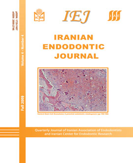
Central Giant Cell Granuloma: A Potential Endodontic Misdiagnosis

Safoura Seifi, Ramin Fouroghi

Iranian Endodontic Journal, Vol. 4 No. 4 (2009), 10 October 2009, Page 158-160
https://doi.org/10.22037/iej.v4i4.1493

Central Giant Cell Granulomas (CGCGs) may manifest as radiolucencies anywhere in the mandible or maxilla. In rare cases, it can appear as a localized periradicular area and mimic an endodontic lesion. This case report presents an uncommon location of CGCG which was not accurately diagnosed nor timely treated. Periodic follow ups of periapical radiolucencies after RCT are necessary. Dentists should include CGCG in differential diagnosis of lesions that are refractory to endodontic treatment. [Iranian Endodontic Journal 2009;4(4):158-60]