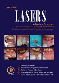
Excision of different oral benign exophitic lesions with diode laser: A clinical Case Series
Journal of Lasers in Medical Sciences,
Vol. 11 No. 4 (2020),
3 October 2020
,
Page 502-508
Abstract
Introduction: Lasers are becoming the standard of care for many dental procedures, and are being introduced as a high-tech instrument. They are also becoming more routine in dentistry through the advent of office-based lasers, which are also simple to use within the oral cavity. Many studies have shown the competencies of laser technology for the management of benign oral lesions as these techniques allow for painless and bloodless oral surgery.
Cases Report: Nine patients attending the Department of oral and maxillofacial Disease, Shahid Beheshti University of Medical Sciences, Tehran, Iran, underwent laser outpatient procedures for the surgical removal of a wide range of benign oral lesions. Regarding the history, present illnesses and clinical figures of all reported cases, the clinician ensured that the lesions were benign and after taking informed consent from every patient, he did complete the excision of lesions with diode lasers. Following the injection of local anesthesia, an 810 nm diode laser was applied for the excisional biopsy of oral lesions. The specimens were sent for histopathological evaluations and the patients were assessed on intraoperative and postoperative complications. The patients were followed up for postoperative complications at one week and 2 weeks post-treatment.
Conclusions: According to our findings, a diode laser can be a choice for the outpatient treatment of oral mucosal benign lesions as this technique provides the painless and almost bloodless treatment.
- Oral lesions
- Benign
- soft tissue
- laser
How to Cite
References
Azma E, Safavi N. Diode laser application in soft tissue oral surgery. J Lasers Med Sci. 2013;4(4):206-11.
D’Arcangelo C, Di Nardo Di Maio F, Prosperi GD, Conte E, Baldi M, Caputi S. A preliminary study of healing of diode laser versus scalpel incisions in rat oral tissue: a comparison of clinical, histological, and immunohistochemical results. Oral Surg Oral Med Oral Pathol Oral Radiol Endod. 2007;103(6):764-73. doi: 10.1016/j.tripleo.2006.08.002.
Brandon MS, Strauss RA. Complications of CO2 laser procedures in oral and maxillofacial surgery. Oral Maxillofac Surg Clin North Am. 2004;16(2):289-99. doi: 10.1016/j.coms.2004.01.005.
Suliman NM, Johannessen AC, Ali RW, Salman H, Astrøm AN. Influence of oral mucosal lesions and oral symptoms on oral health related quality of life in dermatological patients: a cross sectional study in Sudan. BMC Oral Health. 2012;12:19. doi: 10.1186/1472-6831-12-19.
Monteiro LS, Albuquerque R, Paiva A, de la Peña-Moral J, Amaral JB, Lopes CA. A comparative analysis of oral and maxillofacial pathology over a 16-year period, in the north of Portugal. Int Dent J. 2017;67(1):38-45. doi: 10.1111/idj.12258.
Tamarit-Borrás M, Delgado-Molina E, Berini-Aytés L, Gay-Escoda C. Removal of hyperplastic lesions of the oral cavity. A retrospective study of 128 cases. Med Oral Patol Oral Cir Bucal. 2005;10(2):151-62.
Madhu S. Ratre,1 Pratik A. Chaudhari,1 Shaleen Khetarpal,1 and Pratiksha Kumar . Effective management of focal reactive gingival overgrowths by diode laser: A review and report of two cases. Laser Ther. 2019; 28(4): 291–297.
Pai JB, Padma R, Divya, Malagi S, Kamath V, Shridhar A, et al. Mathews A. Excision of fibroma with diode laser: a case series. J Dent Lasers. 2014;8(1):34-38.
Ortega-Concepción D, Cano-Durán JA, Peña-Cardelles JF, Paredes-Rodríguez VM, González-Serrano J, López-Quiles J. The application of diode laser in the treatment of oral soft tissues lesions. A literature review. J Clin Exp Dent. 2017;9(7):e925-e928. doi: 10.4317/jced.53795.
Neukam FW, Stelzle F. Laser tumor treatment in oral and maxillofacial surgery. Phys Procedia. 2010;5:91-100. doi: 10.1016/j.phpro.2010.08.125.
Verma SK, Maheshwari S, Singh RK, Chaudhari PK. Laser in dentistry: An innovative tool in modern dental practice. Natl J Maxillofac Surg. 2012; 3:124-32
Pandurić DG, Bago I, Zore IF, Sušić M, Katanec D, Milenović A, et al. In: Kalantar Motamedi MH, editor. A textbook of advanced oral and maxillofacial surgery. Rijeka, Croatia: InTech; 2013. p. 341-382. doi: 10.5772/3316.
Seyyedi SA, Khashabi E, Falaki F. Laser application in periodontics. J Lasers Med Sci. 2012; 3(1):26-32.
Mazarei Sotoode S, Azimi S, Taheri SA, Asnaashari M, Kahlighi H, Rahmani S, et al. Diode laser in minor oral surgery: a case series of laser removal of different benign exophytic lesions. J Lasers Med Sci. 2015;6(3):133-138. doi:10.15171/jlms.2015.08.
Aoki A, Sasaki KM, Watanabe H, Ishikawa I. Lasers in non-surgical periodontal therapy. Periodontol 2000. 2004;36:59-97.
Ishikawa I, Aoki A, Takasaki AA, Mizutani K, Sasaki KM, Izumi Y. Application of lasers in periodontics: true innovation or myth? Periodontol 2000. 2009;50:90-126. doi: 10.1111/j.1600-0757.2008.00283.x.
Schindl M, Kerschan K, Schindl A, Schon H, Heinzl H, Schindl L. Induction of complete wound healing in recalcitrant ulcers by low-intensity laser irradiation depends on ulcer cause and size. Photodermatol Photoimmunol Photomed. 1999;15(1):18-21. doi: 10.1111/j.1600-0781.1999.tb00047.x.
Najafi S, Sheykhbahaei N, khayamzadeh M, Gholizadeh N. The effect of low-level laser on the number of Candida albicans colonies in-vitro: a new finding. BMC Oral Health. 2019;19:4. doi: 10.1186/s12903-019-0814-5.
Wyman A, Duffy S, Sweetland HM, Sharp F, Rogers K. Preliminary evaluation of a new high power diode laser. Lasers Surg Med. 1992;12(5):506-9. doi: 10.1002/lsm.1900120509.
- Abstract Viewed: 682 times
- PDF Downloaded: 436 times