Original Article


Prevalence of Referred Pain with Pulpal Origin in the Head, Face and Neck Region

Siamak Mardani, Mohammad Jafar Eghbal, Maryam Baharvand

Iranian Endodontic Journal, Vol. 3 No. 2 (2008), 2 April 2008, Page 8-10
https://doi.org/10.22037/iej.v3i2.384

INTRODUCTION: This study was designed to evaluate the prevalence of referred pain with pulpal source in the head, face and neck region among patients referred to Dental School of Shahid Beheshti University MC, Tehran, Iran in 2004. MATERIALS AND METHODS: In this cross-sectional study, 100 patients (55 males and 45 females) referred to oral medicine department of Shahid Beheshti Dental School evaluated via clinical and radiographic examination to seek their pain sources and sites. Inclusion criteria were report of pain and a dental clinician accomplished detection of pain origin. Exclusion criteria were non-odontogenic painful diseases, advanced periodontal disease, and substantial carious lesions. Visual analogue scale (VAS) was used to score pain intensity; meanwhile the patients were asked to mark the painful sites on an illustrated head and neck mannequin. RESULTS: Sixty-five percent of patients reported pain in sites which diagnostically differed from the pain source. According to statistical analysis, duration (P<0.01), spontaneity (P<0.001) and quality (P<0.01) of pain influenced its referral nature, while sex and age of patients, kind of stimulus, throbbing and intensity of pain had no considerable effect on pain referral (P>0.05). CONCLUSION: The prevalence of referred pain with pulpal origin in the head, face and neck region is moderately high which requires precise diagnosis by dental practitioners. Some hallmarks of irreversible pulpitis (e.g. spontaneous and persistent pain after elimination of stimulus) are related to pain referral.
Comparison of Canal Preparation Pattern of K3 and ProTaper Rotary Files in Curved Resin Blocks

Nahid Mohammadzade Akhlaghi, Zohreh Khalilak, Ladan Baradaran Mohajeri, Mahshid Sheikholeslami, Saeed Saedi

Iranian Endodontic Journal, Vol. 3 No. 2 (2008), 2 April 2008, Page 11-16
https://doi.org/10.22037/iej.v3i2.402

INTRODUCTION: The purpose of this study was to evaluate and compare canal preparation pattern of K3 and ProTaper rotary files in curved resin blocks. MATERIALS AND METHODS: Twenty-four resin blocks were used in this experimental study and randomly divided into two groups. Their initial images were scanned. After preparation, their images were scanned again in the same position. Pre and post preparation images were superimposed by Photoshop software and the removed resin was measured in 5 different points, and then analyzed statistically by ANOVA and T-test. RESULTS: At O point (orifice), significantly (P<0.05) more outer canal wall was removed in the ProTaper group than in the K3 group. There was no significant difference at any other points of outer wall. Removed material of inner canal wall was not significantly different between the two groups. CONCLUSION: Under the condition of this study, both systems performed acceptable preparation pattern except at the beginning of the curve.

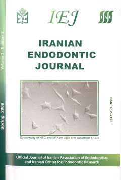
Cytotoxic effect of a new endodontic cement and mineral trioxide aggregate on L929 line culture

Jamileh Ghoddusi, Jalil Tavakkol Afshari, Zakiyeh Donyavi, Azam Brook, Reza Disfani, Mohammad Esmaeelzadeh

Iranian Endodontic Journal, Vol. 3 No. 2 (2008), 2 April 2008, Page 17-23
https://doi.org/10.22037/iej.v3i2.403

Introduction: The aim of this study was to compare the cytotoxicity of Mineral Trioxide Aggregate (MTA) and a New Endodontic Cement (NEC) on L929 mouse fibroblasts. Materials and Methods: Different dilutions (Neat, 1/2, 1/10, 1/100) of fresh and set materials placed adjacent flasks of L929 in DMEM medium. Cellular viability was assessed using MTT assay in three time intervals (24, 48, and 72 h after mixing). Differences in mean cell viability values between materials were assessed by using the One-way ANOVA and Bonferoni post-test. Optical microscopic analysis of morphology of the untreated control and the cement-treated cell cultures were carried out in all experimental periods. Results: It was indicated that there was not a significant difference in cytotoxicity among the materials of test and between them and the control group. However, there was a statistically significant difference between different time intervals within each group (P< 0.05) and between different concentration of test materials (P<0.05). In all samples, set materials showed better viability than fresh ones. Conclusion: According to results of this study, NEC and MTA have similar cytotoxic effect on L929 cell culture.

A Comparative Study on Rotary Mtwo Versus Passive Step Back of Hand K-File in Preparation of Extracted Curved Root Canals

Safi Laaya, Leila Khojastehpour, Abdol Hosein Layeghnejad, Abdol Hosein Layeghnejad

Iranian Endodontic Journal, Vol. 3 No. 2 (2008), 2 April 2008, Page 24-28
https://doi.org/10.22037/iej.v3i2.404

INTRODUCTION: The preparation of the root canal space includes debridement, shaping and apical preparation. These procedures are challenging and constitute. The recent introduction of automated techniques for canal preparation has created considerable interest. The aim of this study was to test shaping ability of one hand instrumentation (passive step-back technique) and new rotary NiTi system (Mtwo instrumentation) in curved root canals. MATERIALS AND METHODS: A total number of 40 curved molars selected for this in vitro study, 20 canals for each group. The mean of canal curvature in studied groups was similar. After preparation of access cavity, samples were divided into two groups. A custom made block was used for standardization of radiograph beam angel before and after preparation. In group 1, samples instrumented with passive step-back technique using stainless steel K-file, Gates Glidden and Pesso reamers. Mtwo NiTi rotary file and Endo IT power driven motor were used for instrumentation of samples in group 2. Transportation of canals curvature (loss of primary curvature) after canal preparation was assessed for each sample with the aid of AutoCAD 2007 software. Stereomicroscope provided data for measurement of changes in working length after canal preparation. RESULTS: The mean of changes in canals curvature (canal transportation) and mean of working length changes in group 1 (passive step-back technique) were 11.77 degree and 0.202 mm (P<0.001) and in group 2 (rotary Mtwo) were 5.58 degree and 0.202 mm (P<0.001). According to the present results, the original canal curvature was maintained significantly better with automated Mtwo files than with hand instrumentation. CONCLUSION: These can be hypothesized that the instrumentation with Mtwo NiTi rotary file preserves the canal curvature adequately.

A Comparison of Mtwo Rotary File with K-File on Negotiation of Second Mesiobuccal Canal in Maxillary First Molar: A Clinical Study

Kiamars Honardar, Nahal Vesal, Faeze Hamze, Kiumars Nazarimoghadam, Hossein Labaf, Laila Shakeri

Iranian Endodontic Journal, Vol. 3 No. 2 (2008), 2 April 2008, Page 29-32
https://doi.org/10.22037/iej.v3i2.405

INTRODUCTION: This study was carried out to compare the capability of K-files with Mtwo rotary file in terms of negotiation as well as depth of penetration in second mesiobuccal (MB2) canal of maxillary first molar. MATERIALS AND METHODS: A total of 32 MB2 canal of maxillary first molars having different root curvatures (not more than 30 degree) and root lengths were selected and then detected by K-file #10, #8 and #6. Based upon file penetration the samples were assigned into four groups. Group A: K-file #10 penetrate>2mm into coronal third, group B: K-file #10 could penetrate<2mm into coronal third, continue proceeding with #8 which had>2mm penetration, group C: K-files #10 and 8 could penetrate<2mm, continue proceeding with #6 for deeper penetration, and group D: Mtwo file #10/.04 was applied into all three above groups until resistance was felt. Finally, accurate working lengths at each group after K-file #10 insertions into the canal were determined radiographically. The mean depths of root canal penetration were analyzed statistically using Duncan test by SAS software (version 9.1) in GLM procedure. RESULTS: The mean of initial penetration for #10 Mtwo files was 19.16 mm, whilst it was 7.72 mm for K-File #6, 10.72 mm for K-File #8, and 12 mm for K-File #10. The difference between Mtwo rotary files and hand K-Files was statistically significant (P<0.01). CONCLUSION: Mtwo rotary files could be an efficient substitute for hand files to negotiate MB2 canal both more easily and rapidly. To our knowledge, it is the first time that a NiTi rotary file showed success in negotiation of any canal.

Antimicrobial Activity of Three Root Canal Irrigants on Enterococcus Faecalis: An in Vitro Study

Zohreh Ahangari, Mohammad samiee, Gita Eslami, Mohammad Amin Yolmeh

Iranian Endodontic Journal, Vol. 3 No. 2 (2008), 2 April 2008, Page 33-37
https://doi.org/10.22037/iej.v3i2.406

INTRODUCTION: The aim of this study was to compare the antimicrobial effects of 2.5% Sodium hypochlorite (NaOCl), 2% Chlorhexidine Gluconate (CHX) and BioPure MTAD (MTAD) on Enterococcus (E) faecalis-contaminated root canals of human extracted teeth. MATERIALS AND METHODS: Seventy human intact extracted single-rooted teeth with straight root canal randomly divided into 5 groups: positive control (n=5), negative control (n=5), 2.5% NaOCl (n=20), 2% CHX (n=20), and MTAD (n=20). Each tooth was instrumented using the passive step-back technique hand and rotary instruments. E. faecalis incubated into the canals and grew for 4 weeks. Canals irrigated using three mentioned solutions for 5 minutes. Samples were taken from canal walls and transferred into Brain Heart Infusion (BHI) culture medium and placed in an incubator at 37ºC for 96 hours and bacteriological evaluations were done. Chi- Square test and SPSS software were used for the statistical analysis of the results. RESULTS: Bacterial growth was seen in only one sample of MTAD group (5%), but in 4 of CHX group (20%) and 5 of NaOCl group (25%). Chi-Square test showed no statistically difference between groups. CONCLUSION: Based on the results of this study, it seems that all three solutions have acceptable antimicrobial effect on E. faecalis.

Case Report


Non-Surgical Root Canal Treatment of Dens Invaginatus 3 in a Maxillary Lateral Incisor

Saeed Moradi, Zakyeh Donyavi, Mohammad Esmaealzade

Iranian Endodontic Journal, Vol. 3 No. 2 (2008), 2 April 2008, Page 38-41
https://doi.org/10.22037/iej.v3i2.407

The aim of this case report was to describe the clinical management of an unusual dens invaginatus type 3. A case of dens invaginatus in a maxillary lateral incisor with a periapical lesion is reported. The patient presented with pain and localized swelling. Despite the complex anatomy and diagnosis of dens invaginatus, non-surgical root canal treatment was performed successfully. Key Learning Points:- Dens invaginatus may be presented in different forms, and the etiology of this phenomenon is not fully understood.

- Due to abnormal anatomical configuration, dens invaginatus presents technical difficulties in its clinical management.

- Non-surgical root canal treatment can be performed successfully.

Review Article


An update on the antibiotic-based root canal irrigation solutions

Zahed Mohammadi

Iranian Endodontic Journal, Vol. 3 No. 2 (2008), 2 April 2008, Page 1-7
https://doi.org/10.22037/iej.v3i2.383

Abstract: Antibiotics are a valuable addition to health practitioners for the management of bacterial infections. During endodontic treatment and when managing trauma to the teeth, antibiotics may be applied systemically or locally. Due to the potential risk of adverse effects of systemic applications, and the ineffectiveness of systemic prescribed antibiotics in necrotic or pulpless teeth and the periradicular tissues, the local application of antibiotics may be a more effective mode for delivering antibiotics to infected root canals. The purpose of this article is to review the history, rationale, and applications of antibiotics and antibiotic-containing irrigants in endodontics.

Hypothesis