REVIEW


Advanced Treatments in Non-Clear Renal Cell Carcinoma

El Mehdi Tazi, Ismail Essadi, Mohamed Fadl Tazi, Youness Ahellal, Hind M’Rabti, Hassan Errihani

Urology Journal, Vol. 8 No. 1 (2011), 12 March 2011, Page 1-11
https://doi.org/10.22037/uj.v8i1.923

Purpose: To focus on the use of targeted therapies against the non-clear histologic subtypes of renal cell carcinoma (RCC); papillary I and II, chromophobe, and collecting duct. The unique genetic and molecular profiles of each distinct non-clear kidney cancer subtype will be described, as these differences are integral to the development and effectiveness of the novel agents used to treat them. Materials and Methods: On the basis of MEDLINE database searches, we assessed all aspects of targeted therapy in non-clear cell RCC between 2000 and 2010. Trials focusing on non-clear RCC or those that treated clear cell tumors along with significant numbers of non-clear subtypes will be discussed. The role of cytoreductive nephrectomy and the use of neoadjuvant and adjuvant targeted therapy will be reviewed. Lastly, areas of future research will be highlighted. Results: The majority of clinical trials testing novel targeted therapies have excluded non-clear subtypes, providing limited therapeutic options for patients with these diagnoses and their oncologists. Conclusion: Patients presenting with advanced non-clear pathology should undergo a thorough metastatic evaluation and, if appropriate, surgical evaluation to determine if nephrectomy, lymphadenectomy, and/or metastectomy are warranted. Aggressive surgical extirpation is often recommended. Sunitinib also is adequately tolerated and oncologically active in subjects with non-clear histology.
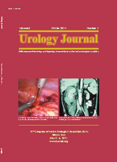
PICTORIAL UROLOGY


ORIGINAL PAPER (ENDOUROLOGY AND STONE DISEASE)


Role of Tamsulosin in Clearance of Upper Ureteral Calculi After Extracorporeal Shock Wave Lithotripsy: A Randomized Controlled Trial

Santosh Kumar Singh, Devendra Singh Pawar, Mahavir Singh Griwan, Jag Mohan Indora, Sachit Sharma

Urology Journal, Vol. 8 No. 1 (2011), 12 March 2011, Page 14-20
https://doi.org/10.22037/uj.v8i1.926

Purpose: To evaluate the role of tamsulosin in stone clearance in patients with upper ureteral stone after extracorporeal shock wave lithotripsy (SWL). Materials and Methods: This randomized controlled trial was performed on 117 patients with a single upper ureteral calculus undergoing SWL. The study group received 0.4 mg tamsulosin daily while the control group received hydration and analgesic on demand for a maximum of 3 months. Follow-up visits were performed at 1, 2, and 3 months after SWL. Efficiency of tamsulosin was evaluated in terms of success rate, time for expulsion of fragments, number of SWL sessions, incidence of steinstrasse, and pain intensity. Results: The clearance rate after 1, 2, and 3 months were higher in tamsulosin group than the control group (85%, 89.8%, and 91.5% versus 70.6%, 79.3%, and 86.2%; P = .01, P = .11, and P = .34, respectively). The mean time for expulsion of the fragments was 26.78 ± 11.96 days and 31.28 ± 18.31 days in tamsulosin and control groups, respectively (P = .138). Steinstrasse developed in 8 patients in tamsulosin group and in 13 patients in control group (P = .167). Visual analogue scale pain score was 24.92 ± 7.57 in tamsulosin group and 41.81 ± 17.24 in control group (P = .000). Conclusion: Tamsulosin helps in clearance of upper ureteral stones after 1 month of SWL, particularly stones with size of 11 to 15 mm with less

requirement of SWL sessions and analgesics.

Does Bleeding During Percutaneous Nephrolithotomy Necessitate keeping the Nephrostomy Tube? A Randomized Controlled Clinical Trial

Masoud Etemadian, Mohammad Javad Soleimani, Ramin Haghighi, Mohammad Reza Zeighami, Neda Najimi

Urology Journal, Vol. 8 No. 1 (2011), 12 March 2011, Page 21-26
https://doi.org/10.22037/uj.v8i1.927

Purpose: To compare outcomes in two groups of patients with kept and discarded nephrostomy tube after percutaneous nephrolithotomy (PCNL) complicated with bleeding. Materials and Methods: Two hundred patients who had undergone PCNL complicated with hemorrhage were recruited in this study. Patients were randomly allocated to two groups: group A, who underwent tubeless PCNL and tract port was packed for 3 to 4 minutes after removing Amplatz sheath, and group B, for whom a 24-F nephrostomy tube was left in place at the end of the procedure. Patients were followed up for 3 months to check if bleeding occurred. Results: The mean operation time was 68 ± 4.3 minutes in group A and 74 ± 5.6 minutes in group B (P = .098). The mean stone size was similar in groups A and B (36.26 ± 5.3 mm versus 35.35 ± 5.85 mm; P = .613). The mean hemoglobin drop was 3.65 ± 1.20 g/dL in group A and 3.13 ± 1.06 g/dL in group B. There was no significant difference between the mean of stonefree rate in groups A and B (92.58% ± 5.97 versus 89.60% ± 8.3; P = .210). Patients in group A experienced a significantly less duration of hospitalization than group B (2.42 ± 0.84 days versus 3.70 ± 0.80 days; P < .001). Conclusion: In the absence of clear indication, nephrostomy tube insertion after PCNL does not seem to be beneficial, and its removal does not pose patients at any additional risk.

ORIGINAL PAPER (LAPAROSCOPIC AND ROBOTIC UROLOGY)


Laparoscopic Retroperitoneal Lymph Node Dissection for Stage I Nonseminomatous Germ Cell Testis Tumors: The First Case Series in Iran

Seyed Amir Mohsen Ziaee, Ali Tabibi, Farzaneh Sharifiaghdas, Seyyed Mohammad Ghahestani, Samad Zare, Mohammad Samzadeh

Urology Journal, Vol. 8 No. 1 (2011), 12 March 2011, Page 27-30
https://doi.org/10.22037/uj.v8i1.928

Purpose: To report laparoscopic retroperitoneal lymph node dissection (RPLND) as an approach for management of low-stage nonseminomatous germ cell testis tumors (NSGCT). Materials and Methods: Between August 2002 and December 2008, 19 patients with stage I NSGCT underwent RPLND in our center. Results: Mean operation time was 340 minutes (range, 250 to 360 minutes). Procedure in 2 (10.5%) patients was converted to open. Four (21%) patients had tumors with pure histopathology while other 15 (79%) had mixed histopathology. The mean number of removed lymph nodes was 11 (range, 6 to 14). Pathology revealed lymph node involvement in 8 (42%) patients, including 6 (75%) viable tumors and 2 (25%) teratoma. After on average 47-month follow-up (range, 3 to 70 months), recurrence occurred in 2 (10.5%) patients, who underwent open retroperitoneal lymph node dissection after chemotherapy, and surgical pathology revealed teratoma in one and fibrotic tissue in another. No patient developed systematic metastasis during followup period. Conclusion: Our results show that compared with open surgery, RPLND has same oncologic outcome, but lower, and can be recommended for management of patients with low stage NSGCT.

Laparoscopic Redo Pyeloplasty After Failed Open Surgery

Pejman Shadpour, Ramin Haghighi, Robab Maghsoudi, Masoud Etemedian

Urology Journal, Vol. 8 No. 1 (2011), 12 March 2011, Page 31-37
https://doi.org/10.22037/uj.v8i1.929

Purpose: To report our experience in treating patients with failed previous open pyeloplasty by transperitoneal laparoscopic pyeloplasty. Materials and Methods: Eleven patients with previous failed open pyeloplasty were reviewed, all of whom had undergone transperitoneal laparascopic pyeloplasty. All procedures were performed by a single team. Depending on the anatomic situation, either dismembered or a flap technique was utilized. Subsequent follow-up was by ultrasonography initially, and diuretic renal scintigraphy and/or intravenous urography at least 12 months after the re-operation. Data were collected from the medical records. Results: The study group consisted of 7 men and 4 women with the mean age of 41.4 years (range, 27 to 55 years). Mean operation time was 208 minutes (range, 165 to 250 minutes) and mean hospital stay was 3.6 days (range, 3 to 5 days). Mean follow-up was 24.1 months (range, 12 to 42 months). The overall success rate for these salvage laparoscopic pyeloplasties was 90.9%. Only one female patient developed dull flank pain 3 months after stent removal. There was no conversion to open surgery. None experienced major complications or required blood transfusion. Conclusion: Laparoscopic pyeloplasty can be a valid and feasible option in treating patients with failed prior open pyeloplasty.

ORIGINAL PAPER (PEDIATRIC UROLOGY)


Evaluation of Lower Urinary Tract Symptoms in Children Exposed to Sexual Abuse

Ali Yildirim, Nihat Uluocak, Dogan Atilgan, Mustafa Ozcetin, Fikret Erdemir, Ozgur Boztepe

Urology Journal, Vol. 8 No. 1 (2011), 12 March 2011, Page 38-42
https://doi.org/10.22037/uj.v8i1.930

Purpose: To evaluate the lower urinary tract symptoms (LUTS) in children that are exposed to sexual abuse. Materials and Methods: Fifty-two patients, including 8 male and 44 female children/adolescents presented with sexual abuse to the outpatient clinics were evaluated retrospectively (group 1). In group 1, the subjects were categorized

into sexual touch (n = 35) and sexual penetration (n = 17). All the patients were evaluated with a detailed medical history, physical examination, and a dysfunctional voiding and incontinence scoring system questionnaire. Thirty age-matched children were evaluated as a control group (group 2). Results: The mean age of the patients was 12.2 ± 3.6 years and 12.0 ± 4.5 years in groups 1 and 2, respectively (P = .848). The mean age of the subjects in sexual touch and sexual penetration groups was 10.8 ± 3.6 years and 14.9 ± 1.5 years, respectively. The difference between sexual touch and sexual penetration groups was statistically significant (P = .0001). The incontinence rate was 30.76% and 23.3% in groups 1 and 2, respectively. This difference was not statistically significant (P = .640). The rates of daytime incontinence, nocturnal enuresis, diurnal incontinence, urgency, and continence maneuvers were 25.7%, 17.1%, 22.9%, 42.9%, and 20%, respectively, in sexual touch group, while they were found to be 5.9%, 0%, 0%, 17.6%, and 5.9%, respectively, in sexual penetration group. Conclusion: Although a significant association was not detected between sexual abuse and LUTS, it was seen that LUTS, such as urinary incontinence and urgency, were higher in children exposed to sexual abuse than the control group.

ORIGINAL PAPER(UROLOGICAL ONCOLOGY)


Predictive Factors for Prostatic Involvement by Transitional Cell Carcinoma of the Bladder

Ali Tabibi, Nasser Simforoosh, Mahmoud Parvin, Hamidreza Abdi, Ahmad Javaherforooshzadeh, Farhat Farrokhi, Mohammad Hossein Soltani

Urology Journal, Vol. 8 No. 1 (2011), 12 March 2011, Page 43-47
https://doi.org/10.22037/uj.v8i1.931

Purpose: To evaluate the predictive factors for prostatic involvement according to the bladder transitional cell carcinoma (TCC) characteristics in a prospective study. Materials and Methods: Hundred patients with the bladder TCC who had undergone standard radical cystoprostatectomy were enrolled in this study. A number of factors, including vascular and perineural invasion, number of tumors, maximum diameter of the tumor, presence of carcinoma in situ, distance between the tumor and the bladder neck, grade, and local stage of the tumor were recorded, and their  elationships with prostatic involvement were studied. In addition, hydronephrosis and age of the patients were included in the analysis. Results: The mean age of the patients was 62.6 ± 10.8 years. Of a total of 100 patients, 21 were found to have prostatic involvement with TCC. Univariate statistical analysis showed that vascular invasion and the distance between the tumor and the bladder neck were significantly related to the prostatic involvement (P = .001 and P < .001) and tumor stage had relatively low P value (P = .08). In the logistic regression, only the distance between the tumor and the bladder neck was found to be significantly related to the prostatic involvement (P = .004). Conclusion: This study demonstrated that distance between the tumor and the bladder neck is a predictive factor for prostatic involvement; hence, prostate-sparing or capsule-sparing cystectomy in patients with tumors in short distances from prostatovesical junction is not rationale and should be avoided.

ORIGINAL PAPER (RECONSTRUCTIVE SURGERY)


Dorsal Versus Ventral Oral Mucosal Graft Urethroplasty

Jalil Hosseini, Ali Kaviani, Mokhtar Hosseini, Mohammad Mohsen Mazloomfard, Abdollah Razi

Urology Journal, Vol. 8 No. 1 (2011), 12 March 2011, Page 48-53
https://doi.org/10.22037/uj.v8i1.932

Purpose: To evaluate success rate of dorsal versus ventral oral mucosal graft for anterior urethroplasty. Materials and Methods: In a retrospective study, the results of the ventral and dorsal oral mucosal graft (OMG) anterior urethroplasty were assessed in 24 and 29 patients, respectively. Demographic and clinical characteristics of subjects were gathered from the medical records. Results: Patients were followed up for a mean duration of 32 months (range, 25 to 51 months). The success rates of dorsal and ventral anterior OMG urethroplasty were 83.3% and 75.8%, respectively (P = .5). At penile site, dorsal and ventral OMG were done for 9 and 10 patients with stricture lengths of 3.7 ± 1.1 cm and 3.9 ± 1.2 cm, respectively (P = .7). Success rates of dorsal and ventral penile OMG were 88.9% and 70%, respectively (P = .3). At the bulbar site, dorsal and ventral OMG were performed on 15 and 19 patients with stricture lengths of 4.1 ± 1.1cm and 4.2 ± 1.5 cm, respectively (P = .7). The success rates of dorsal and ventral bulbar OMG were 80% and 79%, respectively (P = .94). Conclusion: Oral mucosal graft is a versatile and an effective procedure for management of strictures throughout the anterior urethra, and in experienced hands, the outcomes are similarly favorable whether a dorsal or ventral approach is taken.

ORIGINAL PAPER (CELLULAR AND MOLECULAR UROLOGY)


Isolation of Human Adult Stem Cells from Muscle Biopsy for Future Treatment of Urinary Incontinence

Farzaneh Sharifiaghdas, Maryam Taheri, Reza Moghadasali

Urology Journal, Vol. 8 No. 1 (2011), 12 March 2011, Page 54-59
https://doi.org/10.22037/uj.v8i1.933

Purpose: To find a suitable and cost-effective technique for isolation and culture of muscle-derived stem cells (MDSCs) obtained from muscle biopsy in large quantities. Materials and Methods: A small muscle biopsy was taken from 10 donor rectus muscles in patients undergoing open abdominal surgery for any reason and transported on ice to the laboratory. The isolation of MDSCs was performed by two techniques; preplate and tissue explants. Initially, the isolation was carried out by preplating technique. However, enzymatic digestion of muscle biopsy in preplate technique compromised the integrity of important surface antigens of resident muscle stem cells and led to dysfunctional sorted cells. Also, many of the cells were lost in this technique and low numbers of MDSCs were yielded upon processing. Thus, we changed condition of centrifuge, but it did not affect cell numbers and their integrities. To overcome these problems, the technique was changed to tissue explants technique. Results: During the first 4 days in explant medium culture, activated satellite cells detached, migrated, and slowly divided. The MDSCs proliferated around the native myofiber and after 2 to 3 weeks, individual muscle cells appeared elongated and fused to create large multinucleated myotubes. On

immunofluorescent staining, these emerged cells were positive for desmin and Pax7 and flow cytometry analysis revealed that these cells were CD45-, CD56+, and variable in CD34. Conclusion: We concluded that tissue explant method is a suitable and costeffective technique for isolation and culture of MDSCs from muscle biopsy in large quantities.

UNCLASSIFIED


Role of Color Doppler Ultrasonography in Evaluation of Scrotal Swellings: Pattern of Disease in 120 Patients With Review of Literature

Syed Amjad Ali Rizvi, Ibne Ahmad, Mohammed Azfar Siddiqui, Samreen Zaheer, Kaleem Ahmad

Urology Journal, Vol. 8 No. 1 (2011), 12 March 2011, Page 60-65
https://doi.org/10.22037/uj.v8i1.934

Purpose: To determine the value of color Doppler ultrasonography (CDUS) as a routine investigational method for diagnosis of scrotal pathologies. Materials and Methods: This prospective observational study (case series)

was carried out over a period of 16 months on 122 patients in the age range of 13 to 70 years old, who presented with scrotal swellings. After adequate history taking and examination, CDUS was performed. The diagnosis of the surgeon and that of radiologist were compared with final outcome, which was based on course and outcome of the disease, fine needle aspiration cytology results, and operative findings. Results: The final diagnoses were epididymitis or epididymo-orchitis (46), hydrocele (26), varicocele (16), testicular malignancy (16), orchitis (6), testicular torsion (4), spermatic cord injury (2), hematocele (2), and pyocele (2). Color Doppler ultrasonography accurately diagnosed all cases of epididymitis or epididymo-orchitis, spermatic cord injury, testicular torsion, varicocele, and hydrocele (sensitivity 100% and specificity 100%). Of 16 subjects diagnosed as testicular malignancy on CDUS, only 14 were subsequently found to have

malignancy. Two cases of orchitis were wrongly diagnosed as malignancy. Similarly, of 6 patients diagnosed as orchitis, 1 was found to have seminoma (sensitivity 87.5% and specificity 66.7%). Overall sensitivity of CDUS in diagnosing scrotal diseases was 98% while specificity was 66.7%. Conclusion: Color Doppler ultrasonography is an excellent, a safe, and reliable method for evaluating patients with scrotal diseases. It aids in diagnosis of testicular tumors and reduces the number of unnecessary exploratory operations. It is especially important in conditions like testicular torsion

where immediate diagnosis is required.

POINT OF TECHNIQUE


CASE REPORT


UROLOGY FOR PEOPLE


NEWS