Iranian Center for Endodontic Research
  • Login
  • Register

Iranian Endodontic Journal

  • Register
  • Home
  • About the journal
  • Issues
    • Current
    • Archives
  • For Authors/Reviewers
    • Submissions
    • Author Guidelines
    • Ethics Policy
    • Editorial & Peer Review Policy
    • Privacy Statement
    • Reviewers Information
  • Indexing/Abstracting
  • Editorial Team
  • Announcements
  • Contact Us
  • Congress Proceedings
Advanced Search
  1. Home
  2. Archives
  3. Vol. 1 No. 2 (2006): SUMMER 2006
  4. Original Article

Vol. 1 No. 2 (2006)

Tir 2006

Scanning Electron Microscopy Study of Dental Gutta-Percha after Cutting

  • Saeed Asgary
  • Masoud Parirokh
  • Mohammad Jafar Eghbal
  • Jamileh Ghoddusi

Iranian Endodontic Journal, Vol. 1 No. 2 (2006), 1 Tir 2006 , Page 57-59
https://doi.org/10.22037/iej.v1i2.413 Published: 2008-12-07

  • View Article
  • Download
  • Cite
  • Statastics
  • Share

Abstract

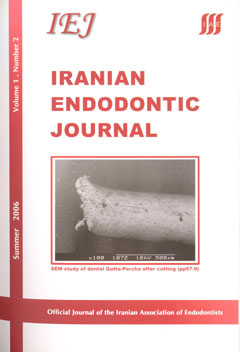
INTRODUCTION: The purpose of this study was to evaluate the morphologic surface of gutta­percha cones after cutting with different methods. MATERIALS AND METHODS: The apical 3 millimeters of forty standardized, gutta-percha cones size 40 were cut off using scissors or a scalpel against a glass slab. The samples were then examined under scanning electron microscopy (SEM) for topographic deformity. RESULTS: According to results, cutting with scissors produced significant topographic deformity in the standardized gutta-percha cone surface but cutting with sharp surgical instrument against a glass slab allowed the development of a smooth and unmodified gutta­percha cone surface. CONCLUSION: Results of this study recommended that cutting the tip of a gutta­percha point with a sharp scalpel against a glass slab would produce more reasonable surface morphology than using scissor for the same procedure.

Keywords:
  • Cutting
  • Gutta-Percha
  • SEM
  • PDF

How to Cite

1.
Asgary S, Parirokh M, Eghbal MJ, Ghoddusi J. Scanning Electron Microscopy Study of Dental Gutta-Percha after Cutting. Iran Endod J [Internet]. 2008 Dec. 7 [cited 2025 May 21];1(2):57-9. Available from: https://journals.sbmu.ac.ir/iej/article/view/413
  • ACM
  • ACS
  • APA
  • ABNT
  • Chicago
  • Harvard
  • IEEE
  • MLA
  • Turabian
  • Vancouver
  • Endnote/Zotero/Mendeley (RIS)
  • BibTeX
  • Abstract Viewed: 264 times
  • PDF Downloaded: 256 times

Download Statastics

  • Linkedin
  • Twitter
  • Facebook
  • Google Plus
  • Telegram

Make a Submission

Make a Submission

Information

  • For Readers
  • For Authors
  • For Librarians

Developed By

Open Journal Systems

Indexing/Abstracting

This journal is indexed in:

  • PubMed
  • Europe PMC
  • Scopus
  • SCImago
  • MIAR
  • CINAHL
  • CAS (Chemical Abstracts)
  • Dimensions
  • DOAJ
  • EBSCO
  • FATCAT
  • Google Scholar
  • IMEMR (Index Medicus for the WHO Eastern Mediterranean Region)
  • Index Copernicus
  • ISC (Islamic World Science Citation Center)
  • Magiran
  • ROAD
  • SID (Scientific Information Database) 
  • SUDOC
  • UC Santa Barbara University
  • WIKIDATA
  • ZDB
    • This journal is Member of: COPE; ICMJ

Current Issue

  • Atom logo
  • RSS2 logo
  • RSS1 logo

Browse

  • Home
  • Archives
  • Submissions
  • About the Journal
  • Editorial Team
  • Contact

   Iranian Endodontic Journal is licensed under the Creative Commons Attribution-NonCommercial-ShareAlike 4.0 International (CC BY-NC-SA 4.0).

Powered by OJSPlus