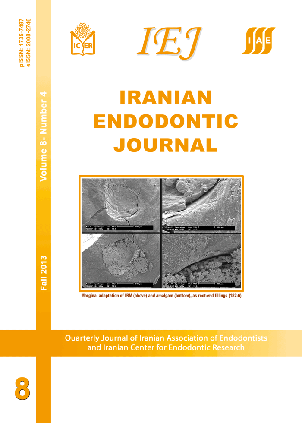
Evaluation of Marginal Adaptation of Root-End Filling Materials Using Scanning Electron Microscopy
Iranian Endodontic Journal,
Vol. 8 No. 4 (2013),
7 Mehr 2013
,
Page 182-186
https://doi.org/10.22037/iej.v8i4.3160
Abstract
Introduction: The importance of perfect apical seal in endodontics, more specifically in periradicular surgery, is the motivation/reason for development of root-end filling materials with favorable physical, chemical and biological characteristics. The aim of this in vitro study was to evaluate the marginal adaptation of root-end filling materials using scanning electron microscopy. Materials and Methods: Twenty five human maxillary anterior teeth were prepared using a K-File #50 to 1 mm short of the apical foramen and filled with gutta-percha and Sealapex using the lateral compaction technique. The apical 3 mm of the roots were sectioned perpendicularly to the long axis of the teeth. A 3-mm-deep root-end cavity was prepared using ultrasonic tips powered by an Enac ultrasonic unit. The teeth were randomly assigned to five groups according to the materials tested including IRM, amalgam, ProRoot MTA, Super-EBA and Epiphany/Resilon. Root-end cavities were filled with the materials prepared according to the manufacturers’ instructions. The root apices were carefully prepared for sputter coating and later evaluation using Scanning Electron Microscope (SEM). The images of root-end fillings were divided into four quadrants and distributed into five categories according to the level of marginal adaptation between the root-end material and the root canal walls. The Fisher exact test with Bonferroni correction was used for statistical analysis. The level of significance was set at P=0.005. Results: SEM images showed the presence of gaps in the root-end filling materials. No significant difference was observed between the tested materials (P>0.005). Conclusion: ProRoot MTA, IRM, amalgam, Super-EBA and Epiphany/Resilon showed similar marginal adaptation as root-end filling materials.
Keywords:
- Marginal Adaptation
- MTA
- Root Canal Filling Materials
- Scanning Electron Microscopy
How to Cite
1.
Oliveira HF, Alencar AHG, Figueiredo JAP, Guedes OA, Decurcio D de A, Estrela C. Evaluation of Marginal Adaptation of Root-End Filling Materials Using Scanning Electron Microscopy. Iran Endod J [Internet]. 2013 Oct. 7 [cited 2026 Sep. 30];8(4):182-6. Available from: https://journals.sbmu.ac.ir/iej/article/view/3160
- Abstract Viewed: 441 times
- PDF Downloaded: 416 times
Download Statastics
Make a Submission
Information
Developed By
Indexing/Abstracting
This journal is indexed in:
- PubMed
- Europe PMC
- Scopus
- SCImago
- MIAR
- CINAHL
- CAS (Chemical Abstracts)
- Dimensions
- DOAJ
- EBSCO
- FATCAT
- Google Scholar
- IMEMR (Index Medicus for the WHO Eastern Mediterranean Region)
- Index Copernicus
- ISC (Islamic World Science Citation Center)
- Magiran
- ROAD
- SID (Scientific Information Database)
- SUDOC
- UC Santa Barbara University
- WIKIDATA
- ZDB


