Cytotoxicity and Effects of a New Calcium Hydroxide Nanoparticle Material on Production of Reactive Oxygen Species by LPS-Stimulated Dental Pulp Cells
Iranian Endodontic Journal,
Vol. 15 No. 4 (2020),
10 October 2020
,
Page 227-235
https://doi.org/10.22037/iej.v15i4.28942
Abstract
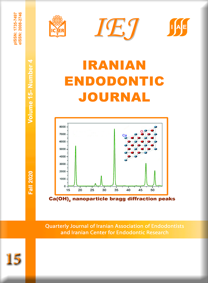
Introduction: The aim of this study was to synthesize and characterize calcium hydroxide (CH) nanoparticles [CH-NP] and compare the cytotoxicity of these materials with that of mineral trioxide aggregate (White MTA) in human dental pulp mesenchymal cells (hDPMCs) stimulated by lipopolysaccharide (LPS). Methods and Materials: The CH-NP were synthesized by the co-precipitation method, and the physical properties were investigated through X-ray diffraction, scanning electron microscopy (SEM) and energy dispersive x-ray spectrometry (EDS). LPS-stimulated hDPMCs were placed in contact with different dilutions of culture media previously exposed to CH-NP and white MTA for 24 h. The groups were tested for cell viability by MTT formazan and Alamar Blue assays, the production of nitric oxide (NO) by Griess method and the production of reactive oxygen species (ROS) by means of the fluorescent oxidant-sensing probe 2’,7’-dichlorodihydrofluorescein diacetate (DCFH-DA). Control groups for viability test were maintained in DMEM (not LPS-stimulated). For NO and ROS production, negative control group was cells in DMEM, and positive control was cells stimulated by LPS. The results were statistically analyzed by two-way ANOVA, Tukey’s test and Dunnett’s test (ɑ=0.05). Results: The results showed that the cell viability remained above 50% in all materials, independent of the dilution in MTT formazan and Alamar Blue tests. MTA showed a reduction in NO production at dilutions of 1:4 to 1:32 compared with the positive control group (P<0.05). The tested materials exhibited lower ROS production by DPMCs than that by cells in the positive control group (P<0.05), and similar ROS production to the negative control group (P>0.05). Conclusion: The outcomes of present in vitro study showed that MTA and [CH-NP] were not cytotoxic materials, with MTA closer to the results of control group (DMEM). MTA and [CH-NP] reduced ROS production at basal levels, with MTA inhibiting NO production at higher dilutions.
- Calcium Hydroxide; Cell Survival; Lipopolysaccharides; Mineral Trioxide Aggregate; Nanoparticles
How to Cite
References
Gandolfi MG, Siboni F, Botero T, Bossù M, Riccitiello F, Prati C. Calcium silicate and calcium hydroxide materials for pulp capping: Biointeractivity, porosity, solubility and bioactivity of current formulations. J Appl Biomater Funct Mater. 2015; 13(1):43-60
Kim D-H, Jang J-H, Lee B-N, Chang H-S, Hwang I-N, Oh W-M, et al. Anti-inflammatory and mineralization effects of ProRoot MTA and Endocem MTA in studies of human and rat dental pulps in vitro and in vivo. J Endod. 2018;44:1534–41.
Parirokh M, Torabinejad M. Mineral Trioxide Aggregate: A Comprehensive Literature Review-Part III: Clinical Applications, Drawbacks, and Mechanism of Action. J. Endod. 2010; 36(3):400-13.
Li Z, Cao L, Fan M, Xu Q. Direct Pulp Capping with Calcium Hydroxide or Mineral Trioxide Aggregate: A Meta-analysis. J. Endod. 2015; 41(9):1412-7.
Ioannidis K, Mistakidis I, Beltes P, Karagiannis V. Spectrophotometric analysis of coronal discolouration induced by grey and white MTA. Int Endod J. 2013; 46(2):137-44.
Lenherr P, Allgayer N, Weiger R, Filippi A, Attin T, Krastl G. Tooth discoloration induced by endodontic materials: A laboratory study. Int Endod J. 2012; 45(10):942-9.
Parirokh M, Torabinejad M, Dummer PMH. Mineral trioxide aggregate and other bioactive endodontic cements: an updated overview – part I: vital pulp therapy. Int. Endod. J. 2018; 51(2):177-205.
Hilton TJ. Keys to Clinical Success with Pulp Capping: A Review of the Literature. Oper Dent. 2009; 34(5):615-25.
L. C, B.I. S. Cytotoxicity and biocompatibility of resin-free and resin-modified direct pulp capping materials: A state-of-the-art review. Dent Mater J. 2017; 36(1):1-7.
Wu T, Xu C, Du R, Wen Y, Chang J, Huan Z, et al. Effects of silicate-based composite material on the proliferation and mineralization behaviors of human dental pulp cells: An in vitro assessment. Dent Mater J. 2018; 37(6):889-896.
Sangwan P, Sangwan A, Duhan J, Rohilla A. Tertiary dentinogenesis with calcium hydroxide: A review of proposed mechanisms. Int Endod J. 2013; 46(1):3-19.
Nowicka A, Wilk G, Lipski M, Kołecki J, Buczkowska-Radlińska J. Tomographic Evaluation of Reparative Dentin Formation after Direct Pulp Capping with Ca(OH)2, MTA, Biodentine, and Dentin Bonding System in Human Teeth. J Endod. 2015; 41(8):1234-40.
Costa E Silva LL, Cosme-Silva L, Sakai VT, Lopes CS, da Silveira APP, Moretti Neto RT, et al. Comparison between calcium hydroxide mixtures and mineral trioxide aggregate in primary teeth pulpotomy: A randomized controlled trial. J Appl Oral Sci. 2019; 27:e20180030.
Dianat O, Saedi S, Kazem M, Alam M. Antimicrobial activity of nanoparticle calcium hydroxide against enterococcus faecalis: An in vitro study. Iran Endod J. 2015; 10(1):39-43.
Ozorio JEV, De Oliveira E Silva Carvalho LF, De Oliveira DA, De Sousa-Neto MD, Da Cruz Perez DE. Standardized propolis extract and calcium hydroxide as pulpotomy agents in primary pig teeth. J Dent Child. 2012; 79(2):53-8.
Louwakul P, Saelo A, Khemaleelakul S. Efficacy of calcium oxide and calcium hydroxide nanoparticles on the elimination of Enterococcus faecalis in human root dentin. Clin Oral Investig. 2017; 21(3):865-871.
Akhavan Zanjani V, Tabari K, Sheikh-Al-Eslamian SM, Abrandabadi AN. Physiochemical properties of experimental nano-hybrid MTA. J Med Life. 2017; 10(3):182-187.
Padovani GC, Feitosa VP, Sauro S, Tay FR, Durán G, Paula AJ, et al. Advances in Dental Materials through Nanotechnology: Facts, Perspectives and Toxicological Aspects. Trends Biotechnol. 2015; 33(11):621-636.
Huang YW, Cambre M, Lee HJ. The Toxicity of Nanoparticles Depends on Multiple Molecular and Physicochemical Mechanisms. Int. J. Mol. Sci. 2017; 18(12). pii: E2702.
Rodrigues EM, Cornélio ALG, Mestieri LB, Fuentes ASC, Salles LP, Rossa-Junior C, et al. Human dental pulp cells response to mineral trioxide aggregate (MTA) and MTA Plus: cytotoxicity and gene expression analysis. Int Endod J. 2017; 50(8):780-789.
Collado-González M, García-Bernal D, Oñate-Sánchez RE, Ortolani-Seltenerich PS, Álvarez-Muro T, Lozano A, et al. Cytotoxicity and bioactivity of various pulpotomy materials on stem cells from human exfoliated primary teeth. Int Endod J. 2017; 50 Suppl 2:e19-e30.
Gordon T, Perlstein B, Houbara O, Felner I, Banin E, Margel S. Synthesis and characterization of zinc/iron oxide composite nanoparticles and their antibacterial properties. Colloids Surfaces A Physicochem Eng Asp. 2011; 347, 1–8.
A.C.A. Silva, M.A.P Zóia, L.I.V. Correia, F.V.P.V. Azevedo, A.T. Paula, L.P. Maia, L.S. Carvalho, L.N. Carvalho, M.P.C. Costa NOD. Biocompatibility of doped semiconductors nanocrystals and nanocomposites. Cytotoxicity. 2018. p. 149–61.
Woźniak A, Malankowska A, Nowaczyk G, Grześkowiak BF, Tuśnio K, Słomski R, et al. Size and shape-dependent cytotoxicity profile of gold nanoparticles for biomedical applications. J Mater Sci Mater Med. 2017; 28(6):92.
Koua L, Sun J, Zhai Y, He Z. The endocytosis and intracellular fate of nanomedicines: Implication for rational design. Asian J Pharm Sci. 2013; 8, 1–10.
Bin C V., Valera MC, Camargo SEA, Rabelo SB, Silva GO, Balducci I, et al. Cytotoxicity and genotoxicity of root canal sealers based on mineral trioxide aggregate. J Endod. 2012; 38(4):495-500.
Mestieri LB, Gomes-Cornélio AL, Rodrigues EM, Salles LP, Bosso-Martelo R, Guerreiro-Tanomaru JM, et al. Biocompatibility and bioactivity of calcium silicatebased endodontic sealers in human dental pulp cells. J Appl Oral Sci. 2015; 23(5):467-71.
Cintra LTA, Benetti F, de Azevedo Queiroz ÍO, Ferreira LL, Massunari L, Bueno CRE, et al. Evaluation of the Cytotoxicity and Biocompatibility of New Resin Epoxy–based Endodontic Sealer Containing Calcium Hydroxide. J Endod. 2017; 43(12):2088-2092.
ISO/EN10993-5. ISO 10993-5 Biological evaluation of medical devices - Part 5: Tests for cytotoxicity: in vitro methods. Int Stand ISO. 2009.
Victoria-Escandell A, Ibañez-Cabellos JS, De Cutanda SBS, Berenguer-Pascual E, Beltrán-García J, García-López E, et al. Cellular responses in human dental pulp stem cells treated with three endodontic materials. Stem Cells Int. 2017; 2017:8920356.
Ledesma-Martínez E, Mendoza-Núñez VM, Santiago-Osorio E. Mesenchymal stem cells derived from dental pulp: A review. Stem Cells Int. 2016; 2016:4709572.
Montoro LA, Turrioni APS, Basso FG, de Souza Costa CA, Hebling J. Infrared LED irradiation photobiomodulation of oxidative stress in human dental pulp cells. Int Endod J. 2014; 47:747–55.
Min KS, Chang HS, Bae JM, Park SH, Hong CU, Kim EC. The Induction of Heme Oxygenase-1 Modulates Bismuth Oxide-induced Cytotoxicity in Human Dental Pulp Cells. J Endod. 2007; 33(11):1342-6.
Mosmann T. Rapid colorimetric assay for cellular growth and survival: application to proliferation and cytotoxicity assays. J Immunol Methods. 1983;65:55–63.
O’Brien J, Wilson I, Orton T, Pognan F. Investigation of the Alamar Blue (resazurin) fluorescent dye for the assessment of mammalian cell cytotoxicity. Eur J Biochem. 2000; 267:5421–6.
Takita T, Hayashi M, Takeichi O, Ogiso B, Suzuki N, Otsuka K, et al. Effect of mineral trioxide aggregate on proliferation of cultured human dental pulp cells. Int Endod J. 2006; 39(5):415-22.
Pedano MS, Li X, Li S, Sun Z, Cokic SM, Putzeys E, et al. Freshly-mixed and setting calcium-silicate cements stimulate human dental pulp cells. Dent Mater. 2018; 34(5):797-808.
Guven EP, Yalvac ME, Sahin F, Yazici MM, Rizvanov AA, Bayirli G. Effect of dental materials calcium hydroxide-containing cement, mineral trioxide aggregate, and enamel matrix derivative on proliferation and differentiation of human tooth germ stem cells. J Endod. 2011; 37(5):650-6.
Poggio C, Ceci M, Dagna A, Beltrami R, Colombo M, Chiesa M. In vitro cytotoxicity evaluation of different pulp capping materials: A comparative study. Arh Hig Rada Toksikol. 2015; 66(3):181-8.
Valdiglesias V, Kiliç G, Costa C, Fernández-Bertólez N, Pásaro E, Teixeira JP, et al. Effects of iron oxide nanoparticles: cytotoxicity, genotoxicity, developmental toxicity, and neurotoxicity. Environ Mol Mutagen. 2015; 56:125–48.
Korhonen R, Lahti A, Kankaanranta H, Moilanen E. Nitric oxide production and signaling in inflammation. Curr Drug Targets Inflamm Allergy. 2005; 4:471–9.
Guzik TJ, Korbut R, Adamek-Guzik T. Nitric oxide and superoxide in inflammation and immune regulation. J. Physiol. Pharmacol. 2003; 54(4):469-87.
da Silva RAB, Leonardo MR, da Silva LAB, Faccioli LH, Medeiros AI de. Effect of a calcium hydroxide-based paste associated to chlorhexidine on RAW 264.7 macrophage cell line culture. Oral Surgery, Oral Med Oral Pathol Oral Radiol Endodontology. 2008; 106(5):e44-51.
Zorov DB, Juhaszova M, Sollott SJ. Mitochondrial reactive oxygen species (ROS) and ROS-induced ROS release. Physiol. Rev. 2014; 94(3):909-50.
Minamikawa H, Yamada M, Deyama Y, Suzuki K, Kaga M, Yawaka Y, et al. Effect of N-acetylcysteine on rat dental pulp cells cultured on mineral trioxide aggregate. J Endod. 2011; 37(5):637-41.
Yamada M, Tsukimura N, Ikeda T, Sugita Y, Att W, Kojima N, et al. N-acetyl cysteine as an osteogenesis-enhancing molecule for bone regeneration. Biomaterials. 2013; 34(26):6147-56.
- Abstract Viewed: 713 times
- PDF Downloaded: 467 times
Download Statastics
Make a Submission
Information
Developed By
Indexing/Abstracting
This journal is indexed in:
- PubMed
- Europe PMC
- Scopus
- SCImago
- MIAR
- CINAHL
- CAS (Chemical Abstracts)
- Dimensions
- DOAJ
- EBSCO
- FATCAT
- Google Scholar
- IMEMR (Index Medicus for the WHO Eastern Mediterranean Region)
- Index Copernicus
- ISC (Islamic World Science Citation Center)
- Magiran
- ROAD
- SID (Scientific Information Database)
- SUDOC
- UC Santa Barbara University
- WIKIDATA
- ZDB


