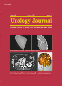
Evaluation of Urinary Stones Ex Vivo With Micro-Computed Tomography: Preliminary Results of an Investigational Technique
Urology Journal,
Vol. 8 No. 3 (2011),
5 September 2011
,
Page 185-190
https://doi.org/10.22037/uj.v8i3.1130
Abstract
Purpose: To evaluate the ultrastructural features of the urinary stones removed with endoscopic stone surgery, using micro computed tomography (micro-CT). Materials and Methods: Patients who had endoscopic surgery for renal or ureteral stones removal were included in this study. After surgery, the stones were classified into three groups and investigated with Skyscan 1174 micro-CT. Group I underwent percutaneous nephrolithotomy (PNL) with ultrasonic lithotripsy; group II had ureteroscopic stone surgery (USS) with pneumatic lithotripsy; and group III (the control group) had stone removal with USS or PNL without lithotripsy. Stone homogeneity, voids, and the internal structure of the stones were evaluated. Chi-square test was used to evaluate the difference statistically. P values less than .05 were considered statistically significant. Results: A total of 24 "calcium oxalate monohydrate" stones from 24 patients were scanned with micro-CT. Stones treated with ultrasonic lithotripsy (group I) were more fragile, fragmented, and cracked than those treated with the pneumatic lithotripsy (group II; P = .01). Stones in group II were more homogeneous and smooth than those in group I and resembled those of the control group (P = .02). Homogeneous, non-fragile stones and heterogeneous, fragile calculi were seen in all groups. Conclusion: The stone fragility could be confirmed by micro-CT investigation. Ultrasonic lithotripters increase the stone fragility, which is demonstrated with increased heterogeneity by micro-CT.
How to Cite
- Abstract Viewed: 317 times
- ES Downloaded: 338 times