Original Article


The Effect of Different Suture Removal Time Intervals on Surgical Wound Healing

Masoud Parirokh, Saeed Asgary, Mohammad Jafar Eghbal

Iranian Endodontic Journal, Vol. 1 No. 3 (2006), 1 October 2006, Page 81-86
https://doi.org/10.22037/iej.v1i3.438

INTRODUCTION: This study was carried out to compare the effect of different suture removal time on surgical wound healing. MATERIALS AND METHODS: Twenty-one male albino rabbits were used. Under general and local anesthesia a moucoperiosteal rectangular flap was raised in each animal and then repositioned and sutured. The animals were randomly divided into three experimental groups of seven animals each. In group I and II the sutures were removed after 3 and 5 days respectively and were followed up for 7 and 14 days after surgery. In group III the sutures were removed after 7 days and were followed up for 14 days after surgery. Tissue reactions were observed and recorded using inflammation and gingival indexes at 7 and 14 days after surgery in all three groups. Inflammation and gingival indexes were analyzed by Kurskal Wallis, Firedman and Wilcoxone tests. RESULTS: Results showed that inflammation index was significantly different with two other groups at the day 7 after surgery (P<0.008). Gingival index in group II was significantly different from two other groups at the day 14 (P<0.028); however, there was no significant difference between group II and III at the same interval. CONCLUSION: Based on result of this study, 5 days was recognized to the best time interval for suture removal in comparison with two other time intervals.
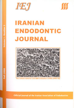
The Cellular Behavior and SEM Evaluation of ProRoot and Root MTAs on Fibroblast L929

Fariborz Moazami, Samira Shahsiah

Iranian Endodontic Journal, Vol. 1 No. 3 (2006), 1 October 2006, Page 87-92
https://doi.org/10.22037/iej.v1i3.440

INTRODUCTION: Mineral trioxide aggregate is being widely used for root end filling, pulp capping, perforation repair, and other endodontic procedures. Recently, a material similar to ProRoot mineral trioxide aggregate (PMTA) was developed in Iran named Root mineral trioxide aggregate (RMTA) with the claim of having the exact result of original MTA. The purpose of this study was to compare the amount of cell cytotoxicity of RMTA with PMTA at three different time periods using scanning electron microscopy (SEM) as well as the amount of cell viability at the above mentioned period. MATERIALS AND METHODS: Three culture plates in each group were packed with a homogenous layer of PMTA and RMTA prepared according to manufacturers instruction. A plate of media without any material was used as control in each group. The material set for 72 h in CO2 incubator and 2x10 of fibroblast L929 was added to each plate. SEM evaluation with x800-3000 magnification and cell viability counting using trepan blue counting method were done after 48, 72, and 168 hours. RESULTS: There was no significant difference between cell viability of PMTA and RMTA, although the amount of cells remained viable in PMTA group was higher at 48 and 168 hours while for RMTA, it was higher after 72 hours. The SEM evaluation showed that PMTA compared with RMTA has less porosity, but relatively similar amount of cell coverage was detected for both materials after 168 hours. CONCLUSION: ProRoot and Root MTAs showed comparative biocompatibility while evaluated in vitro. The results suggest that RMTA can be used as an alternative for PMTA in clinical trials.

Bacterial Leakage of Mineral Trioxide Aggregates and Portland Cement

Mohammad Asnaashari, Saeed Asgary, Asef Khatami

Iranian Endodontic Journal, Vol. 1 No. 3 (2006), 1 October 2006, Page 93-96
https://doi.org/10.22037/iej.v1i3.441

INTRODUCTION: The purpose of this study was to compare the ability of ProRoot mineral trioxide aggregate (PMTA) and Root mineral trioxide aggregate (RMTA) as root-end filling materials, and Portland cement (PC) to prevent bacterial leakage through filled root end cavities. MATERIALS AND METHODS: Fifty-one extracted human single rooted teeth were cleaned and shaped using a step back technique. The root-ends were resected and a 4 mm deep root-end preparation was made with fissure bur. The teeth were randomly divided into three experimental groups (n=15) and a further six teeth served as controls. Three root-end cavities were filled with gutta-percha without a root canal sealer (positive control) and three remaining were filled with sticky wax, covered with two layers of nail polish (negative control). Root-end cavities in each experimental group were filled with PMTA, RMTA or PC. After attaching the teeth to plastic caps of 9 ml vials, the teeth and the caps were sterilized using Gamma ray. Then the caps with teeth were placed on the vials containing Phenol Red Lactose broth. A tenth of microliter of Tripticase Soy broth containing Staphylococcus Epidermidis (SE) was placed into the root canals of the teeth. Every 48 hours inoculation of 0.1 ml of the SE broth culture into each root canal was performed. RESULTS: All positive controls leaked within 3 days, none of the negative controls leaked. Bacterial leakage occurred in 33% of samples in the PMTA group and in 40% of samples in RMTA and PC groups. The results indicated no statistical difference between three test materials after 35 days. CONCLUSION: It was concluded that PMTA, RMTA and PC demonstrated a similar ability to seal root end cavities.

Clinical and Radiographic Evaluation of Procedural Accidents and Errors during Root Canal Therapy

Mohammad Ali Mozayeni, Mohammad Asnaashari, Seyed Jalil Modaresi

Iranian Endodontic Journal, Vol. 1 No. 3 (2006), 1 October 2006, Page 97-100
https://doi.org/10.22037/iej.v1i3.443

INTRODUCTION: Root canal therapy (RCT)_like other dental practices_ can be accompanied with some accidents or unpredictable conditions that are called "procedural accidents". Having the knowledge about these accidents and their etiology is essential to have RCT completion and to prevent the repeat of these accidents. This study was designed to evaluate accidents occurring during RCT in patients referred to endodontic department of Shahid Beheshti dental school during 2002. MATERIALS AND METHODS: This study was conducted via descriptive method. Data were collected from observation, clinical examination and oral radiographs, and were recorded in questionnaires, 150 cases from the patients referred to endodontic department were selected randomly and different observed RCT errors were analyzed by SPSS software. The Chi-square and Fisher exact tests were used for analysis. RESULTS: The study showed that 101 patients (67.3%) had one type of RCT errors, and remaining (32.7%) were error free. From the errors studied the most prevalent were "void" which was observed in 41 patients (27.3%), and "ledge" in 39 patients (26%) respectively. The prevalence of other accidents were underfilling in 35 patients (23.3%), poor shaping in 30 patients (20%), overfilling in 23 patients (15.3%), transportation in 13 patients (8.7%), zipping in 3 patients (2%), gouging in 1 patients (0.7%), and strip perforation in 1 patients (0.7%), while no case of broken instruments, vertical fracture, furcation and cervical perforation was observed. CONCLUSION: The most prevalent errors were found in instrumentation and obturation steps, therefore more care and attention must be paid to instructor observation and the education of these stages.

Sealing Ability of Three Commercial Mineral Trioxide Aggregates and an Experimental Root-End Filling Material

Saeed Asgary, Mohammad Jafar Eghbal, Masoud Parirokh, Hassan Torabzadeh

Iranian Endodontic Journal, Vol. 1 No. 3 (2006), 1 October 2006, Page 101-105
https://doi.org/10.22037/iej.v1i3.446

INTRODUCTION: The purpose of this study was to compare the sealability of three different commercial types of mineral trioxide aggregate (MTA) and calcium enrichment mixture (CEM) cement as an experimental root-end filling material. MATERIALS AND METHODS: Forty-six single rooted teeth were cleaned, shaped, and obturated. The apical 3 mm of each root was resected and root-end cavities with 3 mm depth were prepared. The samples were randomly divided into 4 experimental groups comprised of 10 roots each. The cavities were filled with CEM cement and MTAs. Six roots were used as positive and negative controls. Samples were prepared and then immersed in 1% methylene blue dye for 3 days. Roots were split longitudinally and examined under stereomicroscope. RESULTS: Positive and negative controls responded as expected. CEM cement showed the least mean dye penetration value. ANOVA revealed no statistically significant differences among experimental groups. CONCLUSION: It was concluded that the experimental CEM cement exhibited similar sealing property as commercial types of MTA.

An Evaluation of MTA Cements as Coronal Barrier

Zahed Mohammadi, Abbasali Khademi

Iranian Endodontic Journal, Vol. 1 No. 3 (2006), 1 October 2006, Page 106-108
https://doi.org/10.22037/iej.v1i3.449

INTRODUCTION: Coronal leakage seems to play an important role in the failure of endodontic treatment. A double seal over root canal filling has been suggested as a means of improving the coronal seal. Several restorative materials have been used in an attempt to produce a coronal barrier. The purpose of this study was to assess gray-coloured mineral trioxide aggregate (GMTA), white-coloured mineral trioxide aggregate MTA (WMTA), and Principle (a resin-modified glass ionomer) as coronal barriers to bacterial leakage. MATERIALS AND METHODS: Fifty-one human anterior teeth were cleaned and obturated with gutta-percha and sealer. In group 1, teeth received a 3 mm barrier of GMTA. In groups 2 and 3, samples received WMTA and Principle, respectively. Obturated teeth without barrier were used as positive control and obturated teeth covered with epoxy resin were used as negative control. A leakage model utilizing Enterococcus faecalis used for the evaluation. Leakage was recorded when turbidity was observed. RESULTS: All controls behaved as expected. Three samples in group 1, three samples in group 2, and four samples leaked in group 3. There was no statistically significant difference in leakage between GMTA and WMTA or between GMTA and Principle. CONCLUSION: It seems that GMTA, WMTA and Principle can be recommended as a coronal barrier for up to 90 days.

INTRODUCTION: Along with introduction of dentin bonding agents (DBA), their clinical use as lining materials is increasing rapidly. Since remaining dentinal thickness (RDT) has always been a concern for cytopathic effect of restorative materials, its effect on reduction of cytotoxicity of these materials especially DBAs is critical. The purpose of this study was to evaluate and compare the cytotoxicity of three dentin bonding systems, belonged to the 4th, 5th and 6th generation of DBAs on L929 cell line. MATERIALS AND METHODS: Thirty human premolar teeth were included. Class I cavity preparations were prepared on occlusal surfaces. After crown separation, a flat dentinal surface was provided and RDT (remaining dentinal thickness) was adjusted at 0.5 and 1.5 mm. Then, cavities were treated in three groups with experimental DBAs: Group 1: Scotchbond multipurpose, Group 2: Excite, Group 3: AdheSE. Blue inlay wax sealed the cavities. Crowns were immersed in culture medium for 24 hours and the cytotoxicity of the resultant toxic medium was measured quantitatively with MTT assay in 4 serial dilutions. Data were analyzed with ANOVA and Tukey’s test at 95% significance level. RESULTS: MTT assay determined that only in neat dilution of 0.5 mm RDT, cell changes were significantly different from control. Besides, no significant differences were found between the three experimental DBAs regarding cytotoxic effect on L929 cell line. CONCLUSION: Considering the limitations of an in vitro study, if the RDT is less than 0.5 mm in vivo, regardless of the type of DBA, destructive cellular changes in pulp tissue can be expected.

Case Report


Endodontic Treatment of a Taurodontism Tooth: Report of a Case

Shahrzad Nazari, Farshid MirMotalebi

Iranian Endodontic Journal, Vol. 1 No. 3 (2006), 1 October 2006, Page 114-116
https://doi.org/10.22037/iej.v1i3.451

Taurodontism is a rare dental anomaly in which the involved tooth has an enlarged and elongated body and pulp chamber with apical displacement of the pulpal floor. Endodontic treatment of these teeth is challenging, because it is hard to identify the number of root canals. In this article a case of bilateral involvement of maxillary first molars is presented. Endodontic treatment of right maxillary first molar with taurodontism was indicated due to irreversible pulpitis. This article describes the procedures of root canal therapy.