Original Article


Accuracy of Conventional and Digital Radiography in Detecting External Root Resorption

Abbas Mesgarani, Sina Haghanifar, Maryam Ehsani, Samereh Dokhte Yaghub, Ali Bijani

Iranian Endodontic Journal, Vol. 9 No. 4 (2014), 1 October 2014, Page 241-245
https://doi.org/10.22037/iej.v9i4.5055

Introduction: External root resorption (ERR) is associated with physiological and pathological dissolution of mineralized tissues by clastic cells and radiography is one of the most important methods in its diagnosis. The aim of this experimental study was to evaluate the accuracy of conventional intraoral radiography (CR) in comparison with digital radiographic techniques, i.e. charge-coupled device (CCD) and photo-stimulable phosphor (PSP) sensors, in detection of ERR. Methods and Materials: This study was performed on 80 extracted human mandibular premolars. After taking separate initial periapical radiographs with CR technique, CCD and PSP sensors, the artificial defects resembling ERR with variable sizes were created in apical half of the mesial, distal and buccal surfaces of the teeth. Ten teeth were used as control samples without any resorption. The radiographs were then repeated with 2 different exposure times and the images were observed by 3 observers. Data were analyzed using SPSS version 17 and chi-squared and Cohen’s Kappa tests with 95% confidence interval (CI=95%). Result: The CCD had the highest percentage of correct assessment compared to the CR and PSP sensors, although the difference was not significant (P=0.39). It was shown that the higher dosage of radiation increases the accuracy of diagnosis; however, it was only significant for CCD sensor (P=0.02). Also, the accuracy of diagnosis increased with the increase in the size of lesion (P=0.001). Conclusion: Statistically significant difference was not observed for accurate detection of ERR by conventional and digital radiographic techniques.
Push-Out Bond Strength of Dorifill, Epiphany and MTA-Fillapex Sealers to Root Canal Dentin with and without Smear Layer

Mohammad Frough Reyhani, Negin Ghasemi, Saeed Rahimi, Amin Salem Milani, Hadi Mokhtari, Sahar Shakouie, Hossein Safarvand

Iranian Endodontic Journal, Vol. 9 No. 4 (2014), 1 October 2014, Page 246-250
https://doi.org/10.22037/iej.v9i4.5655

Introduction: The aim of the present experimental study was to evaluate the push-out bond strength of Dorifill, Epiphany and MTA-Fillapex sealers to root canal dentin in presence and absence of smear layer (SL). Methods and Materials: Sixty human single-rooted teeth were selected and divided into six groups (n=10). The canal irrigation protocol in groups 1, 3 and 5 consisted of 2.5% NaOCl during instrumentation and normal saline at the end of preparation plus a 5-min irrigation with 17% ethylenediaminetetraacetic acid (EDTA). In the remaining groups, normal saline was used for canal irrigation. The root canals were filled with Epiphany/Resilon (groups 1 and 2), Dorifill/gutta-percha (groups 3 and 4) and MTA-Fillapex/gutta-percha (groups 5 and 6). After two weeks of storage in 95% relative humidity at 37ºC, 2 mm-thick dentin disks were prepared from coronal third of each root. The push-out bond strength test was carried out using a universal testing machine. Data were analyzed with the two-way ANOVA and post hoc Tukey’s tests. Statistical significance was defined at 0.05. Results: The highest (3.06±0.38 MPa) and lowest (1.16±0.32 MPa) push-out bond strength values were recorded in Epiphany/Resilon-NaOCl/EDTA and Dorifill/gutta-percha/normal saline groups, respectively. There were significant differences in the bond strength of sealers (P<0.05). In addition, elimination of the SL significantly increased the bond strength of all sealers (P<0.05). Conclusion: The Epiphany/Resilon group exhibited the highest push-out bond strength in the presence and absence of the SL. Elimination of the SL resulted in a significant increase in the bond strength of all the sealers to dentin.

General Dental Practitioners’ Knowledge about the Emergency Management of Dental Trauma

Najme Akhlaghi, Nosrat Nourbakhsh, Abbasali Khademi, Leila Karimi

Iranian Endodontic Journal, Vol. 9 No. 4 (2014), 1 October 2014, Page 251-256
https://doi.org/10.22037/iej.v9i4.5272

Introduction: The aim of this descriptive cross-sectional study was to assess the general dental practitioners (GDPs)’s knowledge regarding the emergency management of traumatic dental injuries (TDI) in Isfahan, Iran. Methods and Materials: In this study a two-part questionnaire consisting of 14 questions was distributed among 241 GDPs. Part 1 included seven questions focusing on personal and professional information and part 2 asked questions about seven given cases of dental traumas. One score was dedicated to each correct answer; the total score of 0 to 4 was considered as poor knowledge, while scores 5-8, 9-11 and 12-14 were assigned as moderate, good and excellent knowledge, respectively. The data were analyzed using the Student’s t-test and one-way ANOVA. Spearman’s and Pearson’s correlation coefficient were used to determine the associations between the emergency treatment knowledge and dentists’ professional information. Results: With regards to the level of GDP’s knowledge, the mean score was 7.61±2.68 suggesting a moderate score; a total of 177 (73.2%) of the dentists showed a moderate level of knowledge. A significant association was found between the frequency of dental trauma cases that were encountered and treated by GDPs in their daily practice (P=0.004, r=0.2). Conclusion: The overall knowledge of GDPs about the emergency management of TDI in the selected community was moderate.

The Effect of Acidic pH on Microleakage of Mineral Trioxide Aggregate and Calcium-Enriched Mixture Apical Plugs

Hossein Mirhadi, Fariborz Moazzami, Sareh Safarzdeh

Iranian Endodontic Journal, Vol. 9 No. 4 (2014), 1 October 2014, Page 257-260
https://doi.org/10.22037/iej.v9i4.5850

Introduction: The purpose of this laboratory study was to evaluate the effect of acidic pH on the sealing ability of calcium-enriched mixture (CEM) cement and mineral trioxide aggregate (MTA) apical plugs. Methods and Materials: Seventy single-rooted human maxillary anterior teeth were recruited. The teeth were randomly divided into 4 experimental groups (n=15), and 1 negative and 1 positive control groups of 5. The root canals were cleaned and shaped and the terminal 3 mm of the roots were resected. Then MTA and CEM cement plugs were condensed in apical region with 3 mm thicknesses. The samples were exposed to pH values of 5.5 and 7.4. Leakage was evaluated by the fluid filtration technique at 1, 7, 14, 30 day intervals. Data were analyzed by the repeated measures MANOVA, one-way ANOVA and MANOVA/Bonferroni test. Results: Acidic pH significantly reduced the sealing ability of MTA after 1, 14 and 30 days (P<0.05). The rate of microleakage in CEM cement samples in acidic pH was significantly greater than that in neutral pH in day 30 (P<0.05). There was no significant difference between the sealing property of MTA and CEM cement at both pH levels (P>0.05). Conclusion: It can be concluded that the CEM cement exhibited similar sealing ability as MTA at both pH levels. In addition, an acidic pH environment reduced the sealing ability of MTA and CEM cement after 30 days.


Sealing Ability of Mineral Trioxide Aggregate and Calcium-Enriched Mixture Cement as Apical Barriers with Different Obturation Techniques

Mahdi Tabrizizadeh, Yasin Asadi, Aidin Sooratgar, Saeed Moradi, Hossein Sooratgar, Fatemeh Ayatolahi

Iranian Endodontic Journal, Vol. 9 No. 4 (2014), 1 October 2014, Page 261-265
https://doi.org/10.22037/iej.v9i4.5864

Introduction: Endodontic treatment in pulpless immature teeth is challenging due to the lack of an apical stop. Insertion of an apical plug is an alternative to conventional long-term apexification with calcium hydroxide. The aim of this study was to compare the apical microleakage of mineral trioxide aggregate (MTA) and calcium-enriched mixture (CEM) cement as apical plugs with three different obturation techniques. Methods and Materials: This experimental study was conducted on 130 single rooted human teeth with one canal. Samples were randomly divided into 2 experimental groups (n=60) and two negative and positive control groups containing 5 samples each. After cleaning and shaping, an open apex configuration was prepared in all samples. MTA or CEM cement apical plugs with 5 mm thicknesses were placed. Then, each group was divided to 4 subgroups and the remaining space of root canals were filled with either lateral compaction or thermoplasticized injectable gutta-percha or was obturated by filling the entire canal with apical plug material. In one remaining subgroup the canal space was left unfilled. Microleakage was measured by the fluid filtration method and results were analyzed by means of the two-way ANOVA test. Results: There were no significant differences between microleakage of MTA and CEM cement apical plugs (P=0.92). The difference between three obturation methods was not significant, either (P=0.39). Conclusion: MTA and CEM cement have similar sealing ability as apical plugs and no significant difference was found in microleakage of the three groups.

Effect of Acidic Environment on the Push-Out Bond Strength of Calcium-Enriched Mixture Cement

Fereshte Sobhnamayan, Safoora Sahebi, Misagh Naderi, Nooshin Sadat Shojaee, Najmeh Shanbezadeh

Iranian Endodontic Journal, Vol. 9 No. 4 (2014), 1 October 2014, Page 266-270
https://doi.org/10.22037/iej.v9i4.6150

Introduction: This laboratory study was performed to evaluate the effect of different acidic pH values on the push-out bond strength of calcium-enriched mixture (CEM) cement. Methods and Materials: Forty-eight root dentin slices were obtained from freshly extracted single rooted human teeth and their lumen were instrumented to achieve a diameter of 1.3 mm. Then, CEM cement was mixed according to manufacturers’ instruction and placed in the lumens with minimal pressure. The specimens were randomly divided into four groups (n=12) which were wrapped in pieces of gauze soaked in either synthetic tissue fluid (STF) (pH=7.4) or butyric acid which was buffered at pH values of 4.4, 5.4 and 6.4. They were then incubated for 4 days at 37°C. The push-out test was performed by means of the universal testing machine. Specimens were then examined under a digital light microscope at 20× magnification to determine the nature of the bond failure. The data were analyzed using the Kruskal-Wallis test followed by Dunn’s test for pair-wise comparisons. Results: The highest push-out bond strength (10.19±4.39) was seen in the pH level of 6.4, which was significantly different from the other groups (P<0.05). The values decreased to 2.42±2.25 MPa after exposure to pH value of 4.4. Conclusion: Lower pH value of highly acidic environments (pH=4.4), adversely affects the force needed for displacement of CEM cement; while in higher pH values (pH=6.4) the bond-strength was not affected. CEM cement is recommended in clinical situations where exposure to acidic environment is unavoidable.

Cytotoxic Effects of Various Mineral Trioxide Aggregate Formulations, Calcium-Enriched Mixture and a New Cement on Human Pulp Stem Cells

Zahra Jaberiansari, Seddigheh Naderi, Fahime Sadat Tabatabaei

Iranian Endodontic Journal, Vol. 9 No. 4 (2014), 1 October 2014, Page 271-276
https://doi.org/10.22037/iej.v9i4.5995

Introduction: This in vitro study compared the cytotoxic effects of three commercially available MTA formulations naming ProRoot MTA (PMTA), Angelus MTA (AMTA), and Root MTA (RMTA), with calcium-enriched mixture (CEM) cement and a new nanohybrid MTA (NMTA) on human dental pulp stem cells (DPSC). Methods and Materials: Four disc-shaped specimens of each material were prepared. After completion of setting, 2 different (neat and 1/2) elutes of the test materials were made. Then in each cavity of a 96-well plate, 3000 cells were seeded and incubated in a humidified incubator with 5% CO2 and 95% air at 37°C for 24 h. After this period, the culture medium of each well was replaced with 200 μL of test material elutes. Plain culture medium was used as the negative control and distilled water as the positive control group. Cell viability was assessed using 2, 5-diphenyl-SH-tetrazelium bromide colorimetric assay, aka Mosmann’s tetrazolium toxicity (MTT) assay, at three time intervals (24, 48, and 72 h after mixing). Data were analyzed using the ANOVA and Tukey’s post hoc test (P=0.05). Results: After 24 h, the viability of cells in neat concentration had no significant differences (P>0.05) except for the NMTA. However, CEM and AMTA, at 1/2 concentration exerted significant proliferative effects on cells. At 48 and 72-h intervals, significant proliferation of DPSCs was seen in all samples, except for the NMTA which exerted toxic effects on cells. Conclusion: All of the three commercial MTAs and CEM cement showed comparative biocompatibility. However, NMTA had cytotoxic effects on DPSCs at all the time intervals.


General Dental Practitioners’ Concept towards Using Radiography and Apex-Locators in Endodontics

Maryam Raoof, Maryam Heidaripour, Arash Shahravan, Jahangir Haghani, Arash Afkham, Mahsa Razifar, Sakineh Mohammadalizadeh

Iranian Endodontic Journal, Vol. 9 No. 4 (2014), 1 October 2014, Page 277-282
https://doi.org/10.22037/iej.v9i4.5852

Introduction:Despite being the gold standard as well as a routine technique in endodontics, radiographic working length (WL) determination owns many drawbacks. Electronic apex-locators (EALs) are recommended to complement radiographies. The aim of this study was to evaluate the perceptions of Iranian general dental practitioners (GDPs) towards using radiography and EAL. Methods and Materials: Three hundred and ninety one GDPs attending the 53th Iranian Dental Association Congress completed a questionnaire focusing on the use of radiography and EALs during the various stages of root canal treatment. The data was analyzed with the chi-square test with the level of significance set at 0.05. The results were then calculated as frequencies and percentages. Results: More than half of the GDPs reported using radiographs as the sole method for WL determination. A total of 30.4% of the practitioners were using the combined approach during root canal therapy of a single-rooted tooth, while 38.9% used this method in multi-rooted teeth. Approximately half of the respondents would not order follow-up radiographies after root canal treatment. Conclusion: Radiography continues to be the most common method for WL determination in Iran.

Comparison of CT-Number and Gray Scale Value of Different Dental Materials and Hard Tissues in CT and CBCT

Naghmeh Emadi, Yaser Safi, AliReza Akbarzadeh Bagheban, Saeed Asgary

Iranian Endodontic Journal, Vol. 9 No. 4 (2014), 1 October 2014, Page 283-286
https://doi.org/10.22037/iej.v9i4.5644

Introduction: Computed tomography (CT) and cone-beam CT (CBCT) are valuable diagnostic aids for many clinical applications. This study was designed to compare the gray scale value (GSV) and Hounsfield unit (HU) of selected dental materials and various hard tissues using CT or CBCT. Methods and Materials: Three samples of all test materials including amalgam (AM), composite resin (CR), glass ionomer (GI), zinc-oxide eugenol (ZOE), calcium-enriched mixture (CEM) cement, AH-26 root canal sealer (AH-26), gutta-percha (GP), Coltosol (Col), Dycal (DL), mineral trioxide aggregate (MTA), zinc phosphate (ZP), and polycarbonate cement (PC) were prepared and scanned together with samples of bone, dentin and enamel using two CBCT devices, Scanora 3D (S3D) and NewTom VGi (NTV) and a spiral CT (SCT) scanner (Somatom Emotion 16 multislice spiral CT);. Subsequently, the HU and GSV values were determined and evaluated. The data were analyzed by the Kruskal-Wallis and Mann-Whitney U tests. The level of significance was determined at 0.05. Results: There were significant differences among the three different scanners (P<0.05). The differences between HU/GSV values of 12 selected dental materials using NTV was significant (P<0.05) and for S3D and SCT was insignificant (P>0.05). All tested materials showed maximum values in S3D and SCT (3094 and 3071, respectively); however, bone and dentin showed low/medium values (P<0.05). In contrast, the tested materials and tissues showed a range of values in NTV (366 to15383; P<0.05). Conclusion: Scanner system can influence the obtained HU/GSV of dental materials. NTV can discriminate various dental materials, in contrast to S3D/SCT scanners. NTV may be a more useful diagnostic aid for clinical practice.

An In Vitro Comparison of the Antibacterial Efficacy of Triphala with Different Concentrations of Sodium Hypochlorite

Sahar Shakouie, Mahsa Eskandarinezhad, Negin Gasemi, Amin Salem Milani, Mohammad Samiei, Sara Golizadeh

Iranian Endodontic Journal, Vol. 9 No. 4 (2014), 1 October 2014, Page 287-289
https://doi.org/10.22037/iej.v9i4.5170

Introduction: The antimicrobial efficacy of root canal irrigant plays an important role in increasing the success of root canal treatment (RCT). The aim of the present experimental study was to compare the antimicrobial activity of Triphala (a plant-derived solution) with 0.5, 1, 2.5 and 5% concentrations of sodium hypochlorite (NaOCl), against Enterococcus faecalis (E. faecalis). Methods and Materials: Two hundred plates of cultured E. faecalis, were divided into 5 experimental groups (n=38) and two positive and negative control groups. The antimicrobial activity of the test solutions was determined by measuring the zone of inhibition in the culture media. The mean diameter of inhibited zones between the study groups was compared using the Kruskal-Wallis test and the Mann-Whitney U test was used for the two-by-two comparison of the groups with the level of significance set at 0.05. Results: The Kruskal-Wallis test showed significant differences between the study groups (P<0.05). According to the Mann-Whitney U test the mean diameter of inhibition zones in Triphala group was significantly higher compared to 0.5 and 1% NaOCl (P<0.05). Conclusion: In this study, Triphala exhibited better antimicrobial activity against E. faecalis compared to 0.5 and 1% NaOCl (P<0.05).

The Effect of Adding Fentanyl to Epinephrine-Containing Lidocaine on the Anesthesia of Maxillary Teeth with Irreversible Pulpitis: A Randomized Clinical Trial

Payman Mehrvarzfar, Anahita Poorhashemi, Fatemeh Khodaei, Behnam Bohlouli, Farzin Sarkarat, Mojdeh Kalantar Motamedi, Mohammad Karim Layegh Nejad, Sara Zamaheni

Iranian Endodontic Journal, Vol. 9 No. 4 (2014), 1 October 2014, Page 290-294
https://doi.org/10.22037/iej.v9i4.5855

Introduction: Deep and long-lasting anesthesia is essential throughout endodontic treatment. This study was conducted to compare the effect of adding fentanyl to epinephrine-containing lidocaine on depth and duration of local anesthesia in painful maxillary molars with irreversible pulpitis (IRP). Methods and Materials: This randomized double-blind, clinical trial with parallel design was conducted on 61 healthy volunteers; the control group received a mixture of normal saline and 2% lidocaine with 1:80000 epinephrine and the experimental group received a mixture of fentanyl and 2% lidocaine with 1:80000 epinephrine. The depth and duration of pulpal anesthesia were evaluated by means of electric pulp testing in 5-min intervals during a period of 60 min. Pain intensity was recorded five times: before injection, after injection, during access cavity preparation, initial file placement and pulpectomy using visual analog scale (VAS). All data were analyzed and compared using the chi-square and Mann-Whitney tests. Results: Except for one patient in the control group, all others had deep and long-lasting anesthesia. The difference between pain intensity of the control and experimental groups was not statistically significant (P>0.05). Conclusion: Addition of fentanyl to conventional local anesthetic solution did not increase the effectiveness of infiltration in patients diagnosed with IRP.

Case Report


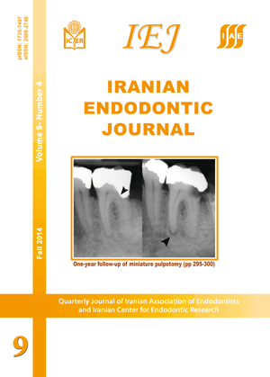
Outcomes of Different Vital Pulp Therapy Techniques on Symptomatic Permanent Teeth: A Case Series

Saeed Asgary, Mahta Fazlyab, Sedigheh Sabbagh, Mohammad Jafar Eghbal

Iranian Endodontic Journal, Vol. 9 No. 4 (2014), 1 October 2014, Page 295-300
https://doi.org/10.22037/iej.v9i4.7303

In modern endodontics, vital pulp therapy (VPT) has been considered an ultra-conservative treatment modality. Based on the level of pulp preservation, VPT includes stepwise excavation, indirect pulp capping (IDPC), direct pulp capping (DPC), miniature pulpotomy (MP), partial/Cvek pulpotomy and coronal/complete pulpotomy (CP). The present article reviews the treatment outcomes of 94 permanent teeth with irreversible pulpitis treated with either IDPC (n=28), DPC (n=28), MP (n=29) or CP (n=9) using calcium-enriched mixture (CEM) cement. After a mean follow-up time of 12.3 months, 93 treated teeth were radiographic/clinically successful; only one radiographic failure was observed in the DPC group.

Root Canal Treatment of a Two-Rooted C-Shaped Maxillary First Molar: A Case Report

Sara Paksefat, Saeed Rahimi

Iranian Endodontic Journal, Vol. 9 No. 4 (2014), 1 October 2014, Page 301-303
https://doi.org/10.22037/iej.v9i4.6292

The most difficult maxillary teeth for endodontic treatment are the maxillary first molars (MFM) due to their complex root canal anatomy. The presence of two roots and C-shaped canals in MFMs has been reported in rare cases. The present case reports root canal treatment of MFM with two roots, where the palatal and buccal roots were joined together in a C-shaped configuration.

Endodontic Treatment of a Double-Rooted Maxillary Second Molar with Four Canals: A Case Report

Hengameh Ashraf, Omid Dianat, Reihaneh Hajrezai, Payam Paymanpour, Sina Azadnia

Iranian Endodontic Journal, Vol. 9 No. 4 (2014), 1 October 2014, Page 304-306
https://doi.org/10.22037/iej.v9i4.6404

A healthy female was referred to Endodontic Department. The referral letter from her dentist expressed that an emergency pulpotomy of tooth #27 had been carried out with probable perforation of the chamber floor which was due to the unusual anatomy of the chamber. Cone-beam computed tomography (CBCT) revealed that the tooth had two mesial and two distal canals. Perforation site was repaired and endodontic treatment was completed. At 24-month follow-up, patient was asymptomatic and clinical and radiographic examinations showed successful outcomes.

Review Article


A Review of the Various Surface Treatments of NiTi Instruments

Zahed Mohammadi, Mohammad Karim Soltani, Sousan Shalavi, Saeed Asgary

Iranian Endodontic Journal, Vol. 9 No. 4 (2014), 1 October 2014, Page 235-240
https://doi.org/10.22037/iej.v9i4.5714

Since the introduction of engine-driven nickel-titanium (NiTi) instruments, attempts have been made to minimize or eliminate their inherent defects, increase their surface hardness/flexibility and also improve their resistance to cyclic fatigue and cutting efficiency. The various strategies of enhancing instrument surface include ion implantation, thermal nitridation, cryogenic treatment and electropolishing. The purpose of this paper was to review the metallurgy and crystal characteristics of NiTi alloy and to present a general over review of the published articles on surface treatment of NiTi endodontic instruments.