Original Article


Antibiotic prescription for endodontic treatment: General dentist knowledge + practice in Shiraz

Mohammad Reza Nabavizadeh, Safoora Sahebi, Ilnaz Nadian

Iranian Endodontic Journal, Vol. 6 No. 2 (2011), 16 March 2011, Page 54-59
https://doi.org/10.22037/iej.v6i2.2091

INTRODUCTION: Diseases of the dental pulp and periapical tissues are chiefly caused by microorganisms. Antibiotics are used in some endodontic cases; however, successful cases can predominantly be achieved by mechanical and chemical cleaning of the canal or surgical intervention.

MATERIALS & METHODS: The aim of this study was to determine the knowledge of General Dental Practitioners (GDPs) in Shiraz in respect to antibiotic prescriptions during and after endodontic treatment. A one-page questionnaire was sent to 200 active general dentists. Of the 120 surveys returned, 93 were accepted. The data were analyzed using t-test, Chi-square, ANOVA and Fisher’s Exact Test.

RESULTS: Only 29% of dentists had full knowledge (correct answers to all questions) of antibiotic prescription protocols in pulpal and periapical disease. Amoxicillin 500 mg capsule was the drug of choice of dentists. Total of 42% of GDPs had full knowledge of antibiotic prescription protocols for persistent or systemic infections cases. GDPs more recently qualified had slightly greater knowledge compared to GDPs with experience; however, this difference was not significant. Also, there was no significant difference between genders.

CONCLUSION: General practitioners’ knowledge about antibiotics seems inadequate and further education is recommended to update the practitioners.
The effect of blood on apical microleakage of Epiphany and AH26: An in vitro study

Zohreh Khalilak, Mehdi Vatanpour, Maryam Javidi, Moravrid Mafi, Farzaneh Afkhami, Farzaneh Daneshvar

Iranian Endodontic Journal, Vol. 6 No. 2 (2011), 16 March 2011, Page 60-64
https://doi.org/10.22037/iej.v6i2.2093

INTRODUCTION: Blood contamination of the canal during preparation can be a major problem in endodontics; this may result in apical microleakage. The aim of this in vitro study was to evaluate the effect of blood on apical microleakage of a resin-based root canal sealer (AH26) and a polymer-based root canal sealer (Epiphany).

MATERIALS & METHODS: In this experimental study, 50 decoronated central incisors and canine teeth were prepared by RaCe rotary system and randomly divided into 4 groups (n=10). Groups A1 and A2 were obturated by Epiphany/Resilon and AH26/Gutta-percha, respectively. The obturations were performed with a single cone technique after drying root canals. In B1 and B2 groups, the test groups, 0.02cc citrated human blood was injected into dried root canals and they were obturated in the same manner. Ten specimens were served as positive and negative controls (n=5).The apical leakage was measured by means of a computerized fluid filtration method after 1 day and 3 weeks. The data was analyzed by One-Sample Kolmogorov-Smirnov, Independent Sample t-test and univariate analysis. Statistical significances were preset at α=0.05.

RESULTS: There was no significant difference in apical microleakage of the two sealers after 1 day and 3 weeks in dry and blood environment (P>0.05). Sealer and environment had no interaction (P>0.05).

CONCLUSION: Blood contamination has no significant effect on the apical microleakage of Epiphny and AH26.

Comparison of the accuracy of NovApex apex locator and radiographic method: in vivo/ex vivo study

Shahriar Shahi, Saeed Rahimi, Amin Salem Milani, Mohammad Asghari Jafarabadi, Gholam Reza Djoorabbaf Naghsh

Iranian Endodontic Journal, Vol. 6 No. 2 (2011), 16 March 2011, Page 65-68
https://doi.org/10.22037/iej.v6i2.2072

INTRODUCTION: Determination of the canal working length with radiographs has many drawbacks. Electronic apex locators have been developed to overcome some of these problems. Recently, a newly designed apex locator called NovApex has been introduced. All the studies conducted to determine the accuracy of NovApex have been carried out in-vitro on extracted teeth. The aim of this in vivo study was to evaluate the accuracy of NovApex compared with traditional radiographic method.

MATERIALS & METHODS: Twenty-five patients whose lower molars that were candidates for extraction were selected. The teeth were accessed, and the radiographic working length was determined by measuring the length of the initial file 0.5mm short of the radiographic apex. Then, NovApex apex locator was used to measure the electronic working length. Subsequently, the tooth was extracted, and the actual working length was measured by introducing a size #15 K-file into each canal until the file tip was visible at the apex, then 1.5mm was subtracted to attain the working length. Cohen's Kappa was computed for each of the methods versus actual working length as a measure of reliability. The accuracies were compared using Chi-square test.

RESULTS: The accuracy of NovApex apex locator and radiographic method in detecting the apical end point within ±0.5mm was 74.7% and 68%, respectively; this was not significantly different (P<0.001).

CONCLUSION: The NovApex apex locator is useful in detecting the apical end point with the accuracy similar to radiographic method. However, neither technique is fully reliable in detecting the apical end point of the canal.

KEYWORDS: Electronic apex locator, NovApex, Radiographic Image Interpretation.


Efficacy of two rotary systems in removing gutta-percha and sealer from the root canal walls

Bahareh Dadresanfar, Payman Mehrvarzfar, Mohammad Ali Saghiri, Sedigh Ghafari, Zohreh Khalilak, Mehdi Vatanpour

Iranian Endodontic Journal, Vol. 6 No. 2 (2011), 16 March 2011, Page 69-73
https://doi.org/10.22037/iej.v6i2.2094

INTRODUCTION: The aim of this in vitro study was to compare the efficacy of two retreatment rotary systems in removal of gutta-percha (GP) and sealer from the root canal walls with and without use of solvent.

MATERIALS & METHODS: Sixty single-canalled distal roots of mandibular molars were prepared and root filled with gutta-percha and AH26. Each canal was randomly allocated to receive one of the retreatment techniques, Mtwo R or ProTaper. The groups were further divided into two subgroups: with or without the use of solvent. The cleanliness of canal walls was determined by stereomicroscope and scanning electron microscopy.

RESULTS: The results showed that Mtwo R without the use of solvent was more efficient in material removal compared to ProTaper D (P<0.05). Most remnants were found in the apical third of the canals (P<0.05).

CONCLUSION: Mtwo R seems to be an efficient rotary system for endodontic retreatment of root canal with GP.

An ex-vivo study on the shaping parameters of two nickel-titanium rotary systems compared with hand instruments

Maryam Ehsani, Samir Zahedpasha, Ali-Akbar Moghadamnia, Jaber Mirjani

Iranian Endodontic Journal, Vol. 6 No. 2 (2011), 16 March 2011, Page 74-79
https://doi.org/10.22037/iej.v6i2.1840

INTRODUCTION: Rotary nickel-titanium (NiTi) instruments are thought to allow shaping of narrow, curved root canals more efficiently and more effectively than stainless steel hand instruments. However, the continued search for even more effective and safer instruments has resulted in new rotary systems being introduced on a regular basis. The aim of this study was to compare shaping parameters of RaCe and Mtwo NiTi rotary files with stainless steel K-Flexofile hand instrument.

MATERIALS & METHODS: A total of 60 mandibular first molars with 20-40 degree of curvature in mesial root were divided in to three groups and each was prepared with one kind of instrument (RaCe, Mtwo, stainless steel K-Flexofile). Using pre and post-radiographs, canal curvature was measured, with the Schneider technique. Preparation time was recorded. For evaluating canal centering and transportation, the tooth was sectioned 3, 6 and 9 mm from the apex. Pre and post- preparation photographs were taken from mesiolingual canal. Data was statistically analyzed using One-way ANOVA and Chi-Square tests.

RESULTS: RaCe and Mtwo maintained canal curvature better than K-Flexofile (P<0.001). Mtwo prepared the canal in a shorter time (P<0.001).

CONCLUSION: Significant statistical difference was not found in the three canal sections between the various systems. RaCe resulted in significantly fewer canal aberrations and better centering ability.


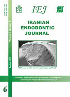
Cyclic fatigue resistance and fractographic analysis of RaCe and ProTaper rotary NiTi instruments

Shahram Azimi, Parisa Delvari, Hamid Cyrus Hajarian, Mohammad Ali Saghiri, Kasra Karamifar, Mehrdad Lotfi

Iranian Endodontic Journal, Vol. 6 No. 2 (2011), 16 March 2011, Page 80-85
https://doi.org/10.22037/iej.v6i2.2095

INTRODUCTION: This study investigated the fatigue and fracture modes of RaCe and ProTaper rotary instruments.

MATERIALS & METHODS: Fatigue resistance was evaluated by rotating the files 30° or 60° and with 2 or 5mm radius of curvature. RaCe taper 06 size 25 and ProTaper F1 files (n=40) were used. The number of rotations to failure was analyzed by two-way ANOVA and independent sample    t-test. Fracture surfaces were examined under a scanning electron microscope.

RESULTS: Both files exhibited significantly more resistance to fracture when angle severity was reduced and increase in curvature radius (P=0.000). ProTaper demonstrated higher number of cycle of failure (P=0.0029) in one group (r=5mm, 60º). SEM observation revealed fatigue mark/features in 17 specimens, shear characteristics in 37 samples, and tensile overload in 26 samples.

CONCLUSION: Radius of curvature was the main factor in torsional and fatigue failures.

Sealing ability promoted by three different endodontic sealers

Graziele Silva, Emmanuel João Nogueira Leal da Silva, Juliana Melo da Silva, Carlos Vieira Andrade-Júnior, Caio César Randi Ferraz

Iranian Endodontic Journal, Vol. 6 No. 2 (2011), 16 March 2011, Page 86-89
https://doi.org/10.22037/iej.v6i2.2083

Introduction: The aim of this study was to evaluate the apical and coronal sealing of endodontic fillings promoted by the associations of Sealer 26/Gutta-percha, Endofill/Gutta-percha and Resilon/ Epiphany. Materials and Methods: It was used 38 human canines, 10 teeth for each test group and 4 teeth for each control group. After teeth conventional endodontic treatment, they were immersed in saline solution for thirty days, sealed and stored in India ink for seven days. Then were submitted to a clearing technique and evaluated for infiltration by stereoscopic microscope. The data were statistically analyzed by ANOVA and Bonferroni tests. Results: The results showed no significant differences (p> 0.05) between the three groups of filling materials used. Conclusion: Considering the limitations of this in vitro study, it seems that no material showed superiority related to the ability of apical and coronary sealing.

Case Report


MTA APICAL PLUG AND CLINICAL APPLICATION OF ANATOMIC POST AND CORE FOR CORONAL RESTORATION: A CASE REPORT

RAHUL kumar, Suvarna Patil, Upendra Hoshing, Ashish Medha, Rushikesh Mahaparale

Iranian Endodontic Journal, Vol. 6 No. 2 (2011), 16 March 2011, Page 90-94
https://doi.org/10.22037/iej.v6i2.2087

ABSTRACT: Apexification with calcium hydroxide is associated with certain difficulties, such as longer treatment time, risk of tooth fracture and incomplete calcification of apical bridge. Mineral trioxide aggregate (MTA) is an alternative material that can be used for apexification of open apices due to its biocompatibility, non-mutagenicity, non-neurotoxicity, regenerative abilities, and good sealing properties. This case report demonstrates application of MTA apical plug and anatomic post and core for the reconstruction of maxillary central incisor. The patient was recalled after six months and no complications were noted. Periapical radiographs demonstrated good adaptation of anatomical post and core to post space and the complete healing of the periapical lesion. This new technique is particularly advantageous in teeth which have open apex, root canals that are not round, wide canals and thin radicular dentin. It is also useful for who request in patients fewer visits.

ABSTRACT: Inflammatory external root resorption (IERR) is a pathological phenomenon of microbial origin. This study reports a case of external apical inflammatory root resorption of maxillary incisors associated with periradicular lesions in a 22 year old female, which was successfully treated by one-visit root canal therapy (RCT). Radiographic investigation revealed periapical radiolucencies in the upper incisors associated with varying degrees of external inflammatory root resorption of teeth 12, 21 and 22. One-visit RCT of all involved teeth was carried out. Access cavities were permanently restored after 1 week. Clinical and radiographic examinations at 1 day, 1 week and 14 month follow-up demonstrated complete resolution of patient's signs/symptoms. The incisors were fully functional, and complete bone healing of the apical radiolucencies had taken place. The treatment outcomes demonstrated that IERR associated with periradicular lesions can respond successfully to one-visit RCT when conducted with adequate disinfection and a satisfactory coronal seal. Hence, one-visit RCT may be a good alternative to multiple-visit RCT involving intra-canal medicaments.