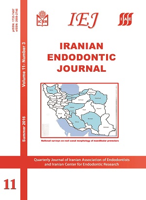
Systematic Review


Root Canal Morphology of Permanent Mandibular Premolars in Iranian Population: A Systematic Review

Sepanta Hosseinpour, Mohammad Javad Kharazifard, Akbar Khayat, Mandana Naseri

Iranian Endodontic Journal, Vol. 11 No. 3 (2016), 30 June 2016, Page 150-156
https://doi.org/10.22037/iej.v11i3.11333

Introduction: It is essential for clinicians to have knowledge about root canal configuration, although its morphology varies largely in different ethnicities and even in different individuals within the same ethnic group. The current study reviewed the root canal configuration of root canals in mandibular first and second premolars among Iranian population based on independent epidemiological studies. Methods and Materials: A comprehensive search was conducted on retrieved articles related to root canal configuration and prevalence of each types of root canal in mandibular premolars based on Vertucci’s classification. An electronic search was conducted in Medline, Scopus and Google Scholar from January 1984 to September 2015. Results: In eleven studies conducted in eight provinces, 1644 mandibular first premolars and 1268 second premolars were investigated. Within mandibular first premolars, 70.9% were Vertucci's type I, followed by 10.4% type III, 7.18% type IV, 5.23% type II and 5.16% type V. In addition, among mandibular second premolars, 82.86% were type I, 6.25 type III, 5.32% type II, 4.27% type IV, and 0.69% type V. Conclusion: These results highlight the necessity of searching for additional possible root canals by clinicians. Moreover, these results indicated the ethnical characteristics of Iranian population regarding the morphology of mandibular premolars compared to other populations.

Keywords: Anatomy; Iranian; Mandibular Premolar; Review; Root Canal
Original Article


Indexing of Iranian Publications in Well-known Endodontic Textbooks: A Scientometric Analysis

Sina Kakooei, Mahshid Mostafavi, Masoud Parirokh, Saeed Asgary

Iranian Endodontic Journal, Vol. 11 No. 3 (2016), 30 June 2016, Page 157-163
https://doi.org/10.22037/iej.v11i3.12632

Introduction: Quoting an article in well-known textbooks is held as a credit for that paper. The numbers of Iranian publications mentioned in endodontic textbooks have increased during recent years. The aim of this investigation was to evaluate the number of Iranian articles quoted in eminent endodontic text books. Methods and Materials: Three known textbooks (Ingle’s Endodontics, Seltzer and Bender’s Dental Pulp and Cohen’s Pathways of the Pulp) were chosen and all the editions of the textbooks since 2000 were investigated for quoted Iranian publications. Only Iranian authors with affiliations from a domestic university were chosen. All references at the end of each chapter were read by hand searching, and results were noted. The trend and percentage of Iranian publications in different editions of the textbooks were also calculated. The number of citations of these publications in Google Scholar and Scopus databases were also obtained. Results: The number of Iranian publications in all well-known textbooks have notably increased since 2000. The number and percentage of Iranian publications in the latest edition of Cohen’s Pathways of the Pulp was higher compared to other textbooks as well as the previous edition of the same text. Conclusion: Number and percentage of Iranian publications in the field of endodontics in all three textbooks have remarkably increased since 2000.

Keywords: Dental Pulp; Endodontics; Index; Ingle; Iranian Publications; Pathways of the Pulp; Quote; Scientometric; Textbooks

Working Length Determination Using Cone-Beam Computed Tomography, Periapical Radiography and Electronic Apex Locator in Teeth with Apical Periodontitis: A Clinical Study

André Luiz Gomide de Morais, Ana Helena Gonçalves de Alencar, Cyntia Rodrigues de Araújo Estrela, Daniel Almeida Decurcio, Carlos Estrela

Iranian Endodontic Journal, Vol. 11 No. 3 (2016), 30 June 2016, Page 164-168
https://doi.org/10.22037/iej.v11i3.5239

Introduction: The purpose of this clinical study was to compare the accuracy of working length (WL) determination using cone-beam computed tomography (CBCT), conventional periapical radiographies and electronic apex locator. Methods and Materials: This study was conducted during root canal treatment of 19 patients with a total of 30 single-rooted teeth diagnosed with apical periodontitis. After taking the initial parallel periapical radiographies, the initial file was advanced into the canal until the WL was detected by the apex locator. Subsequently, the WL was measured and WL radiographies were taken with the file set in the canal. Afterwards, CBCT images were acquired. These three measurements were tabulated and compared and the data were analyzed using the Friedman test. The level of significance was set at 0.05. Results: The mean values for WL determination by electronic apex locator, periapical radiograph and CBCT images were 22.25, 22.43 and 22.65, respectively which was not statistically significant (P>0.05). Conclusion: Working length determination using CBCT images was precise when compared to radiographic method and electronic apex locator.

Keywords: Apical Foramen; Cone-Beam Computed Tomography; Dental Radiography; Electronic Apex Locator; Tooth Apex

Antimicrobial Efficacy and Cytocompatibility of Calcium Hypochlorite Solution as a Root Canal Irrigant: An in Vitro Investigation

Mahdi Sedigh Shams, Ahmad Gholami, Abbas Abbaszadegan, Roohollah Yazdanparast, Milad Saberi Nejad, Azam Safari, Mohammadreza Nabavizadeh, Younes Ghasemi

Iranian Endodontic Journal, Vol. 11 No. 3 (2016), 30 June 2016, Page 169-174
https://doi.org/10.22037/iej.v11i3.9402

Introduction: The purpose of this study was to compare the antimicrobial efficacy of sodium hypochlorite (SH) and calcium hypochlorite (CH) against Enterococcus faecalis (E. faecalis) using quantitative real-time polymerase chain reaction (qPCR) analysis and also to compare their cytocompatibility on L929 murine fibroblasts using Mossman’s tetrazolium toxicity (MTT) assay. Methods and Materials: A broth micro-dilution susceptibility test was used to determine the minimum inhibitory concentration (MIC) of each irrigant against E. faecalis. Then, the root canals of 50 mature extracted human mandibular premolars were contaminated with E. faecalis and were randomly divided into three groups according to the irrigant used (n=20). Canals were irrigated with SH in group I (n=20) and CH in group II (n=20) at their obtained MIC. In group III (n=10), sterile saline was used. Microbial sampling was performed before and after biomechanical preparation. Quantitative PCR was used to quantify E. faecalis in the root canal samples. For cytocompatibility assessment, L929 murine fibroblasts were exposed to various concentrations of the irrigants. Results: Irrigation with test materials resulted in significant reduction in colony forming units (CFU) in post-instrumentation samples (with the MIC values of SH and CH against E. faecalis being 0.5% and 5%, respectively). However, the reduction in the normal saline group was not significant (P=0.203). In addition, 5% CH was more effective than 0.5% SH (P=0.006) in eliminating E. faecalis. Among the different concentrations of tested irrigants, 0.5% CH and 5% SH showed the least and the most cytotoxicity, respectively (P<0.001). The cytotoxicity of 5% CH and 0.5% SH was similar (P=0.99), and lower than 2.5% SH (P<0.001). Conclusion: CH at an MIC of 5% was effective in eliminating E. faecalis in planktonic state and also its biofilm and exhibited comparable cytocompatibility to that of 0.5% SH.

Keywords: Calcium Hypochlorite; Enterococcus faecalis; Polymerase Chain Reaction; Root Canal Irrigant; Sodium Hypochlorite

Comparison of Tooth Discoloration Induced by Calcium-Enriched Mixture and Mineral Trioxide Aggregate

Armita Rouhani, Majid Akbari, Aida Farhai-faz

Iranian Endodontic Journal, Vol. 11 No. 3 (2016), 30 June 2016, Page 175-178
https://doi.org/10.22037/iej.v11i3.10749

Introduction: The aim of this in vitro study was to evaluate the tooth discoloration induced by calcium-enriched mixture (CEM) cement and mineral trioxide aggregate (MTA). Methods and Materials: Forty five endodontically treated human maxillary central incisors were selected and divided into three groups (n=15) after removing the coronal 3 mm of the obturating materials. In the MTA group, white MTA plug was placed in pulp chamber and coronal zone of the root canal. In CEM cement group, CEM plug was placed in the tooth in the same manner. In both groups, a wet cotton pellet was placed in the access cavity and the teeth were temporarily sealed. After 24 h the teeth were restored with resin composite. In the negative control group the teeth were also restored with resin composite. The color change in the cervical third of teeth was measured with a colorimeter and was repeated 3 times for each specimen. The teeth were kept in artificial saliva for 6 months. After this period, the color change was measured again. Data were collected by Commission International de I'Eclairage's L*a*b color values, and corresponding ΔE values were calculated. The results were analyzed using the one-way ANOVA and post-hoc Tukey’s test with the significance level defined as 0.05. Results: There was no significant differences between CEM group and control group in mean discoloration. The mean tooth discoloration in MTA group was significantly greater than CEM and control groups (P<0.05). Conclusion: According to the result of the present study CEM cement did not induce tooth discoloration after six months. Therefore it can be used in vital pulp therapy of esthetically sensitive teeth.

Keywords: Calcium-Enriched Mixture; Dental Cements; Mineral Trioxide Aggregate; Tooth Discoloration; Vital Pulp Therapy

The Antibacterial Efficacy of Photo-Activated Disinfection, Chlorhexidine and Sodium Hypochlorite in Infected Root Canals: An in Vitro Study

Mohammad Samiei, Shahriar Shahi, Ami Ardalan Abdollahi, Mahsa Eskandarinezhad, Ramin Negahdari, Zahra Pakseresht

Iranian Endodontic Journal, Vol. 11 No. 3 (2016), 30 June 2016, Page 179-183
https://doi.org/10.22037/iej.v11i3.11175

Introduction: This study compared the efficacy of light-activated low-power laser, 2% chlorhexidine (CHX) and 2.5% NaOCl in eliminating Enterococcus faecalis (E. faecalis) from the root canal system. Methods and Materials: The root canals of 60 maxillary central incisors were contaminated with E. faecalis and then the bacteria were incubated for 24 h. All the root canals were instrumented in a crown-down manner with #4 and 3 Gates-Glidden drills, followed by RaCe rotary files (40/0.10, 35/0.08, and 30/0.06). The samples were randomly assigned to three experimental groups and one control group (n=15). In the control group no intervention was made. In the photo-activated disinfection (PAD) group, laser therapy was undertaken with diode laser beams (with an output power of 100 mW/cm2) for 120 sec. For the other two experimental groups, root canals were irrigated either with 5 mL of 2% CHX or 2.5% NaOCl solutions, respectively. The Kruskal-Wallis test was used to compare the CFU values of the bacteria and post-hoc Bonferroni test was used for pairwise comparisons. The level of significance was set at 0.05. Results: The inhibition of bacterial growth in all the experimental groups was significantly superior to the control group (P<0.05). There was no significant difference between the effect of PAD and 2% CHX (P=0.05). The effect of 2.5% NaOCl was significantly better than that of the PAD technique (P<0.001). In addition, 2.5% NaOCl was significantly better than 2% CHX (P=0.007). Conclusion: Photodynamic therapy was effective in reducing the E. faecalis counts in comparison with the control group, but 2.5% NaOCl solution was the most effective protocol.

Keywords: Enterococcus faecalis; Laser; Photo-Activated Disinfection; Photodynamic Therapy; Sodium Hypochlorite

Efficacy of ProTaper and Mtwo Retreatment Files in Removal of Gutta-percha and GuttaFlow from Root Canals

Sedigheh Khedmat, Abbas Azari, Ahmad Reza Shamshiri, Mehdi Fadae, Hoorieh Bashizadeh Fakhar

Iranian Endodontic Journal, Vol. 11 No. 3 (2016), 30 June 2016, Page 184-187
https://doi.org/10.22037/iej.v11i3.10918

Introduction: The aim of this in vitro study was to compare the efficacy of ProTaper retreatment (ProTaper R) and Mtwo retreatment (Mtwo R) files in removing gutta-percha and GuttaFlow from endodontically treated straight root canals. Methods and Materials: The root canals of 60 human mandibular single-rooted premolars were prepared and randomly divided into two groups (n=30). In groups A and B the root canals were obturated using lateral condensation of gutta-percha plus AH 26 and GuttaFlow, respectively. The canal orifices were temporarily sealed and the roots were incubated for 3 months at 37ºC and 100% humidity. Primary cone-beam computed tomography (CBCT) images were taken after incubation period. The specimens in each group were randomly divided into two subgroups (n=15). ProTaper R files (D1, D2, and D3) were used in groups A1 and B1 while Mtwo R files (25/0.05 and 15/0.05) were used in groups A2 and B2. The time required to extirpate the root filling was also recorded. After retreatment, another CBCT scan was taken at the same position. The volume of remaining filling materials inside the canals was calculated before and after retreatment. The data was analyzed using the two-way ANOVA and independent t-test. Results: The remaining filling materials in the canals treated with ProTaper were less than Mtwo. The remaining volume of GuttaFlow was less than gutta-percha regardless of the system applied. Mtwo R files removed root fillings faster than ProTaper R. Conclusion: ProTaper R removed filling material more efficiently compared to Mtwo R which required less time to remove root filling material.

Keywords: Gutta-Percha; Mtwo Retreatment Files; ProTaper Retreatment Files; Root Canal Retreatment

Effect of Citric Acid and Ethylenediaminetetraacetic Acid on the Surface Morphology of Young and Old Root Dentin

Miriam Zaccaro Scelza, Fernando de Noronha, Licinio Esmeraldo da Silva, Marcos Maurício, Marco Antonio Gallito, Pantaleo Scelza

Iranian Endodontic Journal, Vol. 11 No. 3 (2016), 30 June 2016, Page 188-191
https://doi.org/10.22037/iej.v11i3.10284

Introduction: The aim of this in vitro study was to evaluate the effect of 10% citric acid and 17% ethylenediaminetetraacetic acid (EDTA) irrigating solutions on the surface morphology of young and old root dentin by determining the number and diameter of dentinal tubules using scanning electron microscopy (SEM). Methods and Materials: Fifty healthy human teeth collected from young (≤30 years) and old (≥60 years) individuals (n=25) were first prepared with a Largo bur #2 to produce smear layer on the root canal surface. Subsequently, the crowns and the root middle and apical thirds were sectioned and removed, and the cervical thirds were sectioned vertically in the buccal-lingual direction into two equal halves. The obtained samples were then immersed in 2.5% sodium hypochlorite for 30 min and randomly separated into two treatment groups for each age group. In each age group, ten samples were selected as controls and did not receive any type of treatment. The rest of the specimens were then rinsed, dried and treated for 4 min with 10% citric acid or 17% EDTA. The samples were then assessed with SEM regarding the number and diameter of dentinal tubules. All data were assessed using Student’s t-test. The level of significance was set at 0.05. Results: Regardless of the type of treatment, no significant differences were observed in the number of open tubules between the young and old root dentin (P>0.05). Nonetheless, the diameter of the tubules in the old root dentin was larger when 17% EDTA was used (P<0.05). Both, young and old root dentin did not differ with the 10% citric acid treatment (P>0.05). Conclusion: The results showed that 17% EDTA treatment induced a significant demineralization in old root dentin.

Keywords: Citric Acid; Dentinal Tubule; Ethylenediaminetetraacetic Acid; Scanning Electron Microscopy; Surface Morphology

Antibacterial Activity of MTA Fillapex and AH 26 Root Canal Sealers at Different Time Intervals

Farnaz Jafari, Hossein Samadi Kafil, Sanaz Jafari, Mohammad Aghazadeh, Tahereh Momeni

Iranian Endodontic Journal, Vol. 11 No. 3 (2016), 30 June 2016, Page 192-197
https://doi.org/10.22037/iej.v11i3.11465

Introduction: The main goal of endodontic treatment is elimination of bacteria and their by-products from infected root canals. This study compared the antibacterial effect of two different sealers, AH 26 and MTA Fillapex, on 4 microorganisms 24, 48 and 72 h and 7 days after mixing. Methods and Materials: The microorganisms used in this study consisted of Lactobacillus acidophilus (ATCC 4356), Lactobacillus casei (ATCC 39392), Staphylococcus aureus (ATCC 25923) and Enterococcus faecalis (ATCC 29212). This test is based on the growth of bacteria and turbidity measurement technique using a spectrophotometer, and direct contact was conducted. Multiple comparisons were carried out using repeated-measures ANOVA followed by Tukey’s test and student’s t-test. The level of significance was set at 0.05. Results: The antibacterial activity in the indirect technique was more than the technique with both sealers. In the direct technique the antibacterial activity on all microorganisms were higher for MTA Fillapex sealer. In the indirect technique, both sealers exhibited similar antibacterial properties. Conclusion: The antibacterial effect of MTA Fillapex sealer was significantly more than that of AH 26 sealer in the direct technique. The antibacterial effects of both sealers were similar in the indirect technique.

Keywords: Antibacterial; Enterococcus faecalis; Lactobacillus; MTA Fillapex; Sealers; Staphylococcus

Comparison of Microleakage of MTA and CEM Cement Apical Plugs in Three Different Media

Fatemeh Ayatollahi, Mahdi Tabrizizadeh, Milad Hazeri Baqdad Abad, Reza Ayatollahi, Fatemeh Zare bidoki

Iranian Endodontic Journal, Vol. 11 No. 3 (2016), 30 June 2016, Page 198-201
https://doi.org/10.22037/iej.v11i3.11297

Introduction: Microleakage is of the causes of endodontic treatment failure. The aim of this in vitro study, was to compare the microleakage of mineral trioxide aggregate (MTA) and calcium-enriched mixture (CEM) cement apical plugs in three environments. Methods and Materials: A total of 130 human extracted single rooted teeth were collected. After decoronation, preparation of the root canal space and simulation of open apices, 5 teeth were selected as positive and negative control groups and the rest of the samples were randomly divided into two groups (MTA plug and CEM cement plug) and each group was divided into 3 subgroups (dry, contaminated with saliva and contaminated with blood). In each group apical plug was placed into the canal. After full setting of the apical plug, microleakage of the samples were evaluated using fluid filtration method and the data were analyzed using two-way analysis of variance. Results: In dry and saliva contaminated environments, the leakage of MTA samples were 40.906±2.081 and 39.608±2.081, respectively which was significantly more than that of CEM cement samples (26.977±2.081 and 27.000±2.081, respectively). However, in blood-contaminated environments, the amount of leakage in MTA group (21.640±2.081) was significantly lower than CEM cement group (44.358±2.081). Conclusion: According to the results of this study, in dry and saliva-contaminated conditions CEM cement provides significantly better seal in comparison to MTA.

Keywords: Calcium-Enriched Mixture Cement; Microleakage; Mineral Trioxide Aggregate

Effect of Amperage and Field of View on Detection of Vertical Root Fracture in Teeth with Intracanal Posts

Yaser Safi, Sepanta Hosseinpour, Alireza Aziz, Masoud Bamedi, Mahsa Malekashtari, Zahra Vasegh

Iranian Endodontic Journal, Vol. 11 No. 3 (2016), 30 June 2016, Page 202-207
https://doi.org/10.22037/iej.v11i3.10529

Introduction: This study sought to assess the effects of amperage (mA) and field of view (FOV) on intracanal metal post artifacts and the diagnostic parameters for detection of vertical root fracture (VRF) in teeth with intracanal posts. Methods and Materials: In this diagnostic study, 80 human single-canal teeth were evaluated by cone-beam computed tomography (CBCT). Nickel chrome cast posts were placed into root canals after root canal therapy and canal preparation. In the test group, fracture was induced by an instron machine while no fracture was induced in the control group. Deterministic and probabilistic sensitivity and specificity values at different exposure settings were statistically analyzed using the one-way ANOVA and pairwise comparisons were performed by Tukey’s test. Results: Significant differences were found between the two groups in terms of deterministic (P<0.0001) and probabilistic (P<0.013) sensitivity and deterministic (P<0.037) and probabilistic (P<0.0001) specificity at different FOV and mA combinations. Conclusion: A smaller FOV and lower mA should be preferably used for detection of VRFs in teeth with intracanal posts.

Keywords: Amperage; Cone-Beam Computed Tomography; Field of View; Metal Artifact; Vertical Root Fracture

A Clinical Update on the Different Methods to Decrease the Occurrence of Missed Root Canals

Zahed Mohammadi, Saeed Asgary, Sousan Shalavi, Paul V. Abbott

Iranian Endodontic Journal, Vol. 11 No. 3 (2016), 30 June 2016, Page 208-213
https://doi.org/10.22037/iej.v11i3.11451

One of the main causes of endodontic treatment failure is the clinician’s inability to localize all the root canals. Due to the complex anatomy of the root canal system, missed canals are not uncommon. There are several strategies to decrease the possibility of missed root canals starting with good pre-operative radiographies. In order to overcome the limitations of conventional radiographies, cone-beam computed tomography (CBCT) can be considered. A correct access cavity preparation is of pivotal importance in localizing the orifices of the root canals. Furthermore, ultrasonics are very important devices to find missed canals. Increasing magnification and illumination enhance the possibility of finding all root canals during root canal treatment. The purpose of the present paper was to review all of the above techniques and devices.

Keywords: Access Cavity; Cone-Beam Computed Tomography; Microscope; Missed Canals; Radiography; Root Canal Morphology; Transillumination; Ultrasonic


Introduction: Adequate seal of iatrogenically perforated area within the root canal system can improve the long term treatment prognosis. This in vitro study evaluated the sealing ability of mineral trioxide aggregate (MTA), calcium-enriched mixture (CEM) cement and Biodentine in repair of furcation perforation in primary molars. Methods and Materials: A total of 61 freshly extracted primary mandibular second molars were randomly divided into three groups (n=17) and 10 teeth were put in negative (without perforation, n=5) and positive (perforated without repair, n=5) control groups. Turbidity was used as the criteria of bacterial leakage, when detected in the model of dual-chamber leakage. Data were analyzed using the Chi-Square and Kaplan-Meier survival analysis in SPSS software. The level of significance was set at 0.05. Results: All positive samples showed turbidity, whereas none of the negative samples allowed bacterial leakage. There was no significant difference between the number of turbidity samples in repaired teeth with all test materials (P=0.13). No significant difference was also detected in the mean survival time (P>0.05). Conclusion: CEM cement and Biodentine showed promising results as perforation repair materials and can be recommended as suitable alternatives of MTA for repair of furcation perforation of primary molars.

Keywords: Biodentine; Biomaterial; Calcium-Enriched Mixture; Furcation Perforation; Mineral Trioxide Aggregate; Perforation Repair; Sealability

Efficacy of a Novel Rotary System in Reduction of Intracanal Bacteria: An in Vitro Study

Maryam Vossoghi, Mitra Vossoghi, Shahriar Shahriari, Farhad Faramarzi, Rasoul Yousefi Mashouf, Maryam Farhadian

Iranian Endodontic Journal, Vol. 11 No. 3 (2016), 30 June 2016, Page 219-222
https://doi.org/10.22037/iej.v11i3.8030

Introduction: This in vitro study aimed to assess the effectiveness of a single-file rotary system (OneShape) in reduction of intracanal bacteria. Methods and Materials: Eighty one single-rooted mandibular first premolars with single canals were used. Six samples were selected as aseptic control group. Seventy five remaining specimens were infected by Enterococcus faecalis and incubated for 72 h at 37°C. Then they were randomly divided into three groups (n=25). In each group, cleaning and shaping procedures were done using either two conventional rotary systems (ProTaper and iRace), or the single-file system (OneShape). Microbial samples from the intracanal environment were taken by paper points in two steps, before and after instrumentation. Then, they were diluted and plated in blood agar. In order to compare bacterial reduction and turbidity, the Kruskal-Wallis test was used followed by the Chi-Square and Mann-Whitney tests for pairwise comparison. The level of significance was set at 0.05. Results: The control group didn’t show any bacterial growth. The pre- and post-instrumentation samples were significantly different between three groups (P=0.02). Hence, there was no significant differences between turbidity of samples (P>0.05). Conclusion: OneShape system is efficient in bacterial reduction. In this regard ProTaper is the most effective system in intracanal bacterial reduction followed by iRaCe and OneShape, respectively.

Keywords: Enterococcus faecalis; iRace; OneShape; ProTaper

In Vitro Evaluation of Different Solvents for Retrieval of Mineral Trioxide Aggregate and Calcium-Enriched Mixture

Nooshin Sadat Shojaee, Alireza Adl, Fereshteh Sobhnamayan, Azam Khademi, Mina Hamedi

Iranian Endodontic Journal, Vol. 11 No. 3 (2016), 30 June 2016, Page 223-227
https://doi.org/10.22037/iej.v11i3.11744

Introduction: The purpose of this study was to evaluate the effect of different solvents; carbonic acid (H2CO3), hydrochloric acid (HCl), chlorhexidine (CHX) and sodium hypochlorite (NaOCl) on the surface hardness of mineral trioxide aggregate (MTA) and calcium-enriched mixture (CEM) cement. Methods and Materials: Plexiglass molds were prepared and filled with Angelus MTA or CEM cement and then exposed to 2% carbonic acid, 37% hydrochloric acid, 2% chlorhexidine, 5.25% sodium hypochlorite and normal saline at intervals of 1 and 21 days, respectively (n=4). Surface microhardness of all specimens was analyzed by a universal testing machine and an electron microscope for some selected samples. Data were analyzed using the three-way ANOVA. Subgroup analysis was performed by Student’s t-test, One-way ANOVA and Tukey’s tests. The level of significance was set at 0.05. Results: On the first day, all solvents and on 21st day HCl, and H2CO3 were more effective in reducing the microhardness of MTA compared to CEM cement (P<0.05). Conclusion: The two experimental cements were differently affected by the solvents at specific time intervals. The solvents were more effective on MTA.

Keywords: Calcium-Enriched Mixture; Microhardness; Mineral Trioxide Aggregate; Retrieval

Shaping Ability of Single-File Systems with Different Movements: A Micro-Computed Tomographic Study

Joedy Santa-Rosa, Manoel Damião de Sousa-Neto, Marco Aurelio Versiani, Giselle Nevares, Felipe Xavier, Kaline Romeiro, Marcely Cassimiro, Graziela Bianchi Leoni, Rebeca Ferraz de Menezes, Diana Albuquerque

Iranian Endodontic Journal, Vol. 11 No. 3 (2016), 30 June 2016, Page 228-233
https://doi.org/10.22037/iej.v11i3.8890

Introduction: This study aimed to perform a rigorous sample standardization and also evaluate the preparation of mesiobuccal (MB) root canals of maxillary molars with severe curvatures using two single-file engine-driven systems (WaveOne with reciprocating motion and OneShape with rotary movement), using micro-computed tomography (micro-CT). Methods and Materials: Ten MB roots with single canals were included, uniformly distributed into two groups (n=5). The samples were prepared with a WaveOne or OneShape files. The shaping ability and amount of canal transportation were assessed by a comparison of the pre- and post-instrumentation micro-CT scans. The Kolmogorov-Smirnov and t-tests were used for statistical analysis. The level of significance was set at 0.05. Results: Instrumentation of canals increased their surface area and volume. Canal transportation occurred in coronal, middle and apical thirds and no statistical difference was observed between the two systems (P>0.05). In apical third, significant differences were found between groups in canal roundness (in 3 mm level) and perimeter (in 3 and 4 mm levels) (P<0.05). Conclusion: The WaveOne and One Shape single-file systems were able to shape curved root canals, producing minor changes in the canal curvature.

Keywords: Micro-Computed Tomography; OneShape; Reciprocating Motion; Rotary System; WaveOne

Case Report


Root Canal Treatment of a Maxillary Second Premolar with Two Palatal Root Canals: A Case Report

Maryam Golmohammadi, Hamid Jafarzadeh

Iranian Endodontic Journal, Vol. 11 No. 3 (2016), 30 June 2016, Page 234-236
https://doi.org/10.22037/iej.v11i3.9543

Accurate diagnosis of the root canal morphology and anatomy is essential for thorough shaping and cleaning of the entire root canal system and consequent successful treatment. This report describes a case of maxillary second premolar with two roots and three root canals (two mesial and distal palatal canals). The case report underlines the importance of complete knowledge about root canal morphology and possible variations, coupled with clinical and radiographic examination in order to increase the ability of clinicians to treat difficult cases.

Keywords: Anatomical Variations; Maxillary Second Premolar; Root Canal Therapy

Removal of a Broken Instrument from a Tooth with Apical Periodontitis Using a Novel Approach

Azar Heydari, Mona Rahmani, Mostafa Heydari

Iranian Endodontic Journal, Vol. 11 No. 3 (2016), 30 June 2016, Page 237-240
https://doi.org/10.22037/iej.v11i3.11178

Instrument breakage during treatment can lead to serious complications and carries the risk of treatment failure. When a file breaks in the canal, bypassing or removal can be difficult and the long-term prognosis of the tooth may be compromised. Sometimes surgery may be indicated for removal of the broken segment. Often some part of the root cannot be cleaned because of blockage by the broken file. This report presents a specific approach in non-surgical removal of a broken file from a maxillary lateral incisor with a buccal sinus tract and a broken instrument in the apical third which was partially over extended into the periapical lesion. The broken file was accessed through the sinus tract and pushed into the canal. The canal was cleaned and shaped, filled with mineral trioxide aggregate (MTA). Conclusion: A sinus tract can be a specific path to reach the root tip and get access to remove the foreign materials pushed beyond the root canal space.

Keywords: Apical Surgery; Broken Instrument; Nonsurgical Retreatment

Conservative Management of Unset Mineral Trioxide Aggregate Root-End Filling: A Case Report

Masoud Parirokh, Sedigheh Farzaneh, Ali Reza Halajmonfared

Iranian Endodontic Journal, Vol. 11 No. 3 (2016), 30 June 2016, Page 241-245
https://doi.org/10.22037/iej.v11i3.12309

This case report presents conservative management of unset mineral trioxide aggregate (MTA) after being placed as a root-end filling material following periapical surgery. Periapical surgery was indicated for a maxillary lateral incisor of a 15-year-old male due to persistent exudate and a large periapical lesion. During surgery Angelus MTA was placed as root-end filling. The next session it was noticed that MTA had failed to completely set. In an orthograde approach, calcium-enriched mixture (CEM) cement was used to obturate the root canal space. The patient was followed up for 27 months and did not exhibit any clinical signs and symptoms. Radiographic images showed complete healing of the lesion.

Keywords: Calcium-Enriched Mixture; Calcium Silicate; CEM Cement; Dens Invaginatus; Mineral Trioxide Aggregate

Single-Visit Pulp Revascularization of a Nonvital Immature Permanent Tooth Using Biodentine

Mohammad Mhd nader Aldakak, Ismail Davut Capar, Mohammad Salem Rekab, Souad Abboud

Iranian Endodontic Journal, Vol. 11 No. 3 (2016), 30 June 2016, Page 246-249
https://doi.org/10.22037/iej.v11i3.11598

An 11-year-old female patient was referred with a chief complaint of pain in the right mandibular second premolar. Clinical and radiographic examinations showed secondary caries under an old composite restoration, a negative response to a pulp test and an immature root with an open apex. After root canal cleaning and shaping, bleeding was invoked in the canal up to 2 mm short of the cemento-enamel junction (CEJ). After 10 min to allow clotting at this level, a plug of Biodentine was placed over the blood clot and the tooth was temporized with glass ionomer cement. At the next visit, the tooth was free of symptoms and a permanent filling was placed. Clinical and radiographic examinations during a two-year follow-up showed complete root maturation and intact supporting soft tissues without sinus tract, pain or swelling. Conclusion: The use of Biodentine in a single-visit apexification protocol to treat an immature permanent tooth with necrotic pulp can create a suitable environment for revascularization, resulting in the completion of root maturation.

Key Words:Biodentine; Immature Apex; Pulp Revascularization